Tag: 3/10/22

A Clue to Breast Cancer Survivors’ Cognitive Problems

Woman receiving mammogram
Source: National Cancer Institute

Why breast cancer survivors experience troubling cognitive problems is a long-standing mystery, and for which inflammation is one possible culprit. A new long-term study of older breast cancer survivors published in the Journal of Clinical Oncology adds evidence to this link.

Higher levels of the inflammatory marker C-reactive protein (CRP) were related to older breast cancer survivors reporting cognitive problems in the new study.

“Blood tests for CRP are used routinely in the clinic to determine risk of heart disease. Our study suggests this common test for inflammation might also be an indicator of risk for cognitive problems reported by breast cancer survivors,” said study lead author Judith Carroll, an associate professor at UCLA. 

The Thinking and Living with Cancer (TLC) Study is one of the first long-term efforts to examine the potential link between chronic inflammation and cognition in breast cancer survivors 60 and older, who make up a majority of the nearly 4 million breast cancer survivors in the United States. Previous research has focused largely on younger women and women immediately after therapy, making it difficult to draw conclusions about CRP’s role in long-term cognitive problems among older breast cancer survivors.   

In TLC, teams of researchers from around the country talked to, and obtained blood samples from, hundreds of breast cancer survivors and women without cancer up to six times over the course of five years. The study was motivated by hearing from survivors and advocates that cognitive problems are one of their major worries. 

“Cognitive issues affect women’s daily lives years after completing treatment, and their reports of their own ability to complete tasks and remember things was the strongest indicator of problems in this study,” said co-senior study author Dr Jeanne Mandelblatt, a professor of oncology at Georgetown University who is the lead of the TLC study.   

“Being able to test for levels of inflammation at the same time that cognition was being rigorously evaluated gave the TLC team a potential window into the biology underlying cognitive concerns,” said Elizabeth C. Breen, a professor emerita of psychiatry and biobehavioral sciences at the Cousins Center for Psychoneuroimmunology at UCLA, who also served as co-senior study author. 

The women’s cognition was evaluated through a commonly used questionnaire. The study found higher CRP levels among survivors were predictive of lower reported cognitive function among breast cancer survivors. There was no similar relationship between CRP levels and reported cognition in the women without cancer. 

Cognitive performance, as measured by standardised neuropsychological tests, failed to show a link between CRP and cognition. The authors say this may indicate women are more sensitive to differences in their everyday cognitive function, self-reporting changes that other tests miss.

The authors said their study supports the need for research on whether interventions that can lower inflammation – including increased physical activity, better sleep, and anti-inflammatory medications – may prevent or reduce cognitive concerns in older breast cancer survivors. 

Source: EurekAlert!

‘SuperAger’ Brains Remain Free of Alzheimer’s Signs

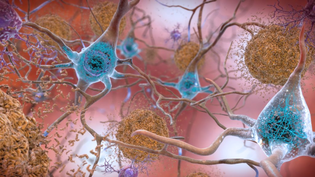
Plaques and neurons. Source: NIAH

According to a study which was published in The Journal of Neuroscience, neurons in the entorhinal cortex (a brain area responsible for memory) were significantly larger and healthier in 80+ year olds who have exceptional memory, also known as ‘SuperAgers’.

Their neurons were larger than those of cognitively average peers, individuals with early-stage Alzheimer’s disease and even those decades younger than SuperAgers. These neurons also did not harbour tau tangles, a hallmark of Alzheimer’s disease.

“The remarkable observation that SuperAgers showed larger neurons than their younger peers may imply that large cells were present from birth and are maintained structurally throughout their lives,” said lead author Tamar Gefen, an assistant professor at Northwestern University Feinberg School of Medicine. “We conclude that larger neurons are a biological signature of the SuperAging trajectory.”

The study of SuperAgers with exceptional memory was the first to show that these individuals carry a unique biological signature that comprises larger and healthier neurons in the entorhinal cortex that are relatively clear of tau tangles.

The Northwestern SuperAging Research Program studies unique individuals known as SuperAgers, 80+ year-olds who show exceptional memory at least as good as individuals 20 to 30 years their junior.

“To understand how and why people may be resistant to developing Alzheimer’s disease, it is important to closely investigate the postmortem brains of SuperAgers,” A/Prof Gefen said. “What makes SuperAgers’ brains unique? How can we harness their biologic traits to help elderly stave off Alzheimer’s disease?”

Scientists studied the entorhinal cortex of the brain because it controls memory and is one of the first locations targeted by Alzheimer’s disease. The entorhinal cortex comprises six layers of neurons. Layer II, in particular, receives information from other memory centres and is a very specific and crucial hub along the brain’s memory circuit.

In the study, scientists show that SuperAgers have large, healthier neurons in layer II of the entorhinal cortex compared to their same-aged peers, individuals with early stages of Alzheimer’s disease and even individuals 20 to 30 years younger. They also showed that these large layer II neurons were spared from the formation of tau tangles.

These findings together suggest that a neuron spared from tangle formation can maintain its structural integrity, and the inverse is true: Tau tangles can lead to neuronal shrinkage.

Participants in the SuperAger study donate their brains for research.

For the study, scientists examined the brains of six SuperAgers, seven cognitively average elderly individuals, six young individuals and five individuals with early stages of Alzheimer’s. Then they measured the size of neurons in layer II of the entorhinal cortex (compared to layers III and V). They also measured the presence of tau tangles in these cases.

For reasons that remain unknown, cell populations in the entorhinal cortex are selectively vulnerable to tau tangle formation during normal aging and in early stages of Alzheimer’s.

“In this study, we show that in Alzheimer’s, neuronal shrinkage (atrophy) in the entorhinal cortex appears to be a characteristic marker of the disease,” Gefen said.

“We suspect this process is a function of tau tangle formation in the affected cells leading to poor memory abilities in older age,” A/Prof Gefen said. “Identifying this contributing factor (and every contributing factor) is crucial to the early identification of Alzheimer’s, monitoring its course and guiding treatment.”

Future studies are needed to understand how and why neuronal integrity is preserved in SuperAgers. A/Prof Gefen wants to focus on probing the cellular environment.

“What are the chemical, metabolic or genetic features of these cells that render them resilient?” she asked. She also plans to investigate other hubs along the memory circuit of the brain to better understand the spread of or resistance to disease.

Source: Northwestern University

Nanoparticles Amplify Benefits of Magnetic Stroke Treatment

Chinese researchers have reported in Materials Today Chemistry that magnetic brain stimulation combined with a nasal spray containing nanoparticles can improve recovery in a mouse model of ischaemic stroke.

The nasal spray non-invasively delivers magnetic nanoparticles into the brain, which serve to amplify the benefits of transcranial magnetic stimulation (TMS). TMS is a method of non-invasive brain stimulation already used clinically or in clinical trials to treat neurological conditions like stroke, Parkinson’s disease, Alzheimer’s disease, depression, and addiction.

Rats that were given combined nanoparticle and TMS treatment every 24 hours for 14 days after an ischaemic stroke had better overall health, put on weight more quickly and had improved cognitive and motor functions compared to those treated with TMS alone. TMS stimulation uses a magnetic field to induces an electrical current in the brain, but is often not able to penetrate deeply enough.

In this new study, the researchers show that magnetic nanoparticles, administered intranasally, can make neurons more responsive and amplify the magnetic signal from TMS to reach deeper brain tissue, aiding recovery. The finding offers new opportunities for treating neurological disorders. 

From impossible to possible

The research answers a key question in nanomedicine – whether it is possible to enhance TMS by using nanoparticles that are non-invasively delivered into the brain. Experts had believed that it was almost impossible because of the blood-brain barrier.

However, the team of researchers overcame this by guiding the magnetic nanoparticles closer to the correct area with a large magnet near the head. 

Dr Gang Ruan, a corresponding author of the study, said: “We were able to overcome the blood-brain barrier and send enough nanoparticles into the brain to use in combination with TMS simulation to improve recovery from stroke. 

“TMS devices are already used for the clinical treatment of neurological disorders but have severe limitations in terms of stimulation strength and depths of the brain they can penetrate. 

“By non-invasively putting magnetic nanoparticles into the brain, we can amplify and enhance the TMS stimulation effects on neurons, making the treatment more effective,” Dr Ruan added.

“Showing it is possible to use nanoparticles in this way paves the way for medical applications of nanoparticles for other neurological disorders.”

Crossing barriers 

The iron oxide nanoparticles, normally used to treat anaemia, were coated various non-toxic substances. 

Dr Ruan explained: “The coating causes the nanoparticles to stick to the blood-brain barrier, increasing their chances of passing through it. Without this coating, the particles just bounce back from the barrier instead of crossing it.

“The modifications of the iron oxide particles also ensure that the nanoparticles can stick to the neurons and increase their responsiveness to TMS stimulation.”

The safety of using the modified nanoparticles needs to be assessed in clinical trials but has the potential to be used in combination with TMS, and other methods such as brain imaging, to gain more insight into how the brain works and improve the treatment of neurological disorders. 

“Many scientists still think it is impossible to non-invasively send enough nanoparticles into the brain to affect brain function. Yet we have shown that it is possible,” said Dr Ruan.

“We combined the expertise on our team in four different disciplines, materials science, biophysics, neuroscience, and medical science, to push the boundaries of our knowledge and challenge what is currently thought in the field.”

Source: EurekAlert!

An Easy-to-use Model for Assessing Hysterectomy Complication Risk

Photo by Piron Guillaume on Unsplash

Researchers have developed easy-to-use online prediction tools that provide personalised risk estimates for patients undergoing hysterectomy for benign disease. The models are described in a study published in the Canadian Medical Association Journal.

Hysterectomy is one of the most common surgical procedures, with one-third of women in Canada undergoing this procedure before age 60. Laparoscopic hysterectomies are more common as they are less invasive than abdominal surgery. Current practice entails that surgeons discuss benefits of the type of procedure and risks of complications with patients.

The researchers developed and tested prediction models with the aim of supplementing a surgeon’s expert opinion on patients’ risks of complications from hysterectomy. Hysterectomy complications can include ureteric, gastrointestinal and vascular injury as well as wound complications. The authors used data from the English National Health Service (NHS) on 68 599 women who had laparoscopic hysterectomies and 125 971 women who had abdominal hysterectomies between 2011 and 2018.

“Historically, a surgeon’s gut feeling has been shown to be a good indicator of postoperative outcomes; however, an expert opinion is the lowest value in evidence-based medicine,” said Dr Krupa Madhvani, Queen Mary University of London. “Although a surgeon’s experience and expert opinion carries utility, it cannot be used solely to guide risk management. In Canada and globally, the overall rate of hysterectomy for benign disease is declining, and more patients are undergoing surgery by lower-volume surgeons, who may not have expertise in every procedure,” write the authors.

Using 11 predictors, such as age, body mass index and diabetes, the researchers also included ethnicity as a potential risk factor.

“Ethnicity has been shown to be an independent factor influencing the route and complications of hysterectomy,” the authors wrote.

They found women of Asian background were at higher risk of major complications after abdominal hysterectomy compared with women who were white, although the risk was not associated with laparoscopy. The most significant risk factor for major complications in both procedures were the presence of adhesions, which is consistent with existing evidence.

“These tools will guide shared decision-making and may lead to referral to centres with greater surgical expertise or to exploration of nonsurgical treatment options,” the authors wrote.

Source: EurekAlert!

Circadian Rhythm and Temperature Link to Cancer

Sleeping man
Photo by Mert Kahveci on Unsplash

Circadian rhythm disruptions have been linked to cancer but the connection has been poorly understood, even though shift workers and others with irregular schedules experience these disruptions regularly. A new Scripps Research article published in Science Advances shows that chronic circadian disruption significantly increased lung cancer growth in animal models.

By identifying the genes implicated, the researchers are illuminating the mysterious link between sleeping patterns and disease, which could help inform everything from developing more targeted cancer treatments to better monitoring high-risk groups.

“There has always been a lot of evidence that shift workers and others with disrupted sleep schedules have higher rates of cancer, and our mission for this study was to figure out why,” said senior author Katja Lamia, PhD, associate professor in the Department of Molecular Medicine.

The researchers used mice with KRAS genes. Half of the mice were housed in a “normal” light cycle, meaning 12 hours of light and 12 hours of darkness. The other half were housed in a light cycle meant to resemble that of shift workers’, where the light hours were moved earlier by eight hours every two or three days.  

The findings agreed with predictions: mice that were exposed to the irregular, shifting light patterns had an increased tumour burden of 68%.

With RNA sequencing, they determined the different genes involved in the cancer growth – and were surprised to discover a subset in the heat shock factor 1 (HSF1) family of proteins.

“This is not the mechanism we were expecting to find here. HSF1 has been shown to increase rates of tumour formation in several different models of cancer, but it has never been linked to circadian disruption before,” Lamia says.

HSF1 genes are responsible for making sure proteins are still made correctly even when a cell is under extreme stress – in this case, when it experiences changes in temperature. The team suspects that HSF1 activity is increased in response to circadian disruption because changes in our sleep cycles disturb the daily rhythms of our bodies’ temperature.

“Normally, our body temperature changes by one or two degrees while we’re sleeping. If shift workers don’t experience that normal drop, it could interfere with how the HSF1 pathway normally operates – and ultimately lead to more dysregulation in the body,” A/Prof Lamia added. She believes cancer cells may exploit the HSF1 pathway to their own benefit and create mutant, misfolded proteins, but says this needs more research.

These findings could potentially point preventative strategies for at-risk groups. By non-invasive monitoring of body temperature, it may be possible to optimise shift workers’ schedules and even halt this type of dysregulation that can lead to cancer. The scientists are now evaluating if HSF1 signalling is required to increase tumour burden and isn’t solely just a correlation.

“Now that we know there’s a molecular link between HSF1, circadian disruption and tumour growth, it’s our job to determine how they’re all connected,” A/Prof Lamia said.

Source: Scripps Research