Tag: 28/6/24

Innovative Cuffless Blood Pressure Device Improves Hypertension Management

A new study led by an investigator from Brigham and Women’s Hospital, evaluated a cuffless monitor that uses optical sensors to record blood pressure continually and efficiently, without disruption to the patient. The study, published in Frontiers in Medicine, highlights promising advancements in hypertension diagnosis, risk assessment and management that may be enabled by use of cuffless devices. 

“The successful management of hypertension depends on patients being able to take blood pressure measurements easily and reliably outside of the traditional doctor’s office setting,” said corresponding author Naomi Fisher, MD, of the Division of Endocrinology, Diabetes and Hypertension at Brigham and Women’s Hospital.  “Cuffless devices have the potential to revolutionise hypertension management.  They provide many more readings than traditional devices, during both the day and night, which can help confirm the diagnosis of hypertension and guide medication titration.” 

Medical guidelines increasingly recommend the incorporation of at-home blood pressure monitoring into hypertension diagnosis and management. This is because isolated blood pressure readings taken at a clinician’s office may be inaccurate: for some, blood pressure tends to rise in medical settings (“white coat hypertension”) while others have normal blood pressure during examination despite hypertensive readings at home (“masked hypertension”).  

Time-in-target-range (TTR) describes how often a patient’s blood pressure is in the normal range, and it is emerging as a promising metric of cardiovascular risk. But TTR requires more frequent blood pressure readings that can feasibly be obtained by patients with traditional blood pressure cuffs, which can be inconvenient, burdensome and sometimes uncomfortable for patients.  

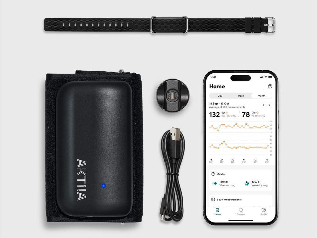
Fisher, who designed and led the study, collaborated with co-authors from Aktiia SA, a Swiss biotechnology company, to analyse over 2.2 million blood pressure readings from 5189 subjects in Europe and the U.K. who wore a cuffless wrist monitor manufactured by Aktiia. On average, the Aktiia device collected 29 readings per day, a substantial increase from the number of blood pressure readings patients typically take with home devices (guidelines recommend four per day, which is more than most patients measure). Over a 15-day period, the researchers obtained an average of 434 readings from each patient.  

By calculating TTR over a 15-day period, the researchers were able to risk stratify participants by percentage of readings in target range and compare these classifications to those generated via traditional measurement patterns, using either 24-hour or week-long daytime monitoring schedules. They found that the traditional methods misclassified 26 and 45 percent of subjects, respectively, compared to the reference TTR. They determined that continual monitoring for seven days is required to obtain 90 percent or greater accuracy in hypertension risk classification, a frequency of measurement that may only be possible with cuffless monitors.  

Though the cuffless device studied here has not been approved by the US Food and Drug Administration, it has been validated in multiple studies and is available for over-the-counter purchase in Europe and the UK. Work to evaluate and set standards for such devices in the U.S. is ongoing. 

“For the first time, by using a cuffless device, we can collect continual out-of-office blood pressure readings and use these data to calculate a new metric, time-in-target-range, which shows great promise as a predictor of risk,” Fisher said. “The use of cuffless devices could create a shift in the paradigm of blood pressure monitoring and hypertension management.” 

Source: Brigham and Women’s Hospital

Using AI, Scientists Discover High-risk Form of Endometrial Cancer

Dr Ali Bashashati observes an endometrial cancer sample on a microscope slide. Credit: University of British Columbia

A discovery by researchers at the University of British Columbia promises to improve care for patients with endometrial cancer, the most common gynaecologic malignancy.  Using artificial intelligence (AI) to spot patterns across thousands of cancer cell images, the researchers have pinpointed a distinct subset of more stubborn endometrial cancer that would otherwise go unrecognised by traditional pathology and molecular diagnostics.

The findings, published in Nature Communications, will help doctors identify patients with high-risk disease who could benefit from more comprehensive treatment.

“Endometrial cancer is a diverse disease, with some patients much more likely to see their cancer return than others,” said Dr Jessica McAlpine, professor at UBC. “It’s so important that patients with high-risk disease are identified so we can intervene and hopefully prevent recurrence. This AI-based approach will help ensure no patient misses an opportunity for potentially lifesaving interventions.”

AI-powered precision medicine

The discovery builds on work by Dr McAlpine and colleagues in the Gynaecologic Cancer Initiative, who in 2013 helped show that endometrial cancer can be classified into four subtypes based on the molecular characteristics of cancerous cells, with each posing a different level of risk to patients.

Dr McAlpine and team then went on to develop an innovative molecular diagnostic tool, called ProMiSE, that can accurately discern between the subtypes. The tool is now used across parts of Canada and internationally to guide treatment decisions.

Yet, challenges remain. The most prevalent molecular subtype, encompassing approximately 50% of all cases, is largely a catch-all category for endometrial cancers lacking discernible molecular features.

“There are patients in this very large category who have extremely good outcomes, and others whose cancer outcomes are highly unfavourable. But until now, we have lacked the tools to identify those at-risk so that we can offer them appropriate treatment,” said Dr McAlpine.

Dr McAlpine turned to long-time collaborator and machine learning expert Dr.Ali Bashashati, an assistant professor of biomedical engineering and pathology and laboratory medicine at UBC, to try and further segment the category using advanced AI methods.

Dr Bashashati and his team developed a deep learning AI model that analyses images of tissue samples collected from patients. The AI was trained to differentiate between different subtypes, and after analysing over 2300 cancer tissue images, pinpointed the new subgroup that exhibited markedly inferior survival rates.

“The power of AI is that it can objectively look at large sets of images and identify patterns that elude human pathologists,” said Dr Bashashati. “It’s finding the needle in the haystack. It tells us this group of cancers with these characteristics are the worst offenders and represent a higher risk for patients.”

Bringing the discovery to patients

The team is now exploring how the AI tool could be integrated into clinical practice alongside traditional molecular and pathology diagnostics.

“The two work hand-in-hand, with AI providing an additional layer on top of the testing we’re already doing,” said Dr McAlpine.

One benefit of the AI-based approach is that it’s cost-efficient and easy to deploy across geographies. The AI analyses images that are routinely gathered by pathologists and healthcare providers, even at smaller hospital sites in rural and remote communities, and shared when seeking second opinions on a diagnosis.

The combined use of molecular and AI-based analysis could allow many patients to remain in their home communities for less intensive surgery, while ensuring those who need treatment at a larger cancer centre can do so.  

“What is really compelling to us is the opportunity for greater equity and access,” said Dr Bashashati. “The AI doesn’t care if you’re in a large urban centre or rural community, it would just be available, so our hope is that this could really transform how we diagnose and treat endometrial cancer for patients everywhere.”

Source: University of British Columbia

Datacentres Form Part of Healthcare Critical Systems – Carrying the Load and so Much More

Photo by Christina Morillo

By Ben Selier, Vice President: Secure Power, Anglophone Africa at Schneider Electric

The adage, knowledge is king couldn’t be more applicable when it comes to the collection and utilisation of data.  And at the heart of this knowledge and resultant information lies the datacentre. Businesses and users count on datacentres, and more so in critical services such as healthcare.

Many hospitals today rely heavily on electronic health records (EHR), and this information resides and is backed up in on-premises datacentres or in the cloud. Datacentres are therefore a major contributor to effective and modernised healthcare.

There are several considerations when designing datacentres for healthcare. For one, hospitals operate within stringent legislation when it comes to the protection of patient information.  The National Health Act (No. 61 of 2003), for example, stipulates that information must not be given to others unless the patient consents or the healthcare practitioner can justify the disclosure.

Datacentres form part of critical systems

To add an extra layer of complexity, in South Africa, datacentres should feature built-in continuous uptime and energy backup due to the country’s unstable power supply.  Hospitals must therefore be designed to be autonomous from the grid, especially when they provide emergency and critical care.

Typically, datacentres are classified in tiers, with the Uptime Institute citing that a Tier-4 datacentre provides 99.995% availability, annual downtime of 0.4 hours, full redundancy, and power outage protection of 96 hours.

In healthcare and when one considers human lives, downtime is simply not an option. And whilst certain healthcare systems and its resultant availability are comparable to a typical Tier-3 or Tier-4 scenario, critical systems in hospitals carry a higher design consideration and must run 24/7 with immediate availability.

In healthcare, the critical infrastructure of a hospital enjoys priority.  What this means is the datacentre is there to protect the IT system which in turn ensures the smooth running of these critical systems and equipment.  There is therefore a delicate balance between the critical systems and infrastructure, and the datacentre, one can’t exist without the other.

Design considerations

To realise the above, hospitals must feature a strong mix of alternative energy resources such as backup generators, uninterrupted power supply (UPS) and renewables such as rooftop solar.

Additionally, like most organisations, storage volume and type and cloud systems will also vary from hospital to hospital. To this end, datacentre design for hospitals is anything but cookie cutter; teams need to work closely with the hospital whilst meeting industry standards for healthcare.

When designing healthcare facilities system infrastructure, the following should also be considered:

  • Software like Building Management Systems (BMS) are not just about building efficiency but also offer benefits such as monitoring and adjusting indoor conditions like temperature control, humidity, and air quality.

The BMS contributes to health and safety and critical operations in hospitals whilst also enabling patient comfort.

  • Maintenance – both building and systems maintenance transcend operational necessity and become a matter of life or death.
  • As mentioned, generators are essential when delivering continuous power which means enough fuel must be stored to run it. Here, hospitals must store fuel safely and in compliance with stringent regulations. In South Africa, proactively managing the refuelling timelines is also critical.  The response times of refuelling these (fuel) bunkers can be severely hindered by issues such as traffic congestion as a result of outages and lights now working.

Selecting the right equipment for hospitals is therefore a delicate balance between technological advancement and safety. For instance, while lithium batteries offer many benefits, when used in hospitals, it is paramount that it is also stored in dry, cool and safe location.

Here, implementing an extinguishing system is a must to alleviate any potential damage from fire or explosions.  That said, lithium batteries are generally considered safe to use but it’s important to be cognisant of its potential safety hazards.

Ultimately, hospitals carry the added weight of human lives which means the design of critical systems require meticulously planning and executed.

How does Oxygen Depletion Disrupt Memory Formation in the Brain?

Scientists identify a positive molecular feedback loop which could explain stroke-induced memory loss.

Ischaemic and haemorrhagic stroke. Credit: Scientific Animations CC4.0

In learning, neurons communicate with each other, and the connections between them getting stronger with repetition. This is known as long-term potentiation or LTP.  

Another type of LTP occurs when the brain is deprived of oxygen temporarily – anoxia-induced long-term potentiation or aLTP. aLTP blocks the former process, thereby impairing learning and memory. Therefore, some scientists think that aLTP might be involved in memory problems seen in conditions like stroke. 

Researchers at the Okinawa Institute of Science and Technology (OIST) and their collaborators have studied the aLTP process in detail. They found that maintaining aLTP requires the amino acid glutamate, which triggers nitric oxide (NO) production in both neurons and brain blood vessels. This process forms a positive glutamate-NO-glutamate feedback loop. Their study, published in iScience, indicates that the continuous presence of aLTP could potentially hinder the brain’s memory strengthening processes and explain the memory loss observed in certain patients after experiencing a stroke.  

The brain’s response to low oxygen 

When there is a lack of oxygen in the brain, the neurotransmitter glutamate is released from neurons in large amounts. This increased glutamate causes the production of NO. NO produced in neurons and brain blood vessels boosts glutamate release from neurons during aLTP. This glutamate-NO-glutamate loop continues even after the brain gets enough oxygen. 

“We wanted to know how oxygen depletion affects the brain and how these changes occur,” stated Dr Han-Ying Wang, a researcher in the former Cellular and Molecular Synaptic Function Unit at OIST and lead author of the study,. “It’s been known that nitric oxide is involved in releasing glutamate in the brain when there is a shortage of oxygen, but the mechanism was unclear.”  

During a stroke, when the brain is deprived of oxygen, amnesia – the loss of recent memories – can be one of the symptoms. Investigating the effects of oxygen deficiency on the brain is important because of the potential medicinal benefits. “If we can work out what’s going wrong in those neurons when they have no oxygen, it may point in the direction of how to treat stroke patients,” Dr Patrick Stoney, a scientist in OIST’s Sensory and Behavioral Neuroscience Unit, explained. 

Brain tissues from mice were placed in a saline solution, mimicking the natural environment in the living brain. Normally, this solution is oxygenated to meet the high oxygen demands of brain tissue. However, replacing the oxygen with nitrogen allowed the researchers to deprive the cells of oxygen for precise lengths of time.  

The tissues were then examined under a microscope and electrodes were placed on them to record electrical activity of the individual cells. The cells were stimulated in a way that mimics how they would be stimulated in living mice. 

Stopping memory and learning activity 

The aLTP process is activated when the brain is deprived of oxygen
The aLTP process is activated when the brain is temporarily deprived of oxygen and glutamate levels increase. If aLTP is maintained for an extended period, this hijacks the normal functioning of the memory strengthening process (LTP), resulting in memory loss. Blocking nitric oxide (NO) synthesis or the molecular pathways that boost glutamate release eventually stops aLTP. Credit: Wang et al., 2024 

The scientists found that maintaining aLTP requires NO production in both neurons and in blood vessels in the brain. Collaborating scientists from OIST’s Optical Neuroimaging Unit showed that in addition to neurons and blood vessels, aLTP requires the activity of astrocytes, another type of brain cell. Astrocytes connect and support communication between neurons and blood vessels. 

“Long-term maintenance of aLTP requires continuous synthesis of nitric oxide. NO synthesis is self-sustaining, supported by the NO-glutamate loop, but blocking molecular steps for NO-synthesis or those that trigger glutamate release eventually disrupt the loop and stop aLTP,” Prof. Tomoyuki Takahashi, leader of the former Cellular and Molecular Synaptic Function Unit at OIST, explained.  

Notably, the cellular processes that support aLTP are shared by those involved in memory strengthening and learning (LTP). When aLTP is present, it hijacks molecular activities required for LTP and removing aLTP can rescue these memory enhancing mechanisms. This suggests that long-lasting aLTP may obstruct memory formation, possibly explaining why some patients have memory loss after a short stroke. 

Prof Takahashi emphasised that the formation of a positive feedback loop formed between glutamate and NO when the brain is temporarily deprived of oxygen is an important finding. It explains long-lasting aLTP and may offer a solution for memory loss caused by a lack of oxygen.  

Source: Okinawa Institute of Science and Technology

Life Healthcare Concludes Agreement to Sub-License “RM2”

Photo by Khwanchai Phanthong on Pexels

Life Healthcare through its wholly owned subsidiary Life Molecular Imaging Limited (LMI), has entered into a contract with Lantheus Holdings Inc. (“Lantheus”), to sub-license one of LMI’s early-stage novel radiotherapeutic and radio diagnostic products (RM2).

“As part of Life Healthcare’s strategy to monetise LMI’s product development portfolio, we are delighted to have found a partner for our RM2 product”, said Pete Wharton-Hood, Life Healthcare, CEO.  “Through this agreement, LMI has secured a partnership for the development of this early-stage diagnostic and therapeutic product through to commercialisation. This exciting opportunity unlocks some of the value in LMI’”, continued Wharton-Hood.

Lantheus will make an upfront payment of $35 million for the sub-licensing rights to RM2, as per the agreement. In addition, several payments will potentially be paid to LMI on the achievement of development and regulatory milestones as well as royalty payments when the product is sold commercially.

The sub-licensing agreement secures Lantheus’ rights to develop the product and complete the early development in collaboration with LMI. “LMI is uniquely positioned to assist in this area, says Wharton -Hood and we are pleased by this development as it showcases and harnesses the specialised, dedicated and focused talent within LMI”. “With Lantheus’ experience in developing and providing access to radiotheranostics in cancer, we are confident in our decision to hand them the reins for this promising theranostic pair and are honored to work with them toward improving the future of people with prostate and breast cancer,” said Ludger Dinkelborg, CEO, Life Molecular Imaging.

Lantheus Holdings, Inc. is listed on NASDAQ in the United States of America and is the leading radiopharmaceutical-focused company committed to delivering life-changing science to enable clinicians to Find, Fight and Follow disease to deliver better patient outcomes. Lantheus has been providing radiopharmaceutical solutions for more than 65 years and has identified value and commercial opportunity in continuing the development of RM2.

LMI is a wholly owned subsidiary in Life Healthcare and is registered in the United Kingdom. The company has a product Neuraceq® which has been approved in many countries and is used to detect amyloid plaque in the brain through a PET-CT Scan and has multiple products in early clinical development. LMI also provides clinical research services for pharmaceutical companies.

Life Healthcare has retained R1bn to provide for funding requirements of LMI as part of the Alliance Medical Group disposal which was concluded earlier this year “This transaction will reduce the quantum required and Life Healthcare will consider distributing a portion of the surplus to shareholders as part of the full year dividend,” stated Wharton-Hood.

About RM2

RM2 is a 9 amino acid peptide that binds to Gastrin Releasing Peptide receptor (GRPr); and can be used to treat multiple malignant tumors like prostate, breast, lung, glioma, and ovarian tumors.

About Life Molecular Imaging

LMI is a wholly owned subsidiary in Life Healthcare and is registered in the United Kingdom. The company has one globally approved product Neuraceq ® that is used to detect amyloid plaque in the brain through a PET-CT scan and has multiple products in early clinical development as well as providing clinical research services for pharmaceutical companies.