Tag: 28/4/22

Obesity in Mice Causes AD Treatments to Backfire

Mouse
Photo by Kanasi on Unsplash

In a new study published in Nature, researchers found that treatments that were effective for atopic dermatitis (AD) in lean mice actually worsened the condition in obese mice.

Tracking the development of AD in obese and lean mice, the researchers found that obese mice developed more inflammation and more severe AD. This increased inflammation was present even after obese mice lost weight. There were similar results in an experimental model of asthma, with obese mice developing more inflammation.

The researchers next looked in detail at immune cells called T cells in lean and obese mice with AD. Lean mice had more TH2 cells, a class of T cells known to play a role in the development of AD. Obese mice had more of a class of T cells called TH17. These cells trigger a different type of inflammation.

Similar trends were seen in blood samples taken from people. Markers of TH17 cell activity increased along with body mass index (BMI) in a database of serum collected from people with AD. Conversely, in samples from patients with severe asthma, TH2 cell activity decreased as BMI increased.

Drugs that block TH2 cell activity are used in the treatment of severe AD as well as asthma and other inflammatory conditions. The researchers tested antibodies to block TH2 cell activity in lean and obese mice with severe AD. While the antibodies reduced skin inflammation in lean mice as expected, they made the condition worse in obese mice. Analysis of immune cells suggested that blocking TH2 cell activity in the obese mice worsened other forms of inflammation.

Obese mice were also found to have less activity of peroxisome proliferator-activated receptor-γ (PPARɣ) in their TH2 cells. When lean mice were engineered to lack PPARɣ, their inflammatory response resembled that of obese mice.

Drugs that increase PPARɣ activity increase insulin sensitivity and are approved for the treatment of type 2 diabetes. The researchers found that giving one of these drugs to obese mice changed their inflammatory response to resemble that of lean mice. It also restored their sensitivity to the antibodies that block TH2 cell activity.

“Our findings demonstrate how differences in our individual metabolic states can have a major impact on inflammation, and how available drugs might be able to improve health outcomes,” said Dr Ronald Evans from the Salk Institute, who helped lead the work.

Source: National Institutes of Health

Gene Mutation in Young Girl May Finally Yield Lupus Treatment

Facial rash characteristic of lupus. Credit: Statpearls

A study published in Nature has identified mutations in an X chromosome gene that senses viral RNA, as a cause of the autoimmune disease lupus, a finding which may explain why the disease is far more common in females, and which might lead to new treatments.

In the study, whole genome sequencing was performed on the DNA of a Spanish child named Gabriela, who was diagnosed with severe lupus at age 7. Such a severe case with early onset of symptoms is rare and suggests a single genetic cause.

In their genetic analysis, the researchers discovered a single point mutation in the TLR7 gene. Referrals from other institutions, they were able to identify other cases of severe lupus where this gene was also mutated.

To confirm that the mutation causes lupus, the team inserted the gene into mice, which went on to develop the disease and showed similar symptoms. The mouse model and the mutation were both named ‘kika’ by Gabriela, the young girl central to this discovery.

Carola Vinuesa, senior author and principal investigator said: “It has been a huge challenge to find effective treatments for lupus, and the immune-suppressors currently being used can have serious side effects and leave patients more susceptible to infection. There has only been a single new treatment approved by the FDA in about the last 60 years.

“This is the first time a TLR7 mutation has been shown to cause lupus, providing clear evidence of one way this disease can arise.”

Professor Nan Shen, co-director of CACPI adds: “While it may only be a small number of people with lupus who have variants in TLR7 itself, we do know that many patients have signs of overactivity in the TLR7 pathway. By confirming a causal link between the gene mutation and the disease, we can start to search for more effective treatments.”

The mutation identified by the researchers makes TLR7 protein bind more readily guanosine and become more active. This in turn increases the sensitivity of the immune cell, making it more likely to incorrectly target healthy tissue.

Interestingly, other studies have shown mutations that cause TLR7 to become less active are associated with some cases of severe COVID infection, highlighting the delicate balance of a healthy immune system.

The findings could also explain why lupus is 10 times more common in females than in males. Because TLR7 is located on the X chromosome, females have two copies of the gene while males have one. Usually, in females one of the X chromosomes is inactive, but in this section of the chromosome, silencing of the second copy is often incomplete. This means females with a mutation in this gene can have two functioning copies.

Study co-author Dr Carmen de Lucas Collantes, said: “Identification of TLR7 as the cause of lupus in this unusually severe case ended a diagnostic odyssey and brings hope for more targeted therapies for Gabriela and other lupus patients likely to benefit from this discovery.”

Gabriela, now a teenager, remains in touch with the research team. She said, “I hope this finding will give hope to people with lupus and make them feel they are not alone in fighting this battle. Hopefully the research can continue and end up in a specific treatment that can benefit so many lupus warriors who suffer from this disease.”

The researchers are now investigating the repurposing of existing treatments which target the TLR7 gene. By targeting this gene, they hope to be able to also help patients with related conditions.

Carola added: “There are other systemic autoimmune diseases, like rheumatoid arthritis and dermatomyositis, which fit within the same broad family as lupus. TLR7 may also play a role in these conditions.”

Source: Francis Crick Institute

GI Issues and Anxiety Linked in Children with Autism

Male doctor with young girl patient
Photo by National Cancer Institute on Unsplash

A new study has found a bi-directional relationship between gastrointestinal (GI) issues and internalised symptoms such as anxiety in children and adolescents with autism, which means the symptoms seem to be affecting each other. The findings could inform future precision medicine research aimed at developing personalised treatments for people with autism experiencing gastrointestinal issues. The study appears in the Journal of Autism and Developmental Disorders.

Autism is known to be often associated with GI issues, and is often overlooked in children despite being a source of pain and anxiety. Food preferences are often for carbohydrates and processed foods. The most common cause of GI issues in children with autism are abdominal pain, constipation, chronic diarrhea and gastroesophageal reflux disease (GERD).

“Research has shown gastrointestinal issues are associated with an increased stress response as well as aggression and irritability in some children with autism,” said Brad Ferguson, an assistant research professor. “This likely happens because some kids with autism are unable to verbally communicate their gastrointestinal discomfort as well as how they feel in general, which can be extremely frustrating. The goal of our research is to find out what factors are associated with gastrointestinal problems in individuals with autism so we can design treatments to help these individuals feel better.”

In the study, Ferguson and his team analysed health data from more than 620 under-18 patients with autism who experience gastrointestinal issues. Then, the researchers examined the relationship between the GI issues and internalised symptoms. Ferguson explained the findings provide more evidence on the importance of the ‘gut–brain axis’ in GI disorders in individuals with autism.

“Stress signals from the brain can alter the release of neurotransmitters like serotonin and norepinephrine in the gut which control gastrointestinal motility, or the movement of stool through the intestines. Stress also impacts the balance of bacteria living in the gut, called the microbiota, which can alter gastrointestinal functioning,” Ferguson said. “The gut then sends signals back to the brain, and that can, in turn, lead to feelings of anxiety, depression and social withdrawal. The cycle then repeats, so novel treatments addressing signals from both the brain and the gut may provide the most benefit for some kids with gastrointestinal disorders and autism.”

Ferguson is collaborating with David Beversdorf, a neurologist who also studies gastrointestinal problems in individuals with autism. Beversdorf had recently helped identify specific RNA biomarkers linked with gastrointestinal issues in children with autism.

“Interestingly, the study from Beversdorf and colleagues found relationships between microRNA that are related to anxiety behaviour following prolonged stress as well as depression and gastrointestinal disturbance, providing some converging evidence with our behavioural findings,” Ferguson said.

Ferguson and Beversdorf are now together investigating the effects of a stress-reducing medication on GI issues in a clinical trial. Ferguson cautioned that treatment could be effective for certain people with autism but not others.

“Our team uses a biomarker-based approach to find what markers in the body are common in those who respond favourably to certain treatments,” Ferguson said. “Our goal is to eventually develop a quick test that tells us which treatment is likely to work for which subgroups of patients based on their unique biomarker signature, including markers of stress, composition of gut bacteria, genetics, co-occurring psychological disorders, or a combination thereof. This way, we can provide the right treatments to the right patients at the right time.”

Source: University of Missouri-Columbia

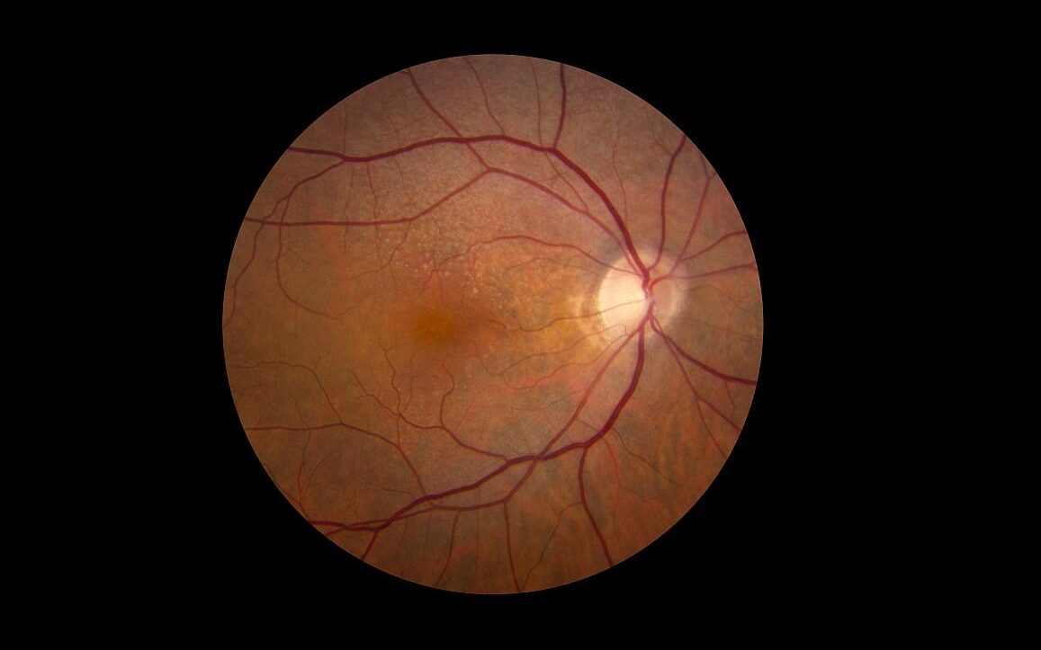
Fenofibrate Confers Modest Risk Reduction for Diabetic Retinal Disease

Retina showing reticular pseudodrusen. Credit: National Eye Institute

Taking the cholesterol-lowering drug fenofibrate had a modest but statistically significant association with reduced risk of vision-threatening diabetic retinopathy (VTDR), according to results of a large study published in JAMA Ophthalmology. In the study, fenofibrate use was associated with an 8% lower risk of progression compared to non-use.

Fenofibrate use had a greater effect on the risk of proliferative diabetic retinopathy, with a 24% decrease in progression (PDR) but did not significantly affect the risk of developing diabetic macular oedema (DME).

The researchers noted that these findings are in line with evidence showing fenofibrate may protect against diabetes-associated breakdown of the blood-retinal barrier, although ophthalmologists rarely use the drug to treat diabetic eye disease.

“Our positive association for progression to PDR coincides with results of previous clinical trials and adds new information with regards to the impact on DME,” the researchers stated.

Protection against progression to PDR “was found without regards to underlying NPDR [nonproliferative diabetic retinopathy] severity level, which is not well coded within the claims database,” the researchers continued. “Understanding this limitation and how the inclusion of NPDR severity levels that may not benefit from fenofibrates would bias our findings to the null means that the positive association seen in our study is actually an underestimate of the true association.”

While fenofibrate’s mechanism of action in diabetic retinopathy is not well understood, “interest in the use of this oral agent has become substantial,” noted Robert N. Frank, MD. author of an accompanying editorial.

“From the point of view of a clinician with a long-time interest in diabetic retinopathy, its causal mechanisms, and its evolving treatments, the possibility that an oral medication originally used for a different disease may be beneficial for the management of diabetic eye disease is exciting,” Dr Frank wrote.

“The evidence that fenofibrate can slow the progression of diabetic retinopathy is growing, but it has not yet become a widely accepted treatment,” he added. “It will be interesting to see how this large population analysis and the results from the ongoing DRCR Retina Network  randomised clinical trial will affect clinical practice in the years to come.”

Two clinical trials that evaluated fenofibrate’s effect on diabetic eye disease, the FIELD study and the ACCORD-Eye trial yielded conflicted findings regarding DME, PDR and progression of diabetic retinopathy. Both trials suggested that only patients with mild nonproliferative eye disease were likely to benefit.

To help inform decision-making on fenofibrate in eye disease, researchers drew on a large health insurer database for 150 252 adults who had NPDR-associated lab values from January 2002 through June 2019. The primary outcomes were a new diagnosis of VTDR (composite of PDR or DME) or DME and PDR individually.

The analysis showed 5835 (3.9%) used fenofibrate. During follow-up, 27 325 patients progressed to VTDR, including 4 086 to PDR and 22 750 to DME. While men accounted for a larger proportion of fenofibrate users (61.1% vs 51.0% of nonusers), patients had similar baseline characteristics.

Study limitations included lack of clinical applicability, not accounting for duration of fenofibrate use, and data being drawn from a single database.

Source: MedPage Today

Shift in Recommendations for Aspirin in CVD Prevention

Anatomical model of a human heart
Photo by Robina Weermeijer on Unsplash

The US Preventive Services Task Force (USPSTF) has issued a recommendation statement on the use of aspirin in the prevention of cardiovascular disease (CVD). The recommendation shifts the use of aspirin to an earlier window, and making it an individualised decision for people in their 40s to 50s with a > 10% 10-year CVD risk.

The previous recommendation from 2016 had called for low-dose aspirin for people in their 50s with a > 10% 10-year CVD risk and individualised decisions for those in their 60s with similar risk. The update comes after new evidence emerged in a number of randomised controlled trials.

For the update, which appears in JAMA Network, a systematic review was conducted on the effectiveness of aspirin to reduce the risk of CVD events (myocardial infarction and stroke), cardiovascular mortality, and all-cause mortality in persons without a history of CVD. The effect of aspirin use on colorectal cancer incidence and mortality was also investigated in primary CVD prevention populations, as well as the harms (particularly bleeding) associated with aspirin use.

The USPSTF concludes with moderate certainty that aspirin use for the primary prevention of CVD events in adults aged 40–59 years with a 10% or greater 10-year CVD risk has a small net benefit, and starting low-dose aspirin use for CVD prevention should be an individual decision for them. Those not at increased risk for bleeding and are willing to take low-dose aspirin daily are more likely to benefit. The USPSTF recommends against initiating low-dose aspirin use for the primary prevention of CVD in adults 60 years or older.

Aspirin’s mechanism of action in CVD protection is well known. Aspirin at low doses is an irreversible cyclooxygenase 1 (COX-1) enzyme inhibitor, and at higher doses, it also inhibits COX-2. By inhibiting platelet function through COX-1, aspirin reduces atherothrombosis risk, and has been used widely for the prevention of CVD events, particularly for secondary prevention. However the COX-1 is also involved with protection of the gastrointestinal mucosa, and inhibition of it can promote gastrointestinal bleeding. The mechanism for the possible antineoplastic effects of aspirin is not as well understood.

Older age is one of the strongest risk factors for CVD, and men have a higher overall CVD disease burden and tend to experience CVD events earlier in life. Race and ethnicity affects CVD burden, with Black persons having the highest prevalence of CVD.

Similar CVD benefits appear for a low aspirin dose (≤ 100mg/d) and for all doses that have been studied in CVD prevention trials (50 to 500mg/d). A pragmatic approach would be to use 81 mg/d, which is the most commonly prescribed dose in the US.

Because CVD risk estimation is imprecise and imperfect at the individual level, the USPSTF suggests using these risk estimates as a starting point to discuss with appropriate candidates their desire for daily aspirin use. The benefits of initiating aspirin use are greater for individuals at higher risk for CVD events (eg, those with > 15% or > 20% 10-year CVD risk).

In addition to age and estimated level of CVD risk, decisions about initiating aspirin use should be based on shared decision-making between clinicians and patients about the potential benefits and harms, based on the relative values the patient places on these (reduced CVD risk vs increased bleeding and stroke risk).

Annual bleeding events in individuals without risk factors for increased bleeding (eg, history of gastrointestinal bleeding risk, history of peptic ulcer disease, or use of nonsteroidal anti-inflammatory drugs or corticosteroids) are rare, but risk for bleeding increases modestly with advancing age. For persons who have initiated aspirin use, the net benefits continue to accrue over time in the absence of a bleeding event. However, benefits shrink with advancing age because of increased bleeding risk, with modelling data suggesting stopping aspirin use around age 75.