Tag: 24/5/24

Inflammatory Bowel Disease may Increase Risk of Heart Failure

Irritable bowel syndrome. Credit: Scientific Animations CC4.0

Inflammatory bowel disease (IBD) is associated with a slightly increased risk of heart failure up to 20 years after diagnosis, according to a comprehensive registry study from Karolinska Institutet published in the European Heart Journal.

The researchers analysed the risk of heart failure in over 80 000 patients with inflammatory bowel disease, that is, Crohn’s disease, ulcerative colitis or unclassified IBD, compared with 400 000 people from the general population, as part of the ESPRESSO study.

The results show that people with IBD have a 19% increased risk of developing heart failure up to 20 years after diagnosis. This corresponds to one extra heart failure case per 130 IBD patients in those 20 years, and the risk increase was seen regardless of the type of IBD. The highest risk of heart failure was seen in older patients, people with lower education and people with pre-existing cardiovascular-related disease at IBD diagnosis.

Contribute to new guidelines

“Both healthcare providers and patients should be aware of this increased risk, and it’s important that cardiovascular health is properly monitored,” says the study’s first author Jiangwei Sun, researcher at the Department of Medical Epidemiology and Biostatistics, Karolinska Institutet. “We hope the results will raise the awareness of health workers as to the increased risk of heart failure in individuals with IBD and contribute to new guidelines for cardiovascular disease management in IBD patients.”

Comparing siblings with and without ABD, the risk increase was slightly lower, 10%, suggesting that genetics and early environmental factors shared within families may play a role. 

“We don’t know if there is a causal relationship, but we will continue to explore genetic factors and the role of IBD medications and disease activities on the risk of heart failure,” says the study’s senior author Professor Jonas F. Ludvigsson from the Department of Medical Epidemiology and Biostatistics, Karolinska Institutet.

Source: Karolinska Institutet

DNA from Ancient Viral Infections Implicated in Some Psychiatric Disorders

Photo by Sangharsh Lohakare on Unsplash

New research led by King’s College London has found that thousands of DNA sequences originating from ancient viral infections are expressed in the brain, with some contributing to susceptibility for psychiatric disorders such as schizophrenia, bipolar disorder, and depression.

Around 8% of the human genome is made up of sequences called Human Endogenous Retroviruses (HERVs), which are products of ancient viral infections that occurred hundreds of thousands of years ago. Until recently, it was assumed that these ‘fossil viruses’ were simply junk DNA, with no important function in the body. However, due to advances in genomics research, scientists have now discovered where in our DNA these fossil viruses are located, enabling us to better understand when they are expressed and what functions they may have.

This new study, published in Nature Communications, builds upon these advances and is the first to show that a set of specific HERVs expressed in the human brain contribute to psychiatric disorder susceptibility, marking a step forward in understanding the complex genetic components that contribute to these conditions.

Dr Timothy Powell, co-senior author on the study and Senior Lecturer at the Institute of Psychiatry, Psychology & Neuroscience (IoPPN), King’s College London, said: “This study uses a novel and robust approach to assess how genetic susceptibility for psychiatric disorders imparts its effects on the expression of ancient viral sequences present in the modern human genome. Our results suggest that these viral sequences probably play a more important role in the human brain than originally thought, with specific HERV expression profiles being associated with an increased susceptibility for some psychiatric disorders.”

The study analysed data from large genetic studies involving tens of thousands of people, both with and without mental health conditions, as well as information from autopsy brain samples from 800 individuals, to explore how DNA variations linked to psychiatric disorders affect the expression of HERVs.

Although most genetic risk variants linked to psychiatric diagnoses impacted genes with well-known biological functions, the researchers found that some genetic risk variants preferentially affected the expression of HERVs. The researchers reported five robust HERV expression signatures associated with psychiatric disorders, including two HERVs that are associated with risk for schizophrenia, one associated with risk for both bipolar disorder and schizophrenia, and one associated with risk for depression.

Dr Rodrigo Duarte, first author and Research Fellow at the IoPPN, King’s College London, said: “We know that psychiatric disorders have a substantial genetic component, with many parts of the genome incrementally contributing to susceptibility. In our study, we were able to investigate parts of the genome corresponding to HERVs, which led to the identification of five sequences that are relevant to psychiatric disorders. Whilst it is not clear yet how these HERVs affect brain cells to confer this increase in risk, our findings suggest that their expression regulation is important for brain function.”

Dr Douglas Nixon, co-senior author on the study and and researcher at the Feinstein Institutes for Medical Research at Northwell Health, in the US, said: “Further research is needed to understand the exact function of most HERVs, including those identified in our study. We think that a better understanding of these ancient viruses, and the known genes implicated in psychiatric disorders, have the potential to revolutionise mental health research and lead to novel ways to treat or diagnose these conditions.”

Source: King’s College London

How Zebrafish Heal from Spinal Cord Injury could Help Humans

Photo by Cottonbro on Pexels

Zebrafish have a remarkable ability to heal their spinal cord after injury. Now, researchers at Karolinska Institutet have uncovered an important mechanism behind this phenomenon – a finding that could have implications for the treatment of spinal cord injury in humans.

In a new study published in Nature Communications, researchers show that the neurons of adult zebrafish immediately start to cooperate after a spinal cord injury, keeping the cells alive and stimulating the healing process.

“We have shown that the neurons form small channels called gap junctions, which create a direct connection between the neurons and enable the exchange of important biochemical molecules, allowing the cells to communicate and protect each other,” explains Konstantinos Ampatzis, a researcher in the Department of Neuroscience at Karolinska Institutet, who led the study.

The researchers will further investigate the exact mechanisms behind this protective strategy in zebrafish and hope this knowledge will lead to new ways of treating spinal cord injury in humans.

“Spinal cord injuries are a major burden for sufferers and their families,” says Konstantinos Ampatzis. “What if we could get human neurons to adopt the same survival strategy and behave like zebrafish neurons after an injury? This could be the key to developing new effective treatments.”

Source: Karolinka Institutet

Milk Samples From the 1940s Reveal Antibiotic Resistance in the Pre-antibiotic Era

Photo by Robin Worrall on Unsplash

Using stored milk samples as a kind of time capsule, veterinary researchers at the University of Connecticut have uncovered insights about the presence of antibiotic resistance even in the pre-antibiotic era.

Sometime in the 1940s or so, someone in what is now the Department of Pathobiology and Veterinary Science got a lyophiliser, a piece of equipment that freeze-dries samples, says Director of the Connecticut Veterinary Medical Diagnostic Laboratory (CVMDL) Dr Guillermo Risatti. Risatti explains that at that time, the microbiology lab was very active in testing milk for the dairy farms in the region. With an exciting new piece of equipment, it seems they started lyophilising hundreds of samples.

The samples have been in storage ever since. Beyond the scant details that these are milk samples containing Streptococcus bacteria from the 1940s, Risatti explains that he and his colleagues – CVMDL Research Associate Dr. Zeinab Helal, Ji-Yeon Hyeon and Dong-Hun Lee – were interested in exploring their microbial history.

Risatti says that over the years, the data was lost, so researchers don’t have precise details of the provenance of the samples. But knowing a bit of history about the department, they can deduce some information.

“We believe that most of them came from Connecticut or perhaps from cases from the region, but we cannot say which parts,” Risatti says. “Most likely, this lab provided a testing service to locals, as this was mainly a pathology lab. Now it’s more like a diagnostic lab, and we receive samples from all over the region, including New York and New Jersey.”

Learning about what these historical samples hold could help with research in unexpected ways, but the first step is piecing together the lost details. To do this, Risatti explains that the team established a workflow using standard techniques to streamline processes to analyse the visual characteristics, called phenotype, and to analyse their genotype with genomic sequencing.

Different species of Streptococcus use different strategies to inflict disease in the organisms they infect. These virulence factors are used to differentiate one species of Streptococcus from another and are one way to distinguish samples through phenotypic analysis. Another phenotypic analysis includes testing bacteria for their susceptibility to antibiotics.

The researchers started with 50 samples collected from 1941 to 1947, and they found that the samples contained seven different Streptococcus species, including two subspecies of S. dysgalactiae. Interestingly, the researchers found some of the samples were resistant to the antibiotic tetracycline and did not carry antibiotic resistance genes typically seen in today’s antibiotic-resistant bacterial strains. Since these samples were collected prior to the antibiotic era, the results add to a growing body of literature showing that antibiotic resistance occurred naturally before humans discovered and began to use antibiotics.

“Antibiotic resistance is a very big area of research, and it has been for many years,” says Risatti. “We did not go any further with our analysis because we don’t have the tools here, but we hope to bring this information to the public. I think it could be the jumpstart for somebody to study further.”

Risatti explains the hope is to partner with large agencies like the CDC and the Department of Public Health to help bolster antibiotic resistance research.

Concussion is Associated with Iron Accumulation in Certain Brain Areas

Photo by Anna Shvets

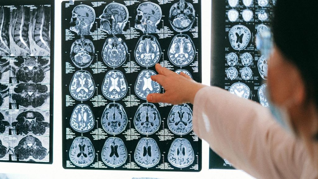
People who suffer from headaches after experiencing concussions may also be more likely to have higher levels of iron in areas of the brain – a sign of injury to brain cells, according to a preliminary study presented at the American Academy of Neurology’s 76th Annual Meeting.

“These results suggest that iron accumulation in the brain can be used as a biomarker for concussion and post-traumatic headache, which could potentially help us understand the underlying processes that occur with these conditions,” said study author Simona Nikolova, PhD, of the Mayo Clinic in Phoenix, Arizona, and a member of the American Academy of Neurology.

The study involved 120 participants, 60 of whom who had post-traumatic headache (PTH) due to mild traumatic brain injury (mTBI), and 60 healthy controls. The injuries were due to a fall for 45% of the people, 30% were due to a motor vehicle accident and 12% were due to a fight. Other causes were the head hitting against or by an object and sports injuries. A total of 46% of the people had one mild traumatic brain injury in their lifetime, 17% had two, 16% had three, 5% had four and 16% had five or more mild traumatic brain injuries.

Participants underwent 3T brain magnetic resonance imaging (T2* maps). T2* differences were determined using age-matched paired t-tests. For the PTH group, scans were done an average of 25 days after injury. T2* correlations with headache frequency, number of lifetime mTBIs, time since most recent mTBI, and Sport Concussion Assessment Tool (SCAT) severity scale scores,

The researchers observed lower T2* values in PTH participants relative to HC in the right supramarginal area, left occipital, bilateral precuneus, right cuneus, right cerebellum, right temporal, bilateral caudate, genu of the corpus callosum, right anterior cingulate cortex and right rolandic operculum (p < 0.001).

Within PTH subjects, there were positive correlations with iron accumulation between lifetime mTBIs, the time since most recent mTBI and headache frequency in certain areas of the brain. For example, T2* levels in headache frequency with T2* in the posterior corona radiata, bilateral temporal, right frontal, bilateral supplemental motor area, left fusiform, right hippocampus, sagittal striatum, and left cerebellum were associated with headache frequency.

“Previous studies have shown that iron accumulation can affect how areas of the brain interact with each other,” Nikolova said. “This research may help us better understand how the brain responds and recovers from concussion.”

Nikolova said that using the indirect measure of iron burden also means that the change in that measure could be due to other factors such as haemorrhage or changes in tissue water rather than iron accumulation.

Source: American Academy of Neurology