Tag: 22/7/22

Monkeypox Symptoms Described in Case Series

Source: Wikimedia CC0

A large case series on monkeypox was published in The New England Journal of Medicine, which will help direct the limited resources such as vaccines to contain the recent spread of the virus. In the study, clinicians led by Queen Mary University of London identified new clinical symptoms of monkeypox infection, which will aid future diagnosis and help to slow the spread of infection. This is the largest case study series to date, reporting on 528 confirmed monkeypox infections at 43 sites between 27 April and 24 June 2022.

Gay and bisexual men make up 98% of infected persons in the current spread of the virus. While in most cases sexual closeness is the most likely route of transmission, researchers stress that the virus can be transmitted by any close physical contact through large respiratory droplets and potentially through clothing and other surfaces.

There is a global shortage of both vaccines and treatments for human monkeypox infection. The findings of this study, including the identification of those most at risk of infection, will help to aid the global response to the virus. Public health interventions aimed at higher risk of exposure could help to detect and slow the spread of the virus. Recognising the disease, contact tracing and advising people to isolate will be key components of the public health response.  

Many of the infected individuals reviewed in the study presented with symptoms not recognised in current medical definitions of monkeypox. These symptoms include single genital lesions and sores on the mouth or anus. The clinical symptoms are similar to those of sexually transmitted infections (STIs) and can easily lead to misdiagnosis.

In some people, anal and oral symptoms have led to people being admitted to hospital for management of pain and difficulties swallowing. Since misdiagnosis can slow detection, hindering efforts to control the spread of the virus, this information is important for clinicians. This information will help increase diagnoses in persons from at-risk groups present with traditional STI symptoms.

Public health measures – such as enhanced testing and education – should be developed and implemented working with at-risk groups to ensure that they are appropriate, non-stigmatising, and to avoid messaging that could drive the outbreak underground.

Source: Queen Mary University of London

How Macrophages Control an Uncooperative Meal

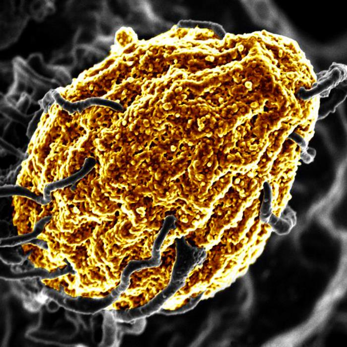
A macrophage digesting a yeast cell (yellow). Credit: NIH

Certain pathogens such as Salmonella have developed strategies to protect themselves from the macrophages’ digesting attempts, causing severe Typhoid infections and inflammations. Scientists report in Nature Metabolism how the inter-organellar crosstalk between phago-lysosomes and mitochondria restricts the growth of such bacteria inside macrophages.

Signals from the digestion cell organelle

As scavenger cells, macrophages have a very prominent digestion organelle, the phago-lysosome, where engulfed microorganisms are commonly degraded into pieces and become inactivated. “It has long been known that the molecule TFEB (Transcription factor EB) is important for the regulation of the phago-lysosomal system. More recent evidence also suggested that TFEB supports the defense against bacteria,” said Max Planck Institute group leader Angelika Rambold.

She and her team wanted to understand how exactly TFEB mediates its anti-bacterial role in macrophages. They confirmed earlier findings showing that a broad range of microbes, bacterial and inflammatory stimuli activate TFEB and thus the phago-lysosomal system.

“It made sense that pathogen signals trigger TFEB as macrophages need a more active digestion system quickly after they devour a meal of bacteria. But, interestingly, the experiments also revealed an additional strong effect of TFEB activation on another intracellular organelle system — mitochondria. This was completely unexpected and novel to us,” said Angelika Rambold.

Instructing mitochondria to increase anti-microbial activity

Composed of inner and outer membranes, mitochondria are the primary sites of cellular respiration and release energy from nutrients. Moreover, the mitochondria in immune cells were recently identified as sources of anti-microbial metabolites.

By using a broad experimental tool set, the investigators identified the pathway controlling an unexpected crosstalk between lysosomes and mitochondria. “Macrophages make use of extensive inter-organellar communication: the lysosome activates TFEB, which shuttles into the nucleus where it controls the transcription of a protein called IRG1. This protein is imported into mitochondria, where it acts as a major enzyme to produce the anti-microbial metabolite itaconate,” explained Angelika Rambold.

Exploiting organelle communication to control bacterial infections

The researchers investigated whether they could make use of this newly identified pathway to control bacterial growth. “We speculated that activating this pathway could be used to target certain bacterial species, such as Salmonella,” said Angelika Rambold. “Salmonella can escape the degradation by the phago-lysosomal system. They manage to grow inside macrophages, which can lead to the spreading of these bacteria to several organs in an infected body,” explained Alexander Westermann, collaborating scientist from the University of Würzburg.

When the researchers activated TFEB in infected macrophages in mice, the TFEB-Irg1-itaconate pathway inhibited the growth of Salmonella inside the cells. These data show that the lysosome-to-mitochondria interplay represents an antibacterial defense mechanism to protect the macrophage from being exploited as a bacterial growth niche.

In light of the increasing emergence of multi-drug resistant bacteria, with more than 10 million expected deaths per year by 2050 according to the various expert groups, it becomes important to identify new strategies to control bacterial infections that escape immune mechanisms. A promising path could be to use the TFEB-Irg1-itaconate pathway or itaconate itself to treat infections caused by itaconate-sensitive bacteria. According to the researchers from more work is needed to assess whether these new intervention points can be successfully applied to humans.

Source: Max Planck Institute of Immunobiology and Epigenetics

Increase in Cardiovascular Disease Diagnoses after COVID

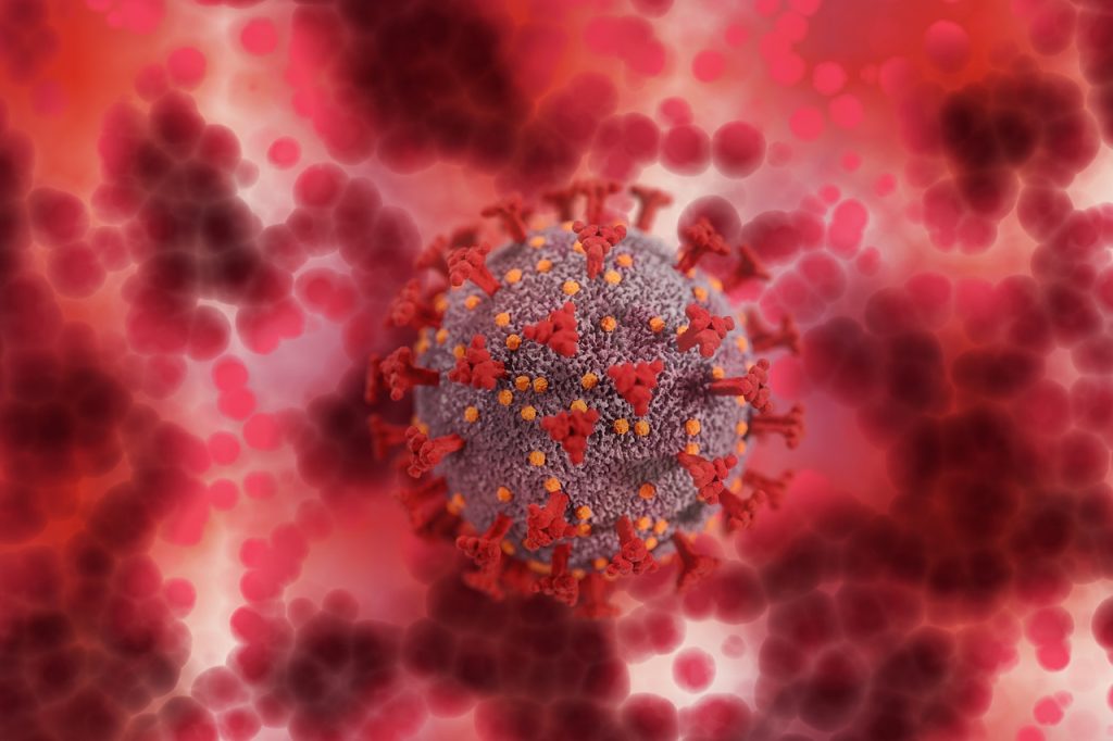
Image from Pixabay

A new study published in PLOS One found that COVID infection is associated with a nearly six-fold increase in cardiovascular disease (CVD) diagnoses over 12 months after the infection. 

The study analysed of UK electronic health records, comparing the risks of new diabetes mellitus (DM) and CVD diagnoses in the 12 months after infection. Researchers matched a cohort of 428 650 COVID patients matched to controls.

There was an 80% increased risk of DM diagnosis in the first month after COVID infection, a trend that has been echoed in previous studies, although those studies’ results seem to indicate a temporary form of the disease resulting from the acute stress of viral infection.

The findings showed that the largest increases were in pulmonary embolism (Relative Risk [RR] 11.51) and in atrial arrhythmias (RR 6.44). New CVD diagnoses rose five weeks after infection and incidence declined within 12 weeks to a year and returned to baseline or showed a net decrease. Increased risk for new DM diagnoses remained elevated by 27% for up to 12 weeks. 

“It’s definitely reassuring that over the longer timeframe, cardiovascular disease and diabetes risk does seem to return to baseline levels,” study author Emma Rezel-Potts, PhD, told The Guardian. “But we do have to be cautious in the acute period with cardiovascular disease and take note that the risk of diabetes seems to be elevated for several months, so that could be a good opportunity for risk prevention.” 

She also stressed that the findings could be explained by many factors. For example, the COVID patients in the study were more likely to be overweight and had more underlying health problems compared to uninfected controls, predisposing them to DM and CVD. Additionally, some may have had underlying conditions which were discovered when they were treated for COVID.

Source: The Guardian

Neural Plastic Changes can Help in Cervical Spinal Cord Injuries

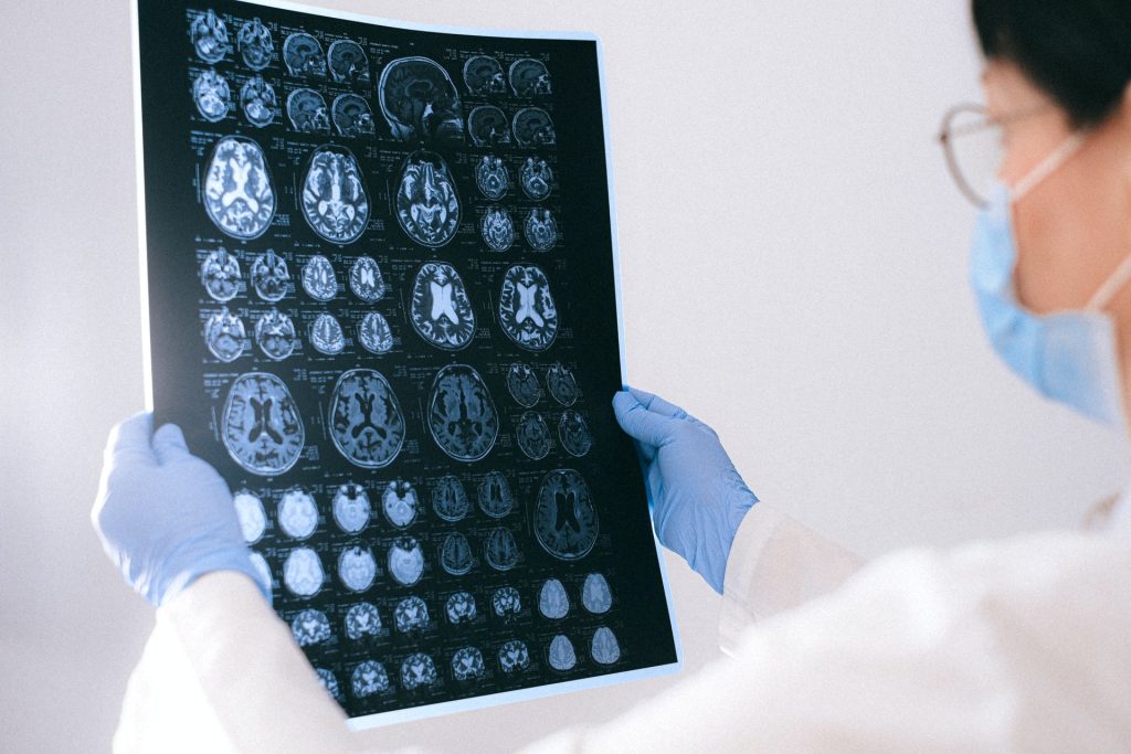
MRI images of the brain
Photo by Anna Shvets on Pexels

By studying damage involving the connection between the brain’s hemispheres, researchers are finding new ways to leverage neural plasticity to promote functional recovery after a spinal cord injury.

In a study published in JCI Insight, the team of researchers used models in the lab to investigate a unilateral spinal cord injury similar to Brown-Sequard Syndrome, a rare neurological condition where damage to the spinal cord in a person results in weakness or paralysis on one side of the body and a loss of sensation on the opposite side.

Assistant Professor Wei Wu at Indiana University School of Medicine, said that the spinal cord injury model damaged the connection between the left hemisphere of the brain and the right side of the body, leading to significant loss of function in the right forelimb.

“The skilled function of upper limbs is very important for the quality of life in the patients with cervical spinal cord injury, but such functional recovery is very difficult to achieve in the severe injury,” said Asst Prof Wu, first author of the paper. “We found that the intact corticospinal system in the opposite side of the brain and spinal cord can be modulated to at least partially take over the control of the forelimb that is damaged by the spinal cord injury, resulting in a forelimb functional improvement.”

Since each hemisphere controls the opposite side of the body, researchers discovered a spontaneous shift of the neural circuits after injury from the left hemisphere to the right. Although there are connections between the right hemisphere of the brain and the right side of the body through some relayed pathways after injury, Asst Prof Wu said that’s not sufficient to support the motor recovery.

Using optogenetics to stimulate the right hemisphere of the brain, the researchers modulated the motor cortex. Additional neural circuits were shifted from the left side to the right side of the brain to dramatically increase and improve forelimb function.

“New circuits in the whisker, jaw forelimb and neck areas in the right hemisphere of the brain are recruited to control the right forelimb,” Asst Prof Wu said. “Interestingly, the beneficial neural plastic changes emerge both in the brain and the distal spinal cord after the optogenetic neuromodulation was applied on the motor cortex.”

Asst Prof Wu said results of the study showed significant improvement to the forelimb; however, there are still many challenges ahead, since complete digital recovery was not achieved.

The research team will continue explore this transhemispheric neural reorganisation to further improve the functional recovery after the spinal cord injury, Asst Prof Wu said. He hopes that these findings will be applied to treatments for spinal cord injuries.

Source: Indiana University School of Medicine

No Consensus on a Definition for ‘Growing Pains’

Photo by Monstera from Pexels

The phrase ‘growing pains’ is often used by people to describe muscle or joint pain in young people and health professionals also use the term. However a broad review of medical literature has found there is no consistent medical definition of the condition behind a diagnosis.

Researchers from the University of Sydney found there is no agreement in the literature on what growing pains really are, what they mean, how they are defined, and how they should be diagnosed.

Growing pains may be a medical misnomer, the researchers said – more than 93% of studies did not refer to growth when defining the condition. Similarly, age was not mentioned in more than 80% of studies’ definitions.

The findings have prompted the researchers to recommend the term growing pains not be used by clinicians and other researchers as a stand-alone diagnosis, until a clear definition backed by evidence has been established.

Growing pains are considered to be one of the most common causes of recurring musculoskeletal pain in children and adolescents. Some studies suggest up to a third of children experience the condition at some point in their life.

The term first arose in 1823 in a book called ‘Maladies de la Croissance’ (‘diseases of growth’).

“Thousands of kids are diagnosed with growing pains by their healthcare professional, but we were curious – what does that diagnosis really mean?” said lead author Dr Mary O’Keeffe from Institute for Musculoskeletal Health at the University of Sydney. 

In order to see how researchers defined the term, and if there were any detailed criteria that led to a diagnosis, the reviewers examined 147 studies that mentioned growing pains. The medical literature spanned many types of research, including systematic reviews, editorials, observational studies, case-control studies, and theses.

“What we found was a little concerning: that there is no consistency in the literature on what ‘growing pains’ means,” said Professor Steven Kamper, at the University of Sydney.  

“The definitions were really variable, vague and often contradictory. Some studies suggested growing pains happened in the arms, or in the lower body. Some said it was about muscles while other studies said joints.”

Only seven studies, less than 10% of the studies examined, mentioned growth related to the pain. More than 80% of the studies did not mention a young person’s age at the time ‘growing pains’ occurred.

There was no widespread agreement or detail on where the pain was located or when the pain happened.

Half of the studies referenced ‘growing pains’ as being located in the lower limb, while 28% reported specifically in the knees.

As for time of occurrence, 48% of studies reported the ‘growing pains’ happens during the evening or night and 42% reported it was recurring.

“What this study uncovered was while ‘growing pains’ is a very popular label used to diagnose musculoskeletal pain, it means very different things to different people,” said senior author Professor Steve Kamper.

“This level of uncertainty means clinicians don’t have a clear guide or criteria to know when the label ‘growing pains might be appropriate for a patient’.”

The study questioned whether growing pains are connected to growth itself in bone or muscle.

“There is a lack of evidence or inconsistent information on growing pains as a condition – and how it is associated with growth, or even the cause of the pain,” said Dr O’Keeffe.

“There is a real opportunity to understand this condition – given how widespread the use of the term is, or whether there is even a need to use this term.”

Source: EurekAlert!