People with irritable bowel syndrome (IBS) have lower bacterial diversity in the intestine than do healthy people, according to research appearing in Microbiology Spectrum. The investigators believe that theirs is the first analysis to find a clear association between IBS and reduced diversity in the microbiota of the gut. The an open-access journal of the American Society for Microbiology.
Normally, “More than 10 000 species of microorganism live in the human intestine,” said corresponding author Jung Ok Shim, MD, PhD, a professor at Korea University College of Medicine. Disruption of the microbiome of the human gastrointestinal tract can trigger IBS. Typically, IBS causes bloating, diarrhoea, and stomach pain or cramps.
Previous studies of gut bacteria in patients with IBS have been controversial, with inconsistent results, due to small sample size and lack of consistent analytical methods used among these studies, said Shim. The investigators combined their own dataset with 9 published, shared datasets, encompassing 576 IBS patients and 487 healthy controls, analysing them with a “unified data processing and analytical method.”
The researchers found that the gut bacterial community is less diverse in IBS patients than in healthy people, said Shim. Additionally, the abundance of 21 bacterial species differed between IBS patients and healthy controls. However, the findings were not statistically significant in the paediatric cohort due to small sample size.
The investigators proved that the disturbed gut bacterial community “is associated with IBS, though this does not mean that the relationship is causal,” said Shim. “Functional studies are needed to prove whether the change in gut micro-organisms contributes to development of IBS.”
Even though IBS is a common disorder, its pathogenesis remains unknown, and as yet there is no effective treatment strategy. “Based on the epidemiological studies of IBS patients, altered gut microbiota was proposed as one of the possible causes of IBS,” the researchers write. “Acute bacterial gastroenteritis can cause chronic, asymptomatic, low-grade intestinal wall inflammation sufficient to alter neuromuscular and epithelial cell function.”
An international study demonstrates for the first time that degradation in epigenetic information can drive ageing in an organism, independently of changes to the genetic code itself. Published in the journal Cell, the work shows that a breakdown in epigenetic information causes mice to age and that restoring the integrity of the epigenome reverses those signs of ageing.
“We believe ours is the first study to show epigenetic change as a primary driver of ageing in mammals,” said the paper’s senior author, David Sinclair, professor of genetics at Harvard Medical School.
The team’s extensive series of experiments provide long-awaited confirmation that DNA changes are not the only, or even the main, cause of ageing. Rather, the findings show, chemical and structural changes to chromatin contribute ageing without changing the genome.
“We expect the findings will transform the way we view the process of ageing and the way we approach the treatment of diseases associated with ageing,” said co-first author Jae-Hyun Yang, research fellow in genetics in the Sinclair lab.
Since it is easier to manipulate epigenetics than DNA, this could lead to a whole new avenue of research. Studies in nonhuman primates are currently underway.
“We hope these results are seen as a turning point in our ability to control aging,” said Sinclair. “This is the first study showing that we can have precise control of the biological age of a complex animal; that we can drive it forwards and backwards at will.”
Beyond mutations
A reigning, decades-old theory of ageing was that it arises from an accumulation of changes to DNA, primarily genetic mutations, which over time prevent more and more genes from functioning properly. Over time, researchers began finding contradictory evidence: in some human and mice, high mutation rates was not accompanied by premature ageing, while many types of aged cells lacked mutations. Some researchers believed that epigenetics could be the true culprit.
A component of epigenetics is the physical structures such as histones that bundle DNA into tightly compacted chromatin and unspool portions of that DNA when needed. Bundled up, genes are inaccessible when but are available to be copied and used to produce proteins when they’re unspooled. Thus, epigenetic factors regulate which genes are active or inactive in any given cell at any given time.
By acting as a toggle for gene activity, these epigenetic molecules help define cell type and function. Since each cell in an organism has basically the same DNA, it’s the on-off switching of particular genes that differentiates a nerve cell from a muscle cell from a lung cell.
“Epigenetics is like a cell’s operating system, telling it how to use the same genetic material differently,” said Yang, who is co-first author with Motoshi Hayano, a former postdoctoral fellow in the Sinclair lab who is now at Keio University School of Medicine in Tokyo.
In the late 1990s and early 2000s, Sinclair’s lab and others showed in yeast and mammals that epigenetic changes were associated with ageing but could not determine whether they caused it or were caused by it. At least, this new study let the scientists disentangle epigenetic causes from genetics.
ICE mice
The team’s main experiment involved creating temporary, fast-healing cuts in the DNA of lab mice, which mimicked those breaks chromosomes that mammalian cells receive on a daily basis from things like breathing, exposure to sunlight and cosmic rays, and contact with certain chemicals. This let the researchers simulate a sped-up life.
Most of the breaks did not happen in the DNA’s coding regions, so did not cause mutations. Rather, the breaks altered the way DNA is folded.
Sinclair and colleagues called their system ICE, short for inducible changes to the epigenome.
At first, epigenetic factors paused their normal job of regulating genes and moved to the DNA breaks to coordinate repairs. Afterward, the factors returned to their original locations.
But as time passed, things changed. The researchers noticed that these factors got ‘distracted’ and did not return home after repairing breaks. The epigenome grew disorganised and began to lose its original information. Chromatin got condensed and unspooled in the wrong patterns, a hallmark of epigenetic malfunction.
As the mice lost their youthful epigenetic function, they began to look and act old. The researchers saw a rise in biomarkers that indicate ageing. Cells lost their identities as, for example, muscle or skin cells. Tissue function faltered. Organs failed.
The team used a tool recently developed by Sinclair’s lab to measure how biologically old the mice were, based on how many sites across the genome lost the methyl groupsnormally attached to them. Compared to untreated mice born at the same time, the ICE mice had aged significantly more.
Young again
Next, the researchers gave the mice a gene therapy that reversed the epigenetic changes they’d caused, which Sinclair likened to rebooting a malfunctioning computer.
The therapy delivered a trio of genes (Oct4, Sox2, and Klf4, together named OSK) that are active in stem cells and can help rewind mature cells to an earlier state. (Sinclair’s lab used OSK to restore sight in blind mice in 2020.)
The ICE mice’s organs and tissues resumed a youthful state.
The therapy “set in motion an epigenetic program that led cells to restore the epigenetic information they had when they were young,” said Sinclair. “It’s a permanent reset.”
How exactly OSK treatment achieved that remains unclear.
At this stage, Sinclair says the discovery supports the hypothesis that mammalian cells maintain a kind of backup copy of epigenetic software that, when accessed, can allow an aged, epigenetically scrambled cell to reboot into a youthful, healthy state.
For now, the extensive experiments led the team to conclude that “by manipulating the epigenome, aging can be driven forwards and backwards,” said Yang.
From here
The ICE method offers researchers a new way to explore the role of epigenetics in ageing and other biological processes.
Because signs of ageing developed in the ICE mice after only six months rather than toward the end of the average mouse life span of two and a half years, the approach also saves time and money for researchers studying aging.
Yang said that researchers can also look beyond OSK gene therapy to other methods such as drugs, to determine how lost epigenetic information might be restored in aged organisms.
Watch the team describe their research in the video below.
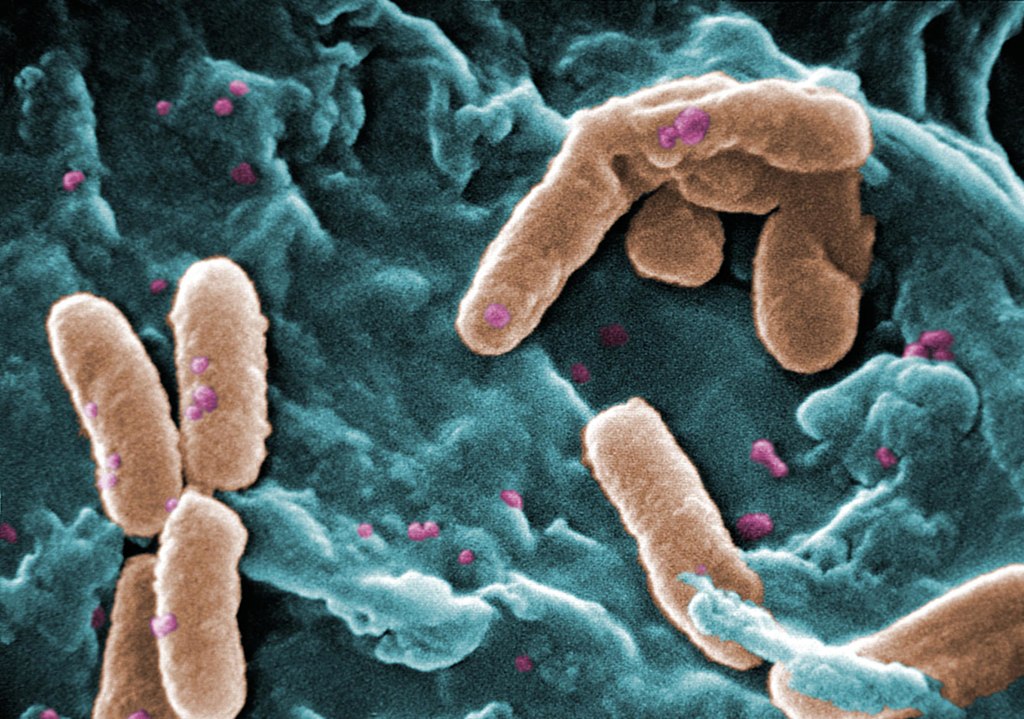
Scanning Electron Micrograph of Pseudomonas aeruginosa. Credit: CDC/Janice Carr
Using a modified version of the bacterium Mycoplasma pneumoniae, researchers have designed the first ‘living medicine’ to treat lung infections. Their method is reported in the journal Nature Biotechnology. The treatment targets Pseudomonas aeruginosa, a common source of hospital-acquired infections and which is naturally multi-drug resistant.
Researchers removed the M. pneumoniae‘s ability to cause disease and repurposing it to attack P. aeruginosa instead. The modified bacterium is used in combination with low doses of antibiotics that would otherwise not work on their own.
Researchers tested the efficacy of the treatment in mice, finding that it significantly reduced lung infections. The ‘living medicine’ doubled mouse survival rate compared to not using any treatment. Administering a single, high dose of the treatment showed no signs of toxicity in the lungs. Once the treatment had finished its course, the innate immune system cleared the modified bacteria in a period of four days.
P. aeruginosa infections are difficult to treat because the bacteria lives in communities that form biofilms. Biofilms can attach themselves to various surfaces in the body, forming impenetrable structures that escape the reach of antibiotics.
P. aeruginosa biofilms can grow on the surface of endotracheal tubes used by critically-ill patients who require mechanical ventilators to breathe. This causes ventilator-associated pneumonia (VAP), a condition affecting 9–27% of patients who require intubation. The incidence exceeds 50% for patients intubated because of severe COVID. VAP can extend the duration in intensive care unit for up to 13 days and kills 9–13% of patients.
The authors of the study engineered M. pneumoniae to dissolve biofilms by equipping it with the ability to produce various molecules including pyocins, toxins naturally produced by bacteria to kill or inhibit the growth of Pseudomonas bacterial strains. To test its efficacy, they collected P. aeruginosa biofilms from the endotracheal tubes of patients in intensive care units. They found the treatment penetrated the barrier and successfully dissolved the biofilms.
“We have developed a battering ram that lays siege to antibiotic-resistant bacteria. The treatment punches holes in their cell walls, providing crucial entry points for antibiotics to invade and clear infections at their source. We believe this is a promising new strategy to address the leading cause of mortality in hospitals,” says Dr María Lluch, co-corresponding author of the study.
With the aim of using the ‘living medicine’ to treat VAP, the researchers will carry out further tests before reaching the clinical trial phase. The treatment is expected to be administered using a nebuliser.
M. pneumoniae is one of the smallest known species of bacteria. Dr Luis Serrano first had the idea to modify the bacteria and use it as a ‘living medicine’ two decades ago. Dr Serrano is a specialist in synthetic biology, a field that involves repurposing organisms and engineering them to have new, useful abilities. With just 684 genes and no cell wall, the relative simplicity of M. pneumoniae makes it ideal for engineering biology for specific applications.
One of the advantages of using M. pneumoniae to treat respiratory diseases is that it is naturally adapted to lung tissue. After administering the modified bacterium, it travels straight to the source of a respiratory infection, where it sets up shop like a temporary factory and produces a variety of therapeutic molecules.
By showing that M. pneumoniae can tackle infections in the lung, the study opens the door for researchers creating new strains of the bacteria to tackle other types of respiratory diseases such as lung cancer or asthma. “The bacterium can be modified with a variety of different payloads – whether these are cytokines, nanobodies or defensins. The aim is to diversify the modified bacterium’s arsenal and unlock its full potential in treating a variety of complex diseases,” says ICREA Research Professor Dr. Luis Serrano.
In addition to designing the ‘living medicine’, Dr. Serrano’s research team are also using their expertise in synthetic biology to design new proteins that can be delivered by M. pneumoniae. The team are using these proteins to target inflammation caused by P. aeruginosa infections.
Though inflammation is the body’s natural response to an infection, excessive or prolonged inflammation can damage lung tissue. The inflammatory response is orchestrated by the immune system, which release mediator proteins such as cytokines. One type of cytokine, IL-10, has well-known anti-inflammatory properties and is of growing therapeutic interest.
Dr Serrano’s research group used protein-design software to engineer new versions of IL-10 purposefully optimised to treat inflammation. The cytokines were designed to be created more efficiently and to have higher affinity, meaning less cytokines are needed to have the same effect.
The researchers engineered strains of M. pneumoniae that expressed the new cytokines and tested its efficacy in the lungs of mice with acute P. aeruginosa infections. They found that engineered versions of IL-10 were significantly more effective at reducing inflammation compared to the wild type IL-10 cytokine.
According to Dr Ariadna Montero Blay, co-corresponding author of that study, “live biotherapeutics such as M. pneumoniae provide ideal vehicles to help overcome the traditional limitations of cytokines and unlock their huge potential in treating a variety of human diseases. Engineering cytokines as therapeutic molecules was critical to tackle inflammation. Other lung diseases such as asthma or pulmonary fibrosis could also stand to benefit from this approach.”
The evolutionary secrets that enable the traditional Chinese medicinal herb known as barbed skullcap to produce cancer fighting compounds have been unlocked by a collaboration of UK and Chinese researchers, who published their research in the journal Molecular Plant.
The researchers used DNA sequencing technology to assemble the genomic sequence of skullcap (Scutellaria barbata) known in China as banzhilian. This gave researchers the genetic information, a microevolutionary history, required to identify how the plant produces the compound scutebarbatine A, which acts against a range of cancer cells.
Professor Cathie Martin, Group Leader at the John Innes Centre, and one of the authors of the study said, “We have found that the primary metabolite has activity against cancer cells but not non-cancer cells which is especially important for an anti-cancer metabolite. Now we are looking to develop synthetic methods for producing more of the lead compound.”
In Traditional Chinese Medicine (TCM), to isolate medicinal chemistry from the plant, the herb is boiled in water for two hours and extract is dried to produce a powder and taken as a decoction (concentrated liquid). Now, with the knowledge of the genes that make up the biochemical pathway behind the anti-cancer activity of the herb, researchers are close to being able to synthesise larger quantities of compounds more rapidly and sustainably by using a host such as yeast.
The research is led by CEPAMS, a partnership between the John Innes Centre and the Chinese Academy of Science and supported by The Royal Society.
“This is a fantastic collaboration about developing interesting drug leads from natural resources and shows the practical value of focusing on the microevolution of a species” said Professor Martin.
The Skullcap genus has been used for centuries in TCM for treatment of different medical conditions. Clinical work has shown that preparations based on Scutellaria barbata during chemotherapy can reduce the risk of metastatic tumours.
CEPAMS Group Leader based at Shanghai Dr Evangelos Tatsis said, “Natural products have long been the lead compounds for the discovery of new drugs. By following the trail of the traditional Chinese plants, we can develop new anti-cancer medicines and this research marks a crucial step in that direction.”
Plant-based traditional medicines have long been used to provide leads for the new drug discovery, leading to drugs such as vinblastine and taxol which are now used clinically as anticancer drugs.
TCM is one of the best catalogued systems with empirical information about the therapeutic properties of herbal remedies.
Anti-cancer drugs obtained from traditional Chinese medicine have higher efficacy than chemical synthetic drugs and with less toxic side effects. The genomes of medicinal skullcaps reveal the polyphyletic origins of clerodane diterpene biosynthesis in the family Laminiaceae, is published in Molecular Plant
Medical science has not yet been able to explain why the Epstein-Barr virus triggers infectious mononucleosis (IM) in some people with initial infections and not in others. But now researchers have identified a unusual T cell response to the virus as the cause, and as a potential target for the development of vaccines. The findings were recently published in the journal Blood.
T cells normally fight the proliferation of the Epstein-Barr virus (EBV) in humans as part of an antiviral immune response. In this response, certain EBV components (peptides) are presented to the T cells by a specific molecule (HLA-E), which is found on the surface of cells infected with EBV. This triggers a non-classical T-cell response that leads to the destruction of the infected cells. Due to a genetic variation (HLA-E*0103/0103), about one third of the population naturally has more HLA-E molecules on EBV-infected cells.
A recently published study has shown that the risk of developing IM following first-time infection with the Epstein-Barr virus depends strongly on this EBV-specific immune response.
“Our research revealed that people with the HLA-E*0103/0103 genetic variation have a lower risk of developing infectious mononucleosis than those who do not have the variation. Our experiments in the lab showed that this gene variation is associated with a highly pronounced EBV-specific -non-classical — immune response,” explained Hannes Vietzen from MedUni Vienna’s Center for Virology, the first author of the study.
Preventive and diagnostic possibilities
EBV is one of the most common viral infections in humans. On initial infection, the virus causes IM in some children and young adults; this disease is characterised by non-specific symptoms, such as fever, as well as exhaustion that in some cases can last for several months. Until now, it was unclear why a first-time EBV infection only leads to IM in a minority of people, while most do not present any symptoms whatsoever. The immune response that the researchers identified could also be a target for research into preventive measures: “This immune response was still measurable years after the initial EBV infection and generally provides long-lasting protection against reinfection with Epstein-Barr, so it might be worth focusing our attention on this mechanism with a view to developing new vaccines in future,” said Hannes Vietzen, looking ahead.
Another finding from the study could also open up new diagnostic options: “The combination of the unfavourable HLA-E genetic variation with certain EBV peptides also appears to play an important role in the development of EBV-associated lymphomas in transplant recipients,” Hannes Vietzen commented. “Analysis of the EBV strains found in these patients could be helpful in identifying high-risk patients at an early stage and treating them in good time.”