Tag: 2/8/22

Research Throws Cold Water on COVID ‘Lab Leak’ Theory

Photo by KOBU Agency on Unsplash

The source of the COVID pandemic likely is down to live animals sold at the Huanan Seafood Wholesale Market, according to an international team of researchers.

The researchers traced the start of the pandemic to the market in Wuhan, China, where animals susceptible to the virus were sold live immediately before the pandemic began. Their findings were published in a pair of papers in the journal Science.

The publications all but rule out other explanations for the start of the pandemic, such as the ‘lab leak’ hypothesis. The authors further conclude that the first spread to humans from animals likely occurred in two separate transmission events in the Huanan market in late November 2019.

The first study looked at the locations of the first known COVID cases, as well as swab samples taken various places in the market. The second study examined genomic sequences of SARS-CoV-2 from samples collected from COVID patients during the first weeks of the pandemic in China.

The first paper, led by University of Arizona virus evolution expert Michael Worobey and Professor Kristian Andersen, was able to determine the locations of almost all of the 174 COVID cases identified by the World Health Organization in December 2019, 155 of which were in Wuhan.

A ‘bullseye’ on the market

Analyses showed that these cases were clustered tightly around the Huanan market, whereas later cases were dispersed widely throughout Wuhan. A striking percentage of early COVID patients had not visited there but turned out to live near the market. This suggests that vendors got infected first and set off a chain of infections among community members in the surrounding area, said Worobey.

“In a city covering more than 3000 square miles, the area with the highest probability of containing the home of someone who had one of the earliest COVID cases in the world was an area of a few city blocks, with the Huanan market smack dab inside it,” said Worobey.

This conclusion was supported by another finding: When the authors looked at the geographical distribution of later COVID cases, from January and February 2020, they found a “polar opposite” pattern, Worobey said. While the cases from December 2019 mapped “like a bullseye” on the market, the later cases coincided with areas of the highest population density in Wuhan.

“This tells us the virus was not circulating cryptically,” Worobey said. “It really originated at that market and spread out from there.”

Worobey and collaborators also addressed the question of whether health authorities found cases around the market simply because that is where they looked.

To rule out bias even more, from the market outwards the team removed cases ran the stats again. They found that even when two-thirds of cases were removed, the findings remained the same.

“Even in that scenario, with the majority of cases, removed, we found that the remaining ones lived closer to the market than what would be expected if there was no geographical correlation between these earliest COVID cases and the market,” Worobey said.

The study also looked at swab samples taken from market surfaces like floors and cages after Huanan market was closed. SARS-CoV-2-positive samples were significantly associated with stalls selling live wildlife.

The researchers determined that mammals now known to be susceptible to SARS-CoV-2, including red foxes, hog badgers and raccoon dogs, were sold live at the Huanan market in the weeks preceding the first recorded COVID cases. The scientists developed a detailed map of the market and showed that SARS-CoV-2-positive samples reported by Chinese researchers in early 2020 showed a clear association with the western portion of the market, where live or freshly butchered animals were sold in late 2019.

“Upstream events are still obscure, but our analyses of available evidence clearly suggest that the pandemic arose from initial human infections from animals for sale at the Huanan Seafood Wholesale Market in late November 2019,” said Prof Andersen at Scripps Research, co-senior author of both studies.

Virus likely jumped from animals to humans more than once

The second study, was an analysis of SARS-CoV-2 genomic data from early cases.

The researchers combined epidemic modeling with analyses of the virus’s early evolution based on the earliest sampled genomes. They determined that the pandemic, which initially involved two subtly distinct lineages of SARS-CoV-2, likely arose from at least two separate infections of humans from animals at the Huanan market in November 2019 and perhaps in December 2019. The analyses also suggested that, in this period, there were many other animal-to-human transmissions of the virus at the market that failed to manifest in recorded COVID-19 cases.

Using molecular clock analysis, which relies on the natural pace with which genetic mutations occur over time, researchers established a framework for the evolution of the SARS-CoV-2 virus lineages. They found that a scenario of a singular introduction of the virus into humans rather than multiple introductions would not align with molecular clock data. Earlier studies had suggested that one lineage of the virus – named A and closely related to viral relatives in bats – gave rise to a second lineage, named B. The more likely scenario in which the two lineages jumped from animals into humans on separate occasions, both at the Huanan market, Worobey said.

“Otherwise, lineage A would have had to have been evolving in slow motion compared to the lineage B virus, which just doesn’t make biological sense,” said Worobey.

The two studies provide evidence that COVID originated via jumps from animals to humans at the Huanan market, likely following transmission to those animals from coronavirus-carrying bats in the wild or on farms in China. Moving forward, the researchers say scientists and public officials should seek better understanding of the wildlife trade in China and elsewhere and promote more comprehensive testing of live animals sold in markets to lower the risk of future pandemics.

Source: University of Arizona

UK Children’s Gender Identity Clinic to Close after Controversies

Photo by Sharon McCutcheon on Unsplash

Following a highly critical independent report and accusations of inadequate and unsafe care, the UK will shut down the Tavistock gender identity clinic for children – the only one in the country. It will be replaced by a number of smaller facilities with closer links with mental health care.

The Tavistock and Portman NHS Foundation Trust clinic, named the Gender Identity Development Service (GIDS), had faced complaints of both long waiting lists for a burgeoning number of referrals, as well as rushing to assign puberty-blocking drugs and cross-sex hormones to children experiencing gender dysphoria.

Concerns had been voiced as early as 2005, when a nurse working at the clinic said that patients were being assessed too quickly and giving in to pressure from interest groups. Nevertheless, demand for its services skyrocketed in later years, from less than 100 per year in 2010 to nearly 2500 by 2018. In 2018, concerns were raised anew, with staff going on to make serious public accusations.

In July 2019, Dr Kirsy Entwhisle, a psychologist at GIDS Leeds hub, said that staff misled patients and made decisions about young people’s “bodies and lives” without “robust evidence”. Some of the children had suffered “very traumatic early experiences” which had not been addressed by the staff. The trust’s safeguarding lead, Sonia Appleby, won a claim from an employment tribunal after trust managers tried to stop her from carrying out her role when staff raised concerns.

One of the loudest critics of Tavistock Centre is Keira Bell, who at 16 was assigned puberty blockers, then cross-sex hormones at 17, and had a double mastectomy at 20 before later de-transitioning.

The former patient, who said she was suffering from anxiety and depression at the time she received treatment, said medics should have considered her mental health issues, “not just reaffirm my naïve hope that everything could be solved with hormones and surgery”.

Along with the unnamed parent of an autistic girl at the clinic, she won a ruling against the NHS assigning cross-sex hormones to children under 16 – but was overturned on appeal.

Helen, a parent of a patient at the clinic, welcomed its closure, but expressed concern for the future of her son’s treatments, according to LGBT site Pink News. While she said her son was treated quickly and received puberty blocking drugs, “From that point on, it felt like it was a little bit like they were winging it,” she said.

During therapy sessions at Tavistock, she said her son was asked a lot of questions and treated “almost like a little bit of an academic curiosity”. She criticised the fact that the same staff evaluated children for medical interventions and also offered therapy session, creating “a fear that they would stop access to medical support”. In contrast to the legal claims of Keira Bell’s and the unnamed patient, she said that GIDS refused to even discuss cross-sex hormones.

Dr David Bell (no relation to Keira Bell) welcomed the closure of Tavistock, telling the BBC: “Some children have got the double problem of living with the wrong treatment, and the original problems weren’t addressed – with complex problems like trauma, depression, large instances of autism.”

Endometriosis Hijacks Foetal Tolerance to Evade the Immune System

Photo by Sora Shimazaki on Pexels

In about 10% of women, endometrium-like tissues (known as lesions) also grow outside of the uterus, leading to endometriosis. Endometriosis is characterised by pain and can cause infertility, but its molecular mechanisms and drivers remain unknown. Now, a comprehensive study reveals how lesions escape immune surveillance, by taking advantage of mechanisms for the body tolerating a foetus during pregnancy.

Definitive diagnosis and clinical response still present significant challenges, with a common treatment being hormonal therapy with surgery. Unfortunately, surgery must be repeated if lesions recur, and they often do. To improve the situation, a better understanding of how and why the lesions grow, their cellular makeup, their microenvironments, and other aspects of their biology is essential.

The Jackson Laboratory’s (JAX) Elise Courtois, PhD, in partnership with UConn Health’s gynaecological surgeon Danielle Luciano, MD, recently completed an important study to develop a comprehensive cell atlas of the disease based on lesions obtained from 14 individuals who had treatment for endometriosis

The paper, published Nature Cell Biology, includes a thorough comparison of healthy endometrium tissue and ectopic (outside their normal site) lesions. The data also describes the endometriosis microenvironment and the conditions that allow the lesions to form and grow in what should be unhospitable regions.

“The study builds a robust foundation for a better understanding of endometriosis and how it grows,” said Dr Luciano.   “It’s exciting progress that we hope leads to earlier diagnosis and the ability to specifically target these abnormal cells for better treatments.”

The research team worked with tissues from individuals who had lesion removal at UConn Health for relief of symptoms. All were also receiving hormone therapy, the most frequent endometriosis management strategy. Not surprisingly, given that lesions are described as endometrial-like tissues growing in the wrong place, the cellular composition of the lesions in the peritoneum were quite similar to that of the normal endometrium. On the other hand, ovarian lesions had extensive differences in both composition and gene expression from the peritoneal ones. So while both ovary and peritoneum are receptive to the formation of lesions, they represent different environments and lead to important cellular and molecular differences between the two sites. The finding indicates that site-specific therapeutic design may be necessary to develop more effective treatments.

Another aspect of endometriosis is that, like cancer, the lesions represent abnormal growth that would typically be eliminated by immune surveillance. The researchers therefore investigated the immune cells in the peritoneal lesion microenvironment to see why they do not eliminate the abnormal lesion cells. They found that macrophages and dendritic cells contribute to conditions that promote immune inhibition and the promotion of immunosurveillance escape. Their specific characteristics are similar to those associated with foetal tolerance during pregnancy, which suggests that endometriosis hijacks a necessary, naturally occurring immune process to allow for lesion formation and persistence.

The paper details other aspects of both normal endometrium and ectopic lesions, including properties of vascularisation and the drivers of regeneration in endometrium and, perhaps, the formation of lesions in endometriosis. Of particular interest were key differences in the vascularisation of peritoneal versus ovarian lesions, further emphasising the site-specific nature of endometriosis. Also of note was the identification of a previously uncharacterised population of epithelial cells that may be progenitor cells for both endometrium and lesion formation, but more work is needed to define their precise role.

“Single cell analyses and hyperplexed antibody-based imaging techniques offer powerful insights into the complexity of the endometriosis microenvironment,” said Dr Courtois. “Understanding this complexity will be key for developing the new, efficient diagnostic and therapeutic tools that are so badly needed.”

Overall, the data captures a full description of endometrium and lesions, laying a strong foundation for understanding the vital cellular players and molecular dynamics of the disease. The data represents an important step forward for research into endometriosis and provides essential information for future therapeutics and diagnostics that can provide relief for those with this under-investigated disease.

Source: University of Connecticut

A Step Closer to a Once-off Treatment for HIV

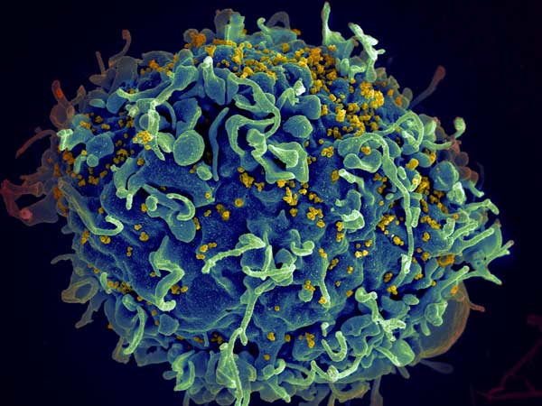
HIV invading a human cell
HIV invading a human cell: Credit NIH

Researchers from Tel Aviv University have demonstrated success of a novel technology that may be developed into a one-time vaccine to treat people with HIV and AIDS. Using CRISPR technology, the researchers engineered B cells that in turn stimulate the immune system to produce HIV-neutralising antibodies.

Published in Nature, the study was led by Dr Adi Barzel and PhD student Alessio Nehmad and conducted in collaboration with additional researchers from Israel and the US.

“Based on this study,” said Dr Barzel, “we can expect that over the coming years we will be able to produce a medication for AIDS, additional infectious diseases and certain types of cancer caused by a virus, such as cervical cancer, head and neck cancer and more.”

He explains that the treatment can become a kind of permanent medication, lingering in the body to fight the virus. “We developed an innovative treatment that may defeat the virus with a one-time injection, with the potential of bringing about tremendous improvement in the patients’ condition. When the engineered B cells encounter the virus, the virus stimulates and encourages them to divide, so we are utilising the very cause of the disease to combat it. Furthermore, if the virus changes, the B cells will also change accordingly in order to combat it, so we have created the first medication ever that can evolve in the body and defeat viruses in the ‘arms race’.”

When they mature, the antibody-generating B cells move into the blood and lymphatic system and from there to the different body parts.

Dr Barzel explained: “Until now, only a few scientists, and we among them, had been able to engineer B cells outside of the body. In this study, we were the first to do this within body and then make those cells generate the desired antibodies. The genetic engineering is conducted with viral carriers derived from viruses that were also engineered. We did this to avoid causing any damage, and solely bring the gene coded for the antibody into the B cells in the body.”

“Additionally, in this case we have been able to accurately introduce the antibodies into a desired site in the B cell genome. All lab models that had been administered the treatment responded, and had high quantities of the desired antibody in their blood. We produced the antibody from the blood and made sure it was actually effective in neutralising the HIV virus in the lab dish.”

Source: Tel Aviv University

CRISPR Editing can Destabilise the Genome, Study Finds

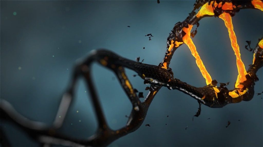
DNA repair
Source: Pixabay/CC0

A new study published in Nature Biotechnology identifies risks in the use of CRISPR gene editing, which is employed in a number of therapies. Looking at its use in T cells, the researchers detected a loss of genetic material in a significant percentage – up to 10% of the treated cells. They explain that such loss can lead to destabilisation of the genome, which might cause cancer.

The study was led by Drs Adi Barzel, Dr Asaf Madi and Dr Uri Ben-David at Tel Aviv University.

Developed about a decade ago, CRISPR cleaves DNA sequences at certain locations in order to delete unwanted segments, or alternately repair or insert beneficial segments. It has already proved impressively effective in treating a range of diseases – cancer, liver diseases, genetic syndromes, and more. In 2020 at the University of Pennsylvania, the first approved clinical trial ever to use CRISPR took T cells from a donor, and expressed an engineered receptor targeting cancer cells, while using CRISPR to destroy genes coding for the original receptor – which otherwise might have caused the T cells to attack cells in the recipient’s body. 

In the present study, the researchers sought to examine whether the potential benefits of CRISPR therapeutics might be offset by risks resulting from the cleavage itself, assuming that broken DNA is not always able to recover.

Dr Ben-David and his research associate Eli Reuveni explained: “The genome in our cells often breaks due to natural causes, but usually it is able to repair itself, with no harm done. Still, sometimes a certain chromosome is unable to bounce back, and large sections, or even the entire chromosome, are lost. Such chromosomal disruptions can destabilise the genome, and we often see this in cancer cells. Thus, CRISPR therapeutics, in which DNA is cleaved intentionally as a means for treating cancer, might, in extreme scenarios, actually promote malignancies.”

To examine the extent of potential damage, the researchers repeated the 2020 Pennsylvania experiment, cleaving the T cells’ genome in exactly the same locations – chromosomes 2, 7, and 14. Using single-cell RNA sequencing, they analysed each cell separately and measured the expression levels of each chromosome in every cell.

They detected a significant loss of genetic material in some of the cells. For example, when chromosome 14 had been cleaved, about 5% of the cells showed little or no expression of this chromosome. When all chromosomes were cleaved simultaneously, the damage increased, with 9%, 10%, and 3% of the cells unable to repair the break in chromosomes 14, 7, and 2 respectively. The three chromosomes did differ, however, in the extent of the damage they sustained. 

Dr Madi and his student Ella Goldschmidt explained: “Single-cell RNA sequencing and computational analyses enabled us to obtain very precise results. We found that the cause for the difference in damage was the exact place of the cleaving on each of the three chromosomes. Altogether, our findings indicate that over 9% of the T-cells genetically edited with the CRISPR technique had lost a significant amount of genetic material. Such loss can lead to destabilisation of the genome, which might promote cancer.”

Based on their findings, the researchers caution that extra care should be taken when using CRISPR therapeutics. They also propose alternative, less risky, methods, for specific medical procedures, and recommend further research into two kinds of potential solutions: reducing the production of damaged cells or identifying damaged cells and removing them before the material is administered to the patient.

Dr Barzel and his PhD student Alessio Nahmad conclude: “Our intention in this study was to shed light on potential risks in the use of CRISPR therapeutics,” adding that as scientists, they “examine all aspects of an issue, both positive and negative, and look for answers.”

Source: EurekAlert!