Tag: 2/11/21

A Trend of Amalgamations is Underway for Medical Schemes

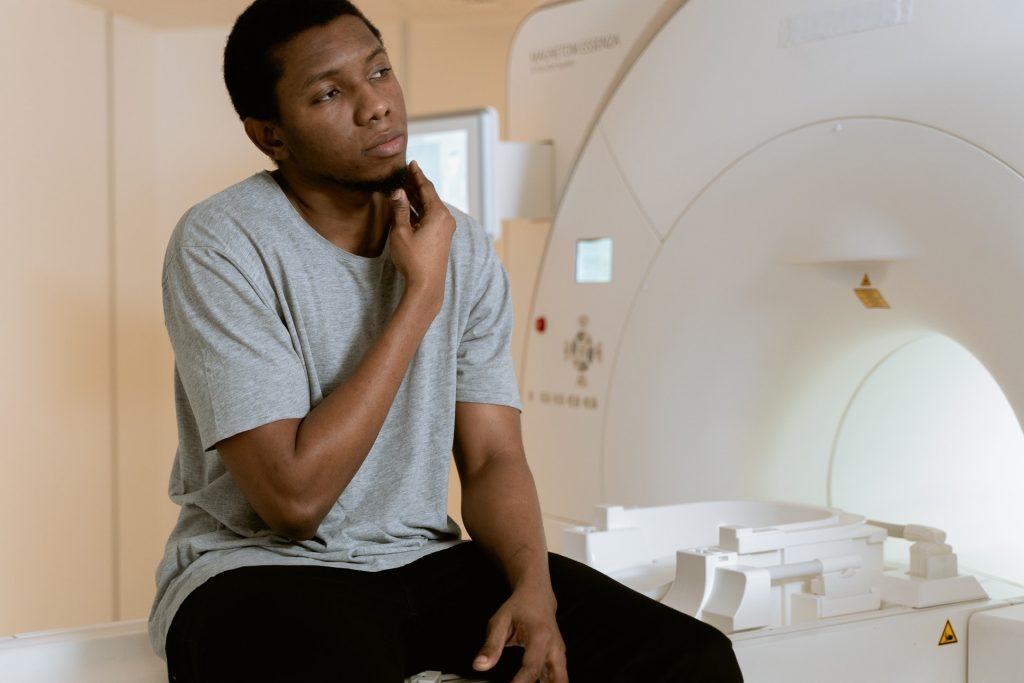
Image by Gustavo Fring on Pexels

A trend of amalgamations is underway in the South African private healthcare industry, in the face of growing challenges and the impending introduction of National Health Insurance (NHI).

Escalating healthcare inflation and costs, a declining and ageing membership, the impact of the COVID pandemic and a growing burden of disease are all impacting the not-for-profit Medical Scheme industry, which is highly regulated.

Medical scheme consolidation is one of the prominent trends, particularly given the prospect of NHI on the horizon, where smaller schemes will not compete, said Lee Callakoppen, principal officer of Bonitas Medical Fund.

The Council for Medical Schemes (CMS) advises that schemes that cannot compete sustainably on price should consider amalgamation partners, Callakoppen said.

“The trend towards amalgamations is not only for the sustainability of the medical scheme but for the benefit of members who ‘own’ the fund’.

“It is not only the call from CMS for schemes to join forces but also strict regulations around minimum solvency ratios and reserves which are more difficult for smaller schemes to maintain.”

It is a requirement of the Medical Schemes Act that medical schemes shall at all times maintain their business in a financially sound condition. They need to have sufficient assets for conducting business, providing for liabilities and having the prescribed solvency requirements of 25%, said Callakoppen.

“It’s a big ask for small schemes in this volatile and uncertain healthcare market,” he said.

A trend of amalgamation for small schemesThe CMS provides regulatory supervision of more than 80 medical schemes registered in the country and oversees amalgamation prospects.

One proviso for amalgamation is that schemes should complement each other and provide a more comprehensive offering to members.

“One clear indicator of risk is the size of the pool of lives being covered,” said Callakoppen. “Schemes with smaller risk pools are struggling to survive and experience more volatile claims”.

“Amalgamation into a bigger scheme means cross-subsidisation of costs. It is a trend I believe will continue, if not accelerate. In fact, in the past decade, we have seen 28 amalgamations approved by the CMS and the Competition Commission.”

The NHI has been criticised for potentially stifling innovation in healthcare, as well as not actually being able to fix the country’s flawed and unequal healthcare system.

Source: BusinessTech

Using The 5:2 Diet for Weight Loss in Gestational Diabetes

Photo by I Yunmai on Unsplash

In a welcome finding, researchers have found that women who have had gestational diabetes can use the popular 5:2 diet for weight loss to help prevent the onset of type 2 diabetes.   

It can be a challenge to lose weight and keep it off, especially for mothers with a new baby. The study by the University of South Australia suggests that the popular 5:2 or intermittent fasting diet is as effective as a conventional energy-restricting diet, giving women greater choice and flexibility for weight loss.

The 5:2 diet allows five days of normal eating each week while substantially restricting calories over two days a week, as opposed to a typical diet that requires moderate energy restrictions daily.

A fifth of pregnancies are affected by gestational diabetes, which carries a ten-fold risk of developing type 2 diabetes later in life, exacerbated even further by being overweight.

A welcome finding for a growing problem
The study’s lead researcher, Dr Kristy Gray, said women looking to lose weight will welcome the finding: “Gestational diabetes is the fastest growing type of diabetes in Australia, affecting 15% of pregnancies.

“Healthy eating and regular physical activity are recommended to manage gestational diabetes, with continuous energy restriction diets – or diets that cut calories by 25–30% being the most common strategy for weight loss and diabetes prevention.

“The trouble is, however, that new mums often put themselves last – they’re struggling with fatigue and juggling family responsibilities – so when it comes to weight loss, many find it hard to stick to a low-calorie diet.

“The 5:2 diet may provide a less overwhelming option. As it only cuts calories over two days, some women may find it easier to adopt and adhere to, as opposed to a consistently low-calorie diet requiring constant management.

“Our research shows that the 5:2 diet is just as effective at achieving weight loss as a continuous energy-restricted diet in women who have had gestational diabetes, which is great, because it provides women with greater choice and control,” she said, adding that women should seek advice from health professionals before starting the diet.

The research investigated the effects of both the 5:2 diet (five days eating normally and two days eating 500 calories) and a continuous energy-restricted diet (1500 calories per day) on weight loss and diabetes risk markers in women with a previous diagnosis of gestational diabetes. Both diets cut energy intake by about 25% a week.

The study was published in the American Journal of Clinical Nutrition.

Source: University of South Australia

How Osteocytes Form Their Networks of Dendrites

Phot by Nino Liverani on Unsplash

Embedded within bone tissue are osteocytes, and the tree-like communication cells known as dendrites. Loss of dendrites to ageing contributes to bone fragility and osteoporosis. 

A study published in Nature Communications has revealed how osteocytes form dendrites – a discovery that might yield strategies to maintain these projections and therefore help maintain people’s bone health throughout their lives.

In their study, the researchers found that deletion of Sp7, a gene linked to both rare and common skeletal diseases, in osteocytes causes severe defects in osteocyte dendrites. This gene codes for a protein called a transcription factor, which controls the expression of other genes. The team found that the Sp7 transcription factor targets a gene called osteocrin, which promotes osteocyte dendrite formation. In mice, activating the osteocrin gene made up for the absence of Sp7 and reversed defects in osteocyte dendrites.

“In this work, we demonstrate key roles for the transcription factor Sp7 and its target osteocrin in orchestrating a gene regulatory network needed to promote healthy connections between bone cells,” said senior author Marc Wein, MD, PhD, an investigator in the endocrine unit at MGH and an assistant professor of medicine at Harvard Medical School. “Understanding how osteocytes maintain this network of connections opens up exciting possibilities for new ways to treat osteoporosis and other diseases where bones are prone to fracture.”

Source: Massachusetts General Hospital

The Need for an African Genetic Library

Source: Mart Production on Pexels

Earlier this year, UCT professor Ambroise Wonkam published the Three Million African Genomes (3MAG) project in Nature, which he said started with a “crazy idea”. Now, it looks like his vision is starting to take shape.

The idea of creating a huge library of genetic information about the population of Africa emerged from his work on how genetic mutations among Africans contribute to conditions like sickle-cell disease and hearing impairments.

African genes contain great genetic variation, more than that seen outside of Africa. As he explained, “We are all African but only a small fraction of Africans moved out of Africa about 20–40 000 years ago and settled in Europe and in Asia.”

Another concern for Prof Wonkam is equity, saying: “Too little of the knowledge and applications from genomics have benefited the global south because of inequalities in health-care systems, a small local research workforce and lack of funding.”

Thus far only about 2% of genomes mapped are African, a good proportion of which are African American. This stes from a lack of prioritising funding, policies and training infrastructure, he says, but it also means the understanding of genetic medicine as a whole is lopsided. By studying African genomes, injustics can be corrected, such as finding that genetic risk profiles based on Europeans could be misleading for those of African descent.

To address these disparities, Prof Wonkam and other scientists are speaking to governments, companies and professional bodies across Africa and internationally, in order to build up capacity over the next decade to make the vision a reality.

He expects three million is the number needed to accurately map genetic variations across Africa. The project will take a decade, he says, costing around $450m per year, with industry already showing interest. 

Biotech firms welcome prospects of new data
The Centre for Proteomic and Genomic Research (CPGR) in Cape Town works with biotech firm Artisan Biomed on a variety of diagnostic tests. Gaps in the applicability of genetic data to the local population are a challenge for the firm, it said.

A genetic mutation in someone could be found but it would be uncertain if that variation is associated with a disease, especially as a marker for that particular population.

“The more information you have at that level, the better the diagnosis, treatment and eventually care can be for any individual, regardless of your ethnicity,” said Dr Lindsay Petersen, the company’s chief operations officer.

Artisan Biomed says the data it collects feeds back into CPGR’s research – allowing them to design a better diagnostic toolkit that is better suited to African populations, for instance.

Dr Judith Hornby Cuff said that the 3MAG project would help streamline processes and improve research, and one day could provide cheaper, more effective and more accessible health care, particularly in the strained South African system.

Prof Wonkam acknowledged that while the costs are huge, the project will “improve capacity in a whole range of biomedical disciplines that will equip Africa to tackle public-health challenges more equitably”.

“We have to be ambitious when we are in Africa. You have so many challenges you cannot see small, you have to see big – and really big,” he said.

Source: BBC News

New Type of Skin Cell Reveals Secrets of Inflammation

The surprise discovery of a new type of cell explains how distress to the skin early in life may prime a person for inflammatory skin disease later, according to a new study in Nature. This finding will likely lead to treatments for autoimmune disorders like scleroderma, and inform understanding of inflammatory disease.

“The results reinforce the idea that what you’re exposed to initially may have lasting ramifications,” said lead researcher Michael Rosenblum, MD, PhD. “It appears that early exposure to inflammation can, through these cells we discovered, imprint an ability for tissues to develop inflammatory disease later in life.”

The team came across this new type of cell while investigating the effects of certain actions known to evoke immune response in mice. One of these actions involved knocking out a group of skin cells that suppress the immune system. Without that regulation, said Dr Rosenblum, a unique cell was observed that seemed to act as a shelter for pathogenic immune cells not typically seen in skin tissues.

“We had to knock out one cell population to see that they were controlling the growth and capacity of these other, unknown cells,” he said, noting that the new cells only became apparent in the tissue exposed to inflammatory triggers. “What normally would be a deserted island on the skin was now inhabited by all these strangers,” he said.

The team dubbed these strangers ‘TIFFs’ (Th2-interacting fascial fibroblasts) after the Th2 immune cells that they help to house. The location of TIFFs in the skin suggests that they belong to a group of cells that make up the fibrous connective tissue that is fascia, said lead author Ian Boothby, a graduate student in Dr Rosenblum’s lab.

“Because most organs have fascia of some sort, what we’re learning about TIFFs in skin may well be widely applicable to the rest of the body, meaning that these cells may play a role in a huge number of inflammatory diseases,” he said.

Boothby and Dr Rosenblum when skin without regulatory cells receives inflammatory triggers, the TIFFs spread like wildfire and become a sort of holding pen for the Th2 immune cells. Later in life, when there is even a small insult to the skin, Dr Rosenblum said, the TIFFs open their floodgates, unleashing the Th2 cells.

It seems that, through these cells, early exposure to inflammalation can leave a life-long imprint.

“All you need to do is push the immune system just a little bit, with a wound or with stress, to unleash all the pathogenic cells living in these TIFFs and create an exaggerated inflammatory response,” he said.

The researchers hypothesise that the exaggerated response may manifest as the creation of fibroses in the fascia, the driving force behind inflammatory skin diseases such as scleroderma.

To confirm the presence of TIFFs in human skin, the team obtained samples from volunteers with eosinophilic fasciitis (EF), a rare inflammatory disorder in which eosinophils build up in the skin fascia, the fibrous tissue between the skin and the muscles below it.

Comparing the EF samples to those of healthy skin, the researchers found TIFFs in both, but looked completely different. In healthy skin, the fascia forms a thin, spidery network between fat cells, while in the EF skin sample, the cells had expanded to form thick bands of fibrous tissue.

Revealing the mysteries of inflammation
TIFFs appear to be present in every organ, said Dr Rosenblum, usually found in the fascia surrounding major organs and serve a role in maintaining structure. They’re also prone to interacting with immune cells. He postulates that TIFFs might have evolved as a sort of emergency brigade in case of injury, able to jump-start repair in the case of internal injury.

“In patients with scleroderma or other fibrosing diseases like EF, that repair program may be kind of co-opted, resulting in this chronic wound-healing response,” said Dr Rosenblum. “If we can understand the biology of these cells, we can come in with drugs that revert them back to what they’re supposed to be doing.”

Source: University of California San Francisco

Atmospheric Plasma Device Boosts Bone Regeneration

Photo by Zoltan Tasi on Unsplash

Scientists in Japan have developed a plasma device that promotes bone regeneration in fractures.

Unlike blood plasma, plasma here refers to the fourth state of matter, effectively a highly ionised gas, which has been long investigated as an effective surgical scalpel which cauterises tissue as it cuts. Other recent applications of plasma technology include surface sterilisation.

Now, a new type of plasma device, termed non-thermal atmospheric pressure plasma (NTAPP), was successfully tested in healing of bone fractures in animal bone defect models. It is cooler than most plasmas that are typically used. In a study published in PLOS ONE, researchers from Osaka City University detailed their findings using the technology in this world-first application.

Acceleration of cell growth
“NTAPP is considered a new therapeutic method,” said first author Akiyoshi Shimatani, “as it has been shown to accelerate cell growth when applied at low enough levels.” He explained that in an ambient atmosphere it can generate highly reactive oxygen and nitrogen species (RONS) which can be directly exposed to tissues.

Indirect treatments have shown the potential advantages of plasma in supporting the creation of stem cells that cause reactive oxygen species and in inducing osteogenic differentiation and bone formation, however, as the team points out there is no report on directly using NTAPP for bone fracture therapy. “Direct exposure of NTAPP is a key part of this study” states Jun-Seok Oh, professor at the OCU Graduate School of Engineering and advisor to the study, “It required a device specifically designed to generate and deliver RONS to areas of the bone defect ‘effectively’.”

The research group developed a pencil-like plasma device that can effectively generate and deliver RONS to an animal model with a well-established critical bone defect, allowing the team to search for the optimal exposure conditions. Comparing groups that were treated with NTAPP for 5, 10, and 15 minutes to control groups with no plasma administered, micro-CT images at eight weeks showed the 10-minute treatment time as the most successful bone regeneration with 1.51 times larger bone volume than the control group.

Since micro-CT images could not determine whether a bone defect has been filled with new bone, tissue or both, the team also ran a histological analysis and confirmed bone defects in the groups treated with plasma were in fact filled with new bone, and had no tissue or gaps like the control groups.

Precision therapy
The biological effect of plasma, like other therapies, depends on the treatment dose delivered into the targets. Although future research will be needed to clarify why the study saw the most bone regeneration during the 10-minute treatment period, surface wettability is understood to promote greater cell spreading and adhesion to biomaterials and implants. Hiroaki Nakamura, professor at the Graduate School of Medicine explained: “We wondered if something similar was occurring where we saw a strong generation of new bone. And we found that compared to the control group, bone surface of the plasma-treated group as statistically and significantly more hydrophilic.”

The research team hopes the plasma device they developed can be applied for surgical use.

Source: Osaka City University