Tag: 19/3/25

The Pupil as a Window into the Sleeping Brain

The eye of the sleeping subject was kept open with a special fixation device to record the pupil movements for several hours.  (Image: Neural Control of Movement Lab / ETH Zurich)

For the first time, researchers have been able to observe how the pupils react during sleep over a period of several hours. A look under the eyelids showed them that more happens in the brain during sleep than was previously assumed.

While eyes are typically closed in sleep, there is a flurry of activity taking place beneath the eyelids: a team of researchers, led by principal investigators Caroline Lustenberger, Sarah Meissner and Nicole Wenderoth from the Neural Control of Movement Lab at ETH Zurich, have observed that the size of the pupil fluctuates constantly during sleep. As they report in Nature Communications, sometimes it increases in size, sometimes it decreases; sometimes these changes occur within seconds, other times over the course of several minutes.

“These dynamics reflect the state of arousal, or the level of brain activation in regions that are responsible for sleep-wake regulation,” says Lustenberger. “These observations contradict the previous assumption that, essentially, the level of arousal during sleep is low.”

Instead, these fluctuations in pupil size show that even during sleep, the brain is constantly switching between a higher and lower level of activation. These new findings also confirm for humans what other research groups have recently discovered in studies on rodents, who also exhibit slow fluctuations in the activation level (known in the field as arousal).

New method for an old mystery

The regions of the brain which control the activation level are situated deep within the brainstem, making it previously difficult to directly measure these processes in humans during sleep. Existing methods are technically demanding and have not yet been established in this context. The ETH researchers’ study therefore relies on pupil measurements. Pupils are known to indicate the activation level when a person is awake. They can therefore be used as markers for the activity in regions situated deeper within the brain.

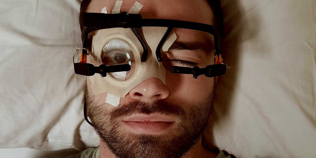
The ETH researchers developed a new method for examining the changes in people’s pupils while asleep: using a special adhesive technique and a transparent plaster, they were able to keep the eyes of the test subjects open for several hours.

“Our main concern was that the test subjects would be unable to sleep with their eyes open. But in a dark room, most people forget that their eyes are still open and they are able to sleep,” explains the study’s lead author, Manuel Carro Domínguez, who developed the technique.

Analysis of the data showed that pupil dynamics is related not just to the different stages of sleep, but also to specific patterns of brain activity, such as sleep spindles and pronounced deep sleep waves – brain waves that are important for memory consolidation and sleep stability. The researchers also discovered that the brain reacts to sounds with varying degrees of intensity, depending on the level of activation, which is reflected in the size of the pupil.

A central regulator of the activation level is a small region in the brainstem, known as the locus coeruleus. In animals, scientists have been able to show that this is important for the regulation of sleep stages and waking. The ETH researchers were unable to prove in this study whether the locus coeruleus is indeed directly responsible for pupil changes. “We are simply observing pupil changes that are related to the level of brain activation and heart activity,” Lustenberger explains.

In a follow-up study, the researchers will attempt to influence the activity of the locus coeruleus using medication, so that they can investigate how this affects pupil dynamics. They hope to discover whether this region of the brain is in fact responsible for controlling the pupils during sleep, and how changes in the level of activation affect sleep and its functions.

Using pupillary dynamics to diagnose illnesses

Understanding pupil dynamics during sleep could also provide important insights for the diagnosis and treatment of sleep disorders and other illnesses. The researchers therefore want to investigate whether pupil changes during sleep can provide indications of dysfunctions of the arousal system. These include disorders such as insomnia, post-traumatic stress disorder and possibly Alzheimer’s. “These are just hypotheses that we want to investigate in the future,” says Lustenberger.

Another goal is to make the technology usable outside of sleep laboratories, such as in hospitals where it could help to monitor waking in coma patients or to diagnose sleep disorders more accurately. The pupil as a window onto the brain could thus pave the way for new opportunities in sleep medicine and neuroscience.

Source: ETH Zurich

Inflammation Pathways are Linked to Changes in Bone Mineral Density over Time

Osteoporosis. Credit: Scientific Animations CC4.0

In one of the first studies of its kind, a team of researchers from Keck School of Medicine of USC has found that proteins and pathways involved in inflammation are associated with changes in bone mineral density (BMD) over time. Findings from the study were published in the Journal of Bone and Mineral Research.

The research, which was supported by the National Institutes of Health, could potentially lead to the identification of biomarkers that would serve as early indicators of a person’s risk for bone health issues later in life.

Bone mineral density is a measure of bone strength quantified by the amount of minerals in bone tissue. It peaks during young adulthood and slowly declines over the rest of the life cycle. BMD serves as an important marker for bone health and is commonly used to predict the risk of osteoporosis and other bone health conditions.

“Proteins are also substantial in the formation and maintenance of bone, and recently more studies have been trying to identify individual proteins associated with bone health,” says Emily Beglarian, the lead author and an epidemiology doctoral candidate in the Department of Population and Public Health Sciences at the Keck School of Medicine.

The study followed 304 obese/overweight Latino adolescents between the ages of 8 to 13 at baseline from the Study of Latino Adolescents at Risk for Type 2 Diabetes over an average period of three years. The researchers examined associations between over 650 proteins and annual measures of BMD, making this one of the first studies to evaluate these associations over years of follow-up. The proteins found to be associated with BMD were then inputted into a protein pathway database.

“The software determined what pathways the proteins were involved in within the human body. Our primary findings were that many of the proteins associated with BMD were involved in inflammatory and immune pathways in adolescent populations. There are other studies that found some of these same pathways were associated in older adult populations,” says Beglarian.

Existing studies suggest chronic inflammation can disrupt normal bone metabolism leading to lower BMD.

Importance of inclusive research

Childhood is a critical period for the development of BMD and this period can predict lifelong bone health.

“Until now, existing studies have centred on very specific populations. Most of them have small sample sizes, include either Chinese or non-Hispanic white populations, and focus on older adults – primarily on women because osteoporosis is four times more common in women than men,” says Beglarian.

“This is one of the first studies to investigate associations between proteins and BMD in younger populations. Investigating bone mineral density in early stages of life is important to determine how to address factors that may prevent people from reaching their potential peak bone density,” says Beglarian.

Advancing the understanding of bone health biomarkers

Additionally, Beglarian examined associations between BMD and a subset of protein markers from the initial proteins, in a separate cohort of young adults. Here she found that several proteins had similar associations with lower BMD. Low BMD is a risk factor for development of adulthood osteopenia and osteoporosis.

The study’s findings could potentially inform the development of biomarkers of bone health to identify people at risk that might benefit from intervention.

“It was interesting to see the way in which our study overlapped and differed with existing studies. Previous research was investigating BMD at the end of life when levels are already much lower,” she says. “Through my research I hope to address factors that decrease BMD earlier in life to help people get to their highest potential peak density, so they are set up over the rest of their lifetime to have a higher BMD.”

Source: Keck School of Medicine of USC

New Strategy to Reduce Tissue Damage from Flesh-eating Bacteria

Streptococcus Pyrogenese bound To human neutrophil. Credit: NIH

A new study published in Nature Communications reveals a novel approach to mitigating tissue damage caused by Streptococcus pyogenes, the flesh-eating bacterium responsible for severe infections such as necrotising fasciitis. The research highlights how disrupting bacterial metabolism can help the body better tolerate infection and heal more effectively. 

The study was led by Wei Xu, PhD, an assistant professor of biomedical sciences at the Marshall University Joan C. Edwards School of Medicine, and colleagues at Washington University School of Medicine and Central China Normal University. The team discovered that S. pyogenes manipulates the body’s immune response through its aerobic mixed-acid fermentation process, which produces metabolic byproducts, such as acetate and formate, that impair immune cell function, delay bacterial clearance and slow wound healing. 

By inhibiting this bacterial metabolic pathway with a pyruvate dehydrogenase inhibitor, the team successfully reduced tissue damage in a mouse model of necrotising skin infection. These findings suggest that reprogramming bacterial metabolism could serve as a novel therapeutic approach, not only to improve host tolerance but also as a potential adjuvant therapy alongside antibiotics. This strategy could enhance the effectiveness of existing treatments, particularly in severe infections where antibiotic resistance or excessive inflammation worsens patient outcomes. 

“This study sheds light on how bacterial metabolism influences the immune system,” Xu said. “By understanding these interactions, we can develop new treatment strategies that protect tissues, enhance antibiotic efficacy and improve patient outcomes.” 

Source: Marshall University Joan C. Edwards School of Medicine

Nature Relieves Physical Pain Signals in the Brain

This effect even occurs with virtual nature – such as nature videos

Photo by Sebastian Unrau on Unsplash

In a new study, an international team of neuroscientists led by the University of Vienna has shown that experiencing nature can alleviate acute physical pain. Surprisingly, simply watching nature videos was enough to relieve pain. Using functional magnetic resonance imaging, the researchers found that acute pain was rated as less intense and unpleasant when watching nature videos – along with a reduction in brain activity associated with pain. The results, published in Nature Communications, suggest that nature-based therapies can be used as promising complementary approaches to pain management.

“Pain processing is a complex phenomenon” explains study lead and doctoral student Max Steininger from the University of Vienna. In order to better understand it and identify treatment options, Steininger and his colleagues investigated how nature exposure influences pain: participants suffering from pain were shown three types of videos: a nature scene, an indoor scene, and an urban scene. The participants rated the pain while their brain activity was measured using functional magnetic resonance imaging. The results were clear: when viewing the nature scene, the participants not only reported less pain but also showed reduced activity in brain regions associated with pain processing.

By analyzing the brain data, the researchers showed that viewing nature reduced the raw sensory signal the brain receives when in pain. “Pain is like a puzzle, made up of different pieces that are processed differently in the brain. Some pieces of the puzzle relate to our emotional response to pain, such as how unpleasant we find it. Other pieces correspond to the physical signals underlying the painful experience, such as its location in the body and its intensity. Unlike placebos, which usually change our emotional response to pain, viewing nature changed how the brain processed early, raw sensory signals of pain. Thus, the effect appears to be less influenced by participants’ expectations, and more by changes in the underlying pain signals,” explains Steininger.

Claus Lamm, head of research in the group, adds: “From another ongoing study, we know that people consistently report feeling less pain when exposed to natural environments. However, the underlying reason for this has remained unclear – until now. Our study suggests that the brain reacts less to both the physical source and the intensity of the pain.”

The current study provides important information on how nature can help alleviate pain and highlights that nature-based therapeutic approaches can be a useful addition to pain treatment. The fact, that this effect was observed by simply watching nature videos suggests that taking a walk outdoors may not be necessary. Virtual nature – such as videos or virtual reality – appears to be effective as well. This opens up a wide range of possible applications in both the private and medical sectors, providing people with a simple and accessible way to relieve their pain.

The study was conducted at the University of Vienna in collaboration with researchers from the Universities of Exeter and Birmingham (UK) and the Max Planck Institute for Human Development.

Source: University of Vienna