Tag: 18/4/24

Bacteria Behind Meningitis in Babies Explains Resurgence
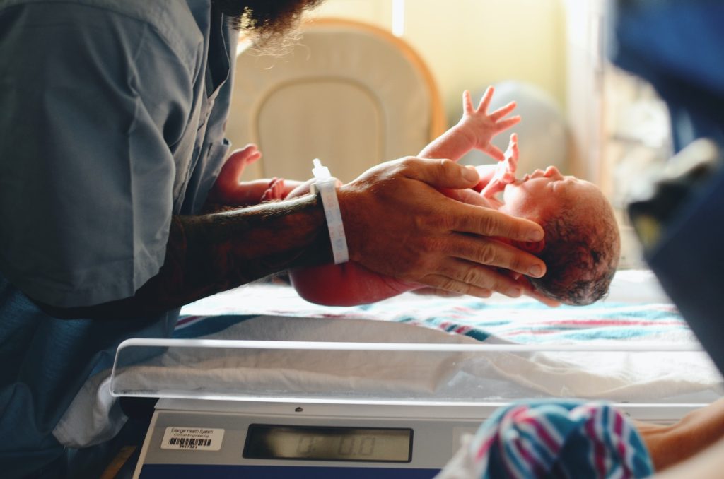
Photo by Christian Bowen on Unsplash

A milestone study led by University of Queensland researchers has identified the main types of E. coli bacteria that cause neonatal meningitis, and revealed why some infections recur despite being treated with antibiotics.

The study, published in eLife, discovered that about 50% of neonatal meningitis infections are caused by two types of E. coli.

E. coli is the most common cause of meningitis in babies born pre-term, but knowing which types allows us to test for those strains and treat them appropriately,” said Professor Mark Schembri, who led the study along with Dr Nhu Nguyen and Associate Professor Adam Irwin.

The study was the largest of its type, examining the genomes of 58 different E. coli bacteria across four continents and using samples collected over 46 years. It found that two types of the bacteria were responsible for the majority of neonatal infections.

Rapid diagnosis and monitoring are key

Associate Professor Irwin, who is also a paediatric infectious disease specialist at the Queensland Children’s Hospital, said speed is of the essence to prevent lasting damage.

“While antibiotics can be effective in treating the infection, this relies on rapid diagnosis. Also, antibiotics don’t always eliminate the bacteria – some of the babies we tracked showed signs of a complete recovery before suffering repeated invasive E. coli infections,” he said.

The researchers discovered the bacteria causing subsequent infections were the same as in the initial infection.

“It’s most likely that bacteria hide out in the intestinal microbiome,” Professor Schembri said. “This tells us we need to keep monitoring these babies after their first infection, as they are at a high risk of subsequent infection.”

Professor Schembri said the E. coli that can lead to meningitis also cause urinary tract infections and colonise the intestinal tract. “There is something about these types of E. coli that equips them to cause both infections,” he said.

“Our next step is to examine the bacteria’s pathway from the intestinal tract or urinary tract into the bloodstream, and then to the brain, so we can consider new ways to stop them.”

Source: University of Queensland

Tracing the Earliest Embryonic Formation of Faces

ES cells can now be used to induce structures with regionalised maxillary and mandibular primordia through the neural crest cell state, allowing for the recapitulation of jaw development in vitro. (POU3F3+ for maxillary and HAND2+ for mandibular)

CREDIT: KyotoU/Mototsugu Eiraku and Yusuke Seto

The highly complex shapes of animal faces originate from their respective transient neural crest cells. These embryonic pluripotent cells within the facial primordium – the early development form – may be necessary for forming proper facial structures. They migrate from their dorsal origin to the ventral craniofacial primordium and contribute to the cartilage, bones, and connective tissues. Analysing the molecular mechanisms in such early stages of development however poses many technical challenges.

Now, a group of Kyoto University researchers have produced neural crest cell-rich aggregates from human pluripotent stem cells and also developed a method to differentiate them in cell populations with a branchial arch-like gene expression pattern. Their research is published in Nature Communications.

“After the cell populations differentiate into precursors of maxillary and mandibular cells in response to external signalling factors, these populations spontaneously form patterns of the facial primordium,” explains Yusuke Seto of KyotoU’s Institute for Life and Medical Sciences.

This cartilage-like structure, reminiscent of Meckel’s cartilage, is formed locally within the aggregates.

“We aim to establish a model for studying early facial development by using the properties of human pluripotent stem cells to generate in vitro tissue resembling the bronchial arch of the primordial face,” adds Ryoma Ogihara, also of the Institute.

Researchers are examining the various developmental processes that cause interspecific and individual differences in facial structure to explain conditions such as craniofacial disorders.

“Using our in vitro model could help us better understand and control signal integration during the fate determination of the branchial arch and cartilage formation in the face and elsewhere. We hope our technology can contribute to the development of cellular materials for new regenerative medicine,” adds Mototsugu Eiraku, also of the Institute.

Source: University of Kyoto

Newly Found Retinal Cells may Paint a Complete Picture of Colour Vision

Photo by Jeffrey Riley on Unsplash

Scientists have long studied just how the eye’s three cone photoreceptor types work together to allow humans to perceive colour. In a new study in the Journal of Neuroscience, researchers at the University of Rochester used adaptive optics to identify rare retinal ganglion cells (RGCs) that could help fill in the gaps in existing theories of colour perception.

The retina has three types of cones to detect colour that are sensitive to either short, medium, or long wavelengths of light. Retinal ganglion cells transmit input from these cones to the central nervous system.

In the 1980s, David Williams, the William G. Allyn Professor of Medical Optics, helped map the “cardinal directions” that explain colour detection. However, there are differences in the way the eye detects colour and how colour appears to humans. Scientists suspected that while most RGCs follow the cardinal directions, they may work in tandem with small numbers of non-cardinal RGCs to create more complex perceptions.

Recently, a team of researchers from Rochester’s Center for Visual Science, the Institute of Optics, and the Flaum Eye Institute identified some of these elusive non-cardinal RGCs in the fovea that could explain how humans see red, green, blue, and yellow.

“We don’t really know anything for certain yet about these cells other than that they exist,” says Sara Patterson, a postdoctoral researcher at the Center for Visual Science who led the study. “There’s so much more that we have to learn about how their response properties operate, but they’re a compelling option as a missing link in how our retina processes colour.”

Adaptive optics peer past the eye’s natural distorations

The team took advantage of adaptive optics, which uses a deformable mirror to overcome light distortion and was first developed by astronomers to reduce atmospheric blurring in ground-based telescopes. In the 1990s, Williams and his colleagues began applying adaptive optics to study the human eye. They created a camera that compensated for distortions caused by the eye’s natural aberrations, producing a clear image of individual photoreceptor cells.

“The optics of the eye’s lens are imperfect and really reduce the amount of resolution you can get with an ophthalmoscope,” says Patterson. “Adaptive optics detects and corrects for these aberrations and gives us a crystal-clear look into the eye. This gives us unprecedented access to the retinal ganglion cells, which are the sole source of visual information to the brain.”

Patterson says improving our understanding of the retina’s complex processes could ultimately help lead to better methods for restoring vision for people who have lost it.

“Humans have more than 20 ganglion cells and our models of human vision are only based on three,” says Patterson. “There’s so much going on in the retina that we don’t know about. This is one of the rare areas where engineering has totally outpaced visual basic science. People are out there with retinal prosthetics in their eyes right now, but if we knew what all those cells do, we could actually have retinal prosthetics drive ganglion cells in accordance with their actual functional roles.”

Source: University of Rochester

To Beat Lung Fibrosis, Researchers Turn to Body’s own Healing Powers

Photo by Robina Weermeijer on Unsplash

The most common type of lung fibrosis is idiopathic – of unknown cause. Researchers are urgently trying to find ways to prevent or slow idiopathic pulmonary fibrosis (IPF) and related lung conditions, which can cause worsening shortness of breath, dry cough, and extreme fatigue. Average survival following diagnosis of IPF is just three to five years, and the disease has no cure.

A recent U-M study from a team led by Sean Fortier, MD and Marc Peters-Golden, MD at University of Michigan Medical School uncovers a pathway used during normal wound healing that has the potential to reverse IPF. They published their research in the Journal of Clinical Investigation.

Using a mouse model, they simulated IPF by administering bleomycin, a chemotherapy agent that causes cell injury and confirmed that the resulting lung scarring resolved itself over the span of about six weeks.

Because of this, “studying fibrosis is kind of tough,” said Fortier. “If we’re going to give experimental drugs to try and resolve fibrosis, we have to do it before it resolves on its own.

Otherwise, we will not be able to tell if the resolution was the action of the drug or natural repair mechanisms of the body.”

However, he said, “there’s actually a lot to learn about how the mouse gets better on its own. If we can learn the molecular mechanisms by which this occurs, we may uncover new targets for IPF.”

The process by which lung injury either leads to healing or fibrosis relies in part on what happens to fibroblasts – cells which forms connective tissue.

During injury or illness, fibroblasts are activated, becoming myofibroblasts that form scar tissue by secreting collagen. When the job is done, these fibroblasts must be deactivated, or de-differentiated, to go back to their quiet state or undergo programmed cell death and be cleared.

“This is the major distinction between normal wound healing and fibrosis – the persistence of activated myofibroblasts,” explained Fortier. That deactivation is controlled by molecular brakes. The study examined one of these brakes, called MKP1 – which the team found was expressed at lower levels in fibroblasts from patients with IPF.

By genetically eliminating MKP1 in fibroblasts of mice after establishing lung injury, the team saw that fibrosis continued uncontrolled.

“Instead of at day 63, seeing that nice resolution, you still see fibrosis,” said Fortier.

“We argued by contradiction: when you knock out this brake, fibrosis that would otherwise naturally disappear, persists and therefore MKP1 is necessary for spontaneous resolution of fibrosis.”

They performed several additional studies using CRISPR techniques to demonstrate how MKP1 applies the brakes, mainly by deactivating the enzyme p38α, which is implicated in a cell’s reaction to stress.

Furthermore, they demonstrated that neither of the two current FDA approved drugs for lung fibrosis, pirfenidone and nintedanib, are able to turn off myofibroblasts.

“That’s totally in keeping with the fact that they do slow the progression, but they don’t halt or reverse disease,” said Fortier.

Fortier hopes the discovery that this pathway reverses fibrosis leads to exploration of additional brakes on fibrosis.

“So much work on fibrosis has focused on how we can prevent it, but when a patient presents to my clinic with a dry cough, shortness of breath, and low oxygen as a result of underlying IPF, the scarring is already present. Of course, we’d love a way to prevent the scarring from getting worse, but the Holy Grail is to reverse it.”

Source: Michigan Medicine – University of Michigan

Epigenetic Changes Drive this Rare Malignant Paediatric Brain Tumour

A healthy neuron. Credit: NIH

A new study published in Life Science Alliance revealed how aberrant epigenetic regulation contributes to the development of atypical teratoid/rhabdoid (AT/RT) tumours, which mainly affect young children. There is an urgent need for more research in this area as current treatment options are ineffective against these highly malignant tumours.

Most tumours take a long time to develop as harmful mutations gradually accumulate in cells’ DNA over time. AT/RT tumours are a rare exception, because the inactivation of one gene gives rise to this highly aggressive form of brain cancer.

AT/RT tumours are rare central nervous system embryonic tumours that predominantly affect infants and young children.

On average, 73 people are diagnosed with AT/RT in the USA each year. However, AT/RT is the most common central nervous system tumour in children under one years old and accounts for 40-50% of diagnoses in this age group. The prognosis for AT/RT patients is grim, with a postoperative median survival of only 11-24 months.

The collaborative study conducted by Tampere University and Tampere University Hospital examined how aberrant DNA methylation distorts cellular developmental trajectories and thereby contributes to the formation of AT/RT. DNA methylation is a normal process of controlling expression whereby methyl groups are added to the DNA strand, adding epigenetic information.

The new study showed that DNA methylation interferes with the activity of multiple regulators, which usually regulate the differentiation and maturation of central nervous system cells during brain development. Disrupted cell differentiation promotes the abnormal, uncontrolled proliferation of cells that eventually form a tumour.

The study also found several genes that regulate cell differentiation or inhibit tumour development and are silenced in AT/RT together with increased DNA methylation.

“These results will provide deeper insights into the development of AT/RTs and their malignancy. In the future, the results will help to accelerate the discovery of new treatments for this aggressive brain tumour,” says senior author Docent Kirsi Rautajoki from Tampere University.

Source: Tampere University