Tag: 17/10/24

Elevated NK Cells Found in Children with Severe RSV

Photo by Andrea Piacquadio on Unsplash

Respiratory syncytial virus (RSV) is the leading cause of hospitalisation in young children due to respiratory complications such as bronchiolitis and pneumonia. Yet little is understood about why some children develop only mild symptoms while others develop severe disease.

To better understand what happens in these cases, clinician-scientists from Brigham and Women’s Hospital, and Boston Children’s Hospital analysed samples from patients’ airways and blood, finding distinct changes in children with severe cases of RSV, including an increase in the number of natural killer (NK) cells in their airways.

The descriptive study, which focuses on understanding the underpinnings of severe disease, may help to lay groundwork for identifying new targets for future treatments. Results are published in Science Translational Medicine.

“As a physician, I help to care for children who have the most severe symptoms, and as a researcher, I’m driven to understand why they become so sick,” said corresponding author Melody G. Duvall, MD, PhD, of the Division of Pulmonary and Critical Care Medicine at Brigham and Women’s Hospital (BWH) and the Division of Critical Care Medicine at Boston Children’s Hospital. “NK cells are important first responders during viral infection – but they can also contribute to lung inflammation. Interestingly, our findings fit with data from some studies in COVID-19, which reported that patients with the most severe symptoms also had increased NK cells in their airways. Together with previous studies, our data link NK cells with serious viral illness, suggesting that these cellular pathways merit additional investigation.”

Duvall and colleagues, including lead author Roisin B. Reilly of the Division of Pulmonary and Critical Care Medicine at BWH, looked at samples from 47 children critically ill with RSV, analysing immune cells found in their airways and peripheral blood. Compared to uninfected children, those with severe illness had elevated levels of NK cells in their airways and decreased NK cells in their blood. In addition, they found that the cells themselves were altered, both in appearance and in their ability to perform their immunological function of killing diseased cells.

Duvall and co-authors have previously described a post-pandemic surge in paediatric RSV infections. While clinicians can only provide supportive care to the most severely sick children, vaccines to prevent RSV are now available for children 19 months and younger, adults 60 years and over, and people who are pregnant.

Source: Brigham and Women’s Hospital

Faulty ‘Fight or Flight’ Drives C. Diff Infections

Clostridioides difficile. Credit: CDC

The portion of our nervous systems responsible for the “fight or flight” response can shape the severity of potentially deadly Clostridioides difficile infections, new research from the School of Medicine reveals in Cell Reports Medicine.

The findings suggest that doctors may be able to save patients from the infections – a plague for hospitals and nursing homes – by using drugs to quiet the hyperactive nervous system response, the researchers say.

“Compared to how much we know about immune system influences in C. difficile infections, the field is just scratching the surface in understanding neuronal contributions to disease,” said researcher William A. Petri Jr., MD, PhD, of UVA Health’s Division of Infectious Diseases and International Health. “Newly identifying components of the nervous system that worsen inflammation will allow us to determine potential therapeutic targets and biomarkers for patients at risk of severe disease.”

C. difficile, is a perpetual burden for healthcare facilities. Extensive antibiotic use, particularly among patients who are hospitalised or in nursing care, can allow it to establish dangerous infections. Further, patients who make it through the severe diarrhoea, nausea, fever and colitis C. difficile can cause are not necessarily in the clear: One in six will develop another C. diff infection within eight weeks, according to the federal Centers for Disease Control and Prevention.

The new UVA research reveals the critical role the nervous system plays in severe C. difficile infections. The researchers found that the “sympathetic” nervous system – the branch that responds to dangerous situations – can be a key driver of serious C. diff.

Normally, our “fight or flight” response is helpful for avoiding danger. It helps us respond quickly, improves our eyesight, boosts our strength. It also can stimulate our immune system and help us recover from injury. But in C. difficile cases, the nervous system can have a hyperactive response that becomes part of the problem, and UVA’s new research explains why.

“Neurons are the first responders that coordinate defences against toxic attacks. Sometimes those responders don’t recruit the right size and kind of artillery and that can make things worse,” said researcher David Tyus, a neuroscience graduate student at UVA. “Interestingly, the receptor we identified as important in C. difficile infection [the alpha 2 adrenergic receptor] has also been linked to irritable bowel syndrome. I’m curious to know if there could be a unifying underlying mechanism between the two disease contexts.”

Promisingly, the researchers found that targeting the receptor in lab mice reduced intestinal inflammation and decreased C. difficile severity and mortality. That suggests that, with further research, doctors may be able to take a similar tact to better treat severe C. diff infections in patients. For example, they may be able to surgically remove a portion of nerves in the gut, or they may be able to develop medicines to target the alpha 2 receptor – as Petri and Tyus are attempting to do.

“Our next step is to determine which cells with the alpha 2 receptor are receiving signals from the sympathetic nervous system and play a role in C. difficile-mediated disease,” Petri said. “We are very excited to think about how our findings translate to clinic and how the sympathetic nervous system might play a role in recurrent infection. I hope that this study sets the foundation for future findings of how neurons affect the course of C. difficile infection outcomes.”

Source: University of Virginia Health System

Alzheimer’s Disease may Damage the Brain in Two Phases

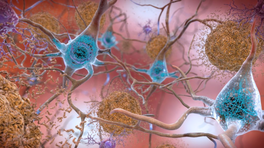
Neurons in the brain of an Alzheimer’s patient, with plaques caused by tau proteins. Credit: NIH

Alzheimer’s disease may damage the brain in two distinct phases, based on new research funded by the National Institutes of Health (NIH) using sophisticated brain mapping tools. According to researchers who discovered this new view, the first, early phase happens slowly and silently – before people experience memory problems – harming just a few vulnerable cell types. In contrast, the second, late phase causes damage that is more widely destructive and coincides with the appearance of symptoms and the rapid accumulation of plaques, tangles, and other Alzheimer’s hallmarks.

“One of the challenges to diagnosing and treating Alzheimer’s is that much of the damage to the brain happens well before symptoms occur. The ability to detect these early changes means that, for the first time, we can see what is happening to a person’s brain during the earliest periods of the disease,” said Richard J. Hodes, MD, director, NIH National Institute on Aging. “The results fundamentally alter scientists’ understanding of how Alzheimer’s harms the brain and will guide the development of new treatments for this devastating disorder.”

Scientists analysed the brains of 84 people, and the results, published in Nature Neuroscience, suggest that damage to one type of cell, called an inhibitory neuron, during the early phase may trigger the neural circuit problems that underlie the disease. Additionally, the study confirmed previous findings about how Alzheimer’s damages the brain and identified many new changes that may happen during the disease.

Specifically, the scientists used advanced genetic analysis tools to study the cells of the middle temporal gyrus, a part of the brain that controls language, memory and vision. The gyrus has been shown to be vulnerable to many of the changes traditionally seen during Alzheimer’s. It is also a part of the brain that researchers have thoroughly mapped for control donors. By comparing control donor data with that from people who had Alzheimer’s, the scientists created a genetic and cellular timeline of what happens throughout the disease.

Traditionally, studies have suggested that the damage caused by Alzheimer’s happens in several stages characterized by increasing levels of cell death, inflammation and the accumulation of proteins in the form of plaques and tangles. In contrast, this study suggests that the disease changes the brain in two “epochs” – or phases – with many of the traditionally studied changes happening rapidly during the second phase. This coincides with the appearance of memory problems and other symptoms.

The results also suggest that the earliest changes happen gradually and “quietly” in the first phase before any symptoms appear. These changes include slow accumulation of plaques, activation of the brain’s immune system, damage to the cellular insulation that helps neurons send signals and the death of cells called somatostatin (SST) inhibitory neurons.

The last finding was surprising to the researchers. Traditionally, scientists have thought that Alzheimer’s primarily damages excitatory neurons, which send activating neural signals to other cells. Inhibitory neurons send calming signals to other cells. The paper’s authors hypothesised how loss of SST inhibitory neurons might trigger the changes to the brain’s neural circuitry that underlie the disease.

Recently, a separate NIH-funded brain mapping study by researchers at MIT found that a gene called REELIN may be associated with the vulnerability of some neurons to Alzheimer’s. It also showed that star-shaped brain cells called astrocytes may provide resilience to or resist the harm caused by the disease.

Researchers analysed brains that are part of the Seattle Alzheimer’s Disease Brain Cell Atlas, which is designed to create a highly detailed map of the brain damage that occurs during the disease. The project was led by Mariano I. Gabitto, PhD, and Kyle J. Travaglini, PhD, from the Allen Institute, Seattle. The scientists used tools – developed as part of the NIH’s BRAIN Initiative – Cell Census Network – to study more than 3.4 million brain cells from donors who died at various stages of Alzheimer’s disease.

“This research demonstrates how powerful new technologies provided by the NIH’s BRAIN Initiative are changing the way we understand diseases like Alzheimer’s. With these tools, scientists were able to detect the earliest cellular changes to the brain to create a more complete picture of what happens over the entire course of the disease,” said John Ngai, Ph.D., director of The BRAIN Initiative®. “The new knowledge provided by this study may help scientists and drug developers around the world develop diagnostics and treatments targeted to specific stages of Alzheimer’s and other dementias.”

Source: NIH/National Institute on Aging

Study of Osteoblast-blocking Protein could Help Future Osteoporosis Treatment

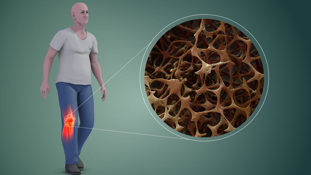
Osteoporosis. Credit: Scientific Animations CC4.0

Scientists have identified a protein that blocks the activity of bone-forming cells (osteoblasts) by stopping them from maturing during the journey to sites of bone formation, finds a new study published in Communications Biology.

A team of researchers led by Dr Amy Naylor and Professor Roy Bicknell along with their team including Dr Georgiana Neag from the University of Birmingham have found that protein CLEC14A, which is found on endothelial cells in bone, block the function of bone development cells called osteoblasts.

During bone development, the endothelial cell’s job is to transport immature osteoblasts to sites where new bone is needed. However, when the protein CLEC14A is also present on the outside of the endothelial cell, osteoblasts are prevented from maturing to the point where they can form bone tissue.

This additional understanding of how blood vessel cells control bone-forming osteoblasts under normal, healthy conditions provide an avenue to develop treatments for patients who have insufficient bone formation

Dr Amy Naylor

In this study, osteoblast cells were taken from transgenic mice that either have been bred to produce CLEC14A or not. The osteoblasts were subsequently used in vitro in an induction solution, and the team found that cells taken from the protein-free mice reached maturation after 4 days while those in the presence of CLEC14A matured 8 days later. Furthermore, the CLEC14A-free samples saw a significant increase in mineralised bone tissue at day 18 in the study.

Dr Amy Naylor, Associate Professor in the School of Infection, Inflammation and Immunology at the University of Birmingham said:

“In the last decade, a specific type of blood vessel cell was identified within bones. This blood vessel is called ‘type-H’ and is responsible for guiding bone-forming osteoblasts to the places where bone growth is needed. Now we have discovered that a protein called CLEC14A can be found on the surface of type-H blood vessel cells.

“In the experiments we performed, when CLEC14A protein is present the osteoblasts that were sharing a ride on the endothelial cells produce less bone. Conversely, when the protein is removed, they produce more bone.

“This additional understanding of how blood vessel cells control bone-forming osteoblasts under normal, healthy conditions provide an avenue to develop treatments for patients who have insufficient bone formation, for example in patients with fractures that do not heal, osteoporosis or with chronic inflammatory diseases.”

Source: University of Birmingham

Ultrasound Chronic Pain Relief Device Takes a Step Closer

Photo by Pawel Czerwinski on Unsplash

Chronic pain is often caused by faulty signals emerging deep within the brain, giving false alarms about a wound that has since healed, a limb that has since been amputated, or other intricate, hard-to-explain scenarios. Effective treatment options are sorely needed; now, a new device from the University of Utah may represent a practical long-sought solution, using ultrasound to target pain centres deep inside the brain.

Researchers at the University of Utah’s John and Marcia Price College of Engineering and Spencer Fox Eccles School of Medicine have published promising findings about an experimental therapy that has given many participants relief after a single treatment session.

At the core of this research is Diadem, a new biomedical device that uses ultrasound to noninvasively stimulate deep brain regions, potentially disrupting the faulty signals that lead to chronic pain.

The Diadem Device

The findings of a recent clinical trial are published in the journal PainThis study constitutes a translation of two previous studies, published in Nature Communications Engineering and IEEE Transactions on Biomedical Engineering, which describe the unique features and characteristics of the device.

The study was conducted by Jan Kubanek, professor in Price’s Department of Biomedical Engineering (BME), and Thomas Riis, a postdoctoral researcher in his lab, and other collaborators.

The randomised sham-controlled study recruited 20 participants with chronic pain, who each experienced two 40-minute sessions with Diadem, receiving either real or sham ultrasound stimulation. Patients described their pain a day and a week after their sessions, with 60% of the experimental group receiving real treatment reporting a clinical meaningful reduction in symptoms at both points.

“We were not expecting such strong and immediate effects from only one treatment,” says Riis.

“The rapid onset of the pain symptom improvements as well as their sustained nature are intriguing, and open doors for applying these noninvasive treatments to the many patients who are resistant to current treatments,” Kubanek says.

Diadem’s approach is based on neuromodulation, a therapeutic technique that seeks to directly regulate the activity of certain brain circuits. Other neuromodulation approaches are based on electric currents and magnetic fields, but those methods cannot selectively reach the brain structure investigated in the researchers’ recent trial: the anterior cingulate cortex.

After an initial functional MRI scan to map the target region, the researchers adjust Diadem’s ultrasound emitters to correct for the way the waves deflect off of the skull and other brain structures. This procedure was published in Nature Communications Engineering.

The team is now preparing for a Phase 3 clinical, trial which is the final step before FDA approval to use Diadem as a treatment for the general public.

Source: University of Utah