Tag: 16/4/25

Are High Support Bras Bad for the Back?

Photo by John Arano on Unsplash

Research from the University of Portsmouth suggests that bras offering excessive bounce reduction may come with hidden consequences for spinal health.

Sports bras are extremely popular in the health and fitness world, with the bra industry often emphasising “bounce reduction” as a key indicator of a bra’s performance. However, a new study suggests that high-support bras that significantly reduce breast bounce could have a detrimental effect on the spine.

Published in the European Journal of Sport Science, the preliminary research revealed that bras designed to prevent breast bounce during exercise may unknowingly cause potential unseen consequences on the musculoskeletal system.

Dr Chris Mills and a team from the School of Psychology, Sport and Health Sciences at the University of Portsmouth employed advanced tools – including motion capture, force platforms, and a 3D surface scanner – to investigate the effects of breast movement on spinal rotational forces. Using a first-of-its-kind whole-body, female-specific musculoskeletal model, the study examined how varying levels of breast support influenced torso motion, breast forces, and spinal moments during running.

The findings revealed that while sports bras are essential for reducing breast pain during exercise, achieving 100 percent bounce reduction could unintentionally increase loading on the spine. 

Simulated conditions showed that bras eliminating breast movement led to higher spinal moments, which could elevate the risk of lumbar back pain. Researchers emphasised the importance of striking an optimal balance in bra design; reducing breast bounce without overloading the spine.

r Mills said: “While a supportive sports bra is crucial for exercise comfort, excessive bounce reduction may place additional strain on spinal muscles, increasing the risk of back pain.”

The study, built on two decades of research by the University’s Research Group in Breast Health, highlights the need for bra manufacturers to consider the unseen musculoskeletal impacts on the human body in their designs. Professor Wakefield-Scurr, often referred to as the ‘Bra Professor’, added, “These findings suggest that striving for maximum bounce reduction may inadvertently pose challenges to spinal health during activities like running.

“As sports bras evolve, this study challenges industry leaders to innovate designs that balance comfort, breast support, and holistic health, ensuring that bounce reduction doesn’t come at a cost to spinal health.”

The creation of a subject-specific female musculoskeletal model enabled researchers to gain a detailed understanding and approximation of changes in spinal moments, following simulated changes in breast motion during running.

Previous research by the Portsmouth team used the model to predict changes in spinal moments after breast surgery. 

“The musculoskeletal model could become a useful tool in predicting appropriate and personalised rehabilitation recommendations, which could help ease the loading on the spine after breast surgeries”, explained Dr Mills.

“Understanding the individual muscular contributions will help to develop personalised pre-surgical rehabilitation programs as well as bras that work in tandem with each female body to maximise performance and reduce injury risk.

“Moving forward the key goal is to determine what is the optimal amount of bounce reduction to both reduce exercise induced breast pain and also the internal loading on the spine during physical activity.”

Source: University of Portsmouth

Preventing Onset and Development of Heart Failure with Preserved Ejection Fraction

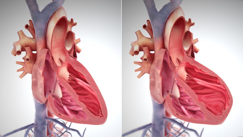
Right side heart failure. Credit: Scientific Animations CC4.0

There is a natural mechanism that protects the heart from heart failure with preserved ejection fraction (HFpEF), according to findings published in Circulation. An international team of researchers at the University of Manchester, Baylor College of Medicine and collaborating institutions discovered that when the cardioprotective mechanism fails, it promotes the development of HFpEF. Restoring the mechanism prevents progression, and presents a promising therapeutic target to prevent and treat this life-threatening disease.

“HFpEF is a complex, multifactorial disease associated with metabolic stress. One of the factors involved is the toxic accumulation of lipids in heart cells,” said co-author Dr Tamer M. A. Mohamed, associate professor of surgery and director of the Laboratory for Cardiac Regeneration Baylor.

The team began by assessing gene changes in hearts from people diagnosed with metabolic stress along with various cardiovascular complications and found alterations in the expression of 488 genes. “We found particularly relevant the simultaneous reduction of the expression of genes XBP1 and EDEM2 in human HFpEF hearts,” Mohamed said.

The link between EDEM2 and other conditions has been studied, but its role in the heart remains unexplored.

“We discovered that the XBP1 protein regulates the Edem2 gene in animal models and that EDEM2 was downregulated in hearts from individuals with metabolic disorders and in mouse models,” Mohamed said. “Further evidence supported the involvement of EDEM2 and XBP1 in cardiovascular diseases and lipid regulation.”

To investigate whether XBP1 and EDEM2 were directly involved in lipid toxicity and HFpEF, the researchers removed the Xbp1 or the Edem2 gene in mice. Consequently, these mice became more vulnerable to metabolic stress-induced heart lipid toxicity and cardiac dysfunction. “It was exciting to see that restoring Xbp1 or Edem2 alleviated lipid overload in the heart and reversed HFpEF,” Mohamed said.

This comprehensive study uncovered that XBP1 and EDEM2 work together to maintain a healthy lipid balance in heart cells.

“Our findings support further study of the XBP1s and EDEM2 pathway as a promising therapeutic target for mitigating cardiac lipotoxicity and progression of HFpEF,” Mohamed said.

Source: Baylor College of Medicine

Africa’s Healthcare Funding Crisis: 3 Strategies to Manage Deadly Diseases

Source: Unsplash CC0

Francisca Mutapi, University of Edinburgh

The increasing trend of reducing foreign aid to Africa is forcing the continent to reassess its approach to healthcare delivery.

African countries face a major challenge of dealing with high rates of communicable diseases, such as malaria and HIV/Aids, and rising levels of non-communicable diseases. But the continent’s health systems don’t have the resources to provide accessible and affordable healthcare to address these challenges.

Historically, aid has played a critical role in supporting African health systems. It has funded key areas, including medical research, treatment programmes, healthcare infrastructure and workforce salaries. In 2021, half of sub-Saharan Africa’s countries relied on external financing for more than one-third of their health expenditures.

As aid dwindles, a stark reality emerges: many African governments are unable to achieve universal health coverage or address rising healthcare costs.

The reduction in aid restricts healthcare services and threatens to reverse decades of health progress on the continent. A fundamental shift in healthcare strategy is necessary to address this crisis.

The well-known maxim that “prevention is better than cure” holds not just for health outcomes but also for economic efficiency. It’s much more affordable to prevent diseases than it is to treat them.

As an infectious diseases specialist, I have seen how preventable diseases can put a financial burden on health systems and households.

For instance, each year, there are global economic losses of over US$33 billion due to neglected tropical diseases. Many conditions, such as lymphatic filariasis, often require lifelong care. This places a heavy burden on families and stretches national healthcare systems to their limits.

African nations can cut healthcare costs through disease prevention. This often requires fewer specialist health workers and less expensive interventions.

To navigate financial constraints, African nations must rethink and redesign their healthcare systems.

Three key areas where cost-effective, preventive strategies can work are: improving water, sanitation, and hygiene; expanding vaccination programmes; and making non-communicable disease prevention part of community health services.

A shift in healthcare delivery

Improving water, sanitation, and hygiene infrastructure

Many diseases prevalent in Africa are transmitted through contact with contaminated water and soil. Investing in safe water, sanitation, and hygiene (WASH) infrastructure is an opportunity. This alone can prevent a host of illnesses such as parasitic worms and diarrhoeal diseases. It can also improve infection control and strengthen epidemic and pandemic disease control.

Currently, WASH coverage in Africa remains inadequate. Millions are vulnerable to preventable illnesses. According to the World Health Organization (WHO), in 2020 alone, about 510 000 deaths in Africa could have been prevented with improved water and sanitation. Of these, 377 000 deaths were caused by diarrhoeal diseases.

Unsafe WASH conditions also contribute to secondary health issues, such as under-nutrition and parasitic infections. Around 14% of acute respiratory infections and 10% of the undernutrition disease burden – such as stunting – are linked to unsafe WASH conditions.

By investing in functional WASH infrastructure, African governments can significantly reduce the incidence of these diseases. This will lead to lower healthcare costs and improved public health outcomes.

Local production of relevant vaccines

Vaccination is one of the most cost-effective health interventions available for preventing infection. Immunisation efforts save over four million lives every year across the continent.

There is an urgent need for vaccines against diseases prevalent in Africa whose current control is heavily reliant on aid. Neglected tropical diseases are among them.

Vaccines can also prevent some non-communicable diseases. A prime example is the human papillomavirus (HPV) vaccine, which can prevent up to 85% of cervical cancer cases in Africa.

HPV vaccination is also more cost-effective than treating cervical cancer. In some African countries, the cost per vaccine dose averages just under US$20. Treatment costs can reach up to US$2,500 per patient, as seen in Tanzania.

It is vital to invest in a comprehensive vaccine ecosystem. This includes strengthening local research and building innovation hubs. Regulatory bodies across the continent must also be harmonised and markets created to attract vaccine investment.

Integrating disease prevention into community healthcare services

Historically, African healthcare systems were designed to address communicable diseases, such as tuberculosis and HIV. This left them ill-equipped to handle the rising burden of non-communicable diseases, such as type 2 diabetes and cardiovascular diseases. One cost-effective approach is to integrate the prevention and management of these diseases into existing community health programmes.

Community health workers currently provide low-cost interventions for health issues such as pneumonia and malaria. They can be trained to address non-communicable diseases as well.

In some countries, community health workers are already filling the service gap. Getting them more involved in prevention strategies will strengthen primary healthcare services in Africa. This investment will ultimately reduce the long-term financial burden of treating chronic diseases.

A treatment-over-prevention approach will not be affordable

Current estimates suggest that by 2030, an additional US$371 billion per year – roughly US$58 per person – will be required to provide basic primary healthcare services across Africa.

Adding to the challenge is the rising global cost of healthcare, projected to increase by 10.4% this year alone. This marks the third consecutive year of escalating costs. For Africa, costs also come from population growth and the rising burden of non-communicable diseases.

By shifting focus from treatment to prevention, African nations can make healthcare accessible, equitable and financially sustainable despite the decline in foreign aid.

Francisca Mutapi, Professor in Global Health Infection and Immunity. and co-Director of the Global Health Academy, University of Edinburgh

This article is republished from The Conversation under a Creative Commons license. Read the original article.

New Method Boosts Cancer Vaccine Potency

Squamous cancer cell being attacked by cytotoxic T cells. Image by National Cancer Institute on Unsplash

The concept of using vaccines to treat cancers has been around for several decades. A vaccine was first approved for prostate cancer in 2010, and another was approved in 2015 for melanoma. Since then, many therapeutic – as opposed to preventive – cancer vaccines have been in development, but none approved. One hurdle is the difficulty in finding antigens in tumours that look foreign enough to trigger an immune response.

Researchers at Tufts have now developed a cancer vaccine that makes tumour antigens more visible to the immune system, leading to a potent response and a lasting immunological memory that helps prevent tumour recurrence. Their vaccine avoids the need to hunt down a specific tumour antigen, instead relying on a digested mix of protein fragments called a lysate that can be generated from any solid tumour. 

The vaccine they produced worked against multiple solid tumours in animal models, including melanoma, triple-negative breast cancer, Lewis lung carcinoma, and clinically inoperable ovarian cancer.

Developed by a team led by postdoctoral scholar Yu Zhao and Qiaobing Xu, professor of biomedical engineering, the method builds on earlier work expressing specific antigens for an enhanced immune response by making lipid nanoparticles that carry mRNA into the lymphatic system. 

“We have significantly improved the cancer vaccine design by making it applicable to any solid tumour from which we can create a lysate, possibly even tumours of unknown origin, without having to select mRNA sequences, and then conjugating another component called AHPC that helps channel the protein fragments from the cancer cells into the immunological response pathway,” said Xu.

Unlike traditional vaccines designed to prevent infectious diseases caused by bacteria or viruses, cancer vaccines work by stimulating the body’s immune system to recognise and attack cancer cells. Unlike most vaccines against pathogens, they are designed to be therapeutic rather than preventive, acting to eliminate an existing disease. Some preventive cancer vaccines do exist, but they are generally targeted to viruses that are linked to cancers, such as HPV.

The key to the increased potency of the new cancer vaccine lies in its ability to direct tumour-derived antigens into a cellular pathway that efficiently presents the antigens to the immune system.

Rounding up the antigens and getting them into an antigen presenting cell like a macrophage or dendritic cell (the police stations, if we continue with the analogy) is generally an inefficient process for tumor antigens. This is where the Tufts research team applied a two-stage method to power up the process.

First, to make sure they round up all tumour proteins-of-interest, they modified the mix of tumour proteins with the AHPC molecule, which in turn recruits an enzyme to put a tag on the protein called a ubiquitin. It allows the cell to identify and process the protein into fragments for presentation to the immune system.

The researchers then packaged the AHPC-modified tumour proteins into tiny lipid nanoparticles, specifically designed to home in on lymph nodes, where most antigen-presenting cells can be found. 

Tested in animal models of melanoma, triple-negative breast cancer, Lewis lung carcinoma, and inoperable ovarian cancer, the vaccine elicited a strong response by cytotoxic T cells, which attack the growing tumours, suppressing further growth and metastasis.

“Fighting cancer has always been an arsenal approach,” said Xu. “Adding cancer vaccines to surgical excision, chemotherapy, and other drugs used to enhance cytotoxic T cell activity could lead to improved patient responses and longer-term prevention of cancer recurrence.”

Source: Tufts University

Head Trauma may Activate Latent Viruses, Leading to Neurodegeneration

Source: CC0

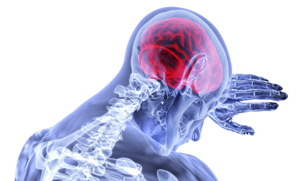
In sports, the connection between head injuries and neurodegenerative diseases such as chronic traumatic encephalopathy, Alzheimer’s disease, and Parkinson’s disease is now well recognised.

Researchers at Tufts University and Oxford University have now uncovered mechanisms that may connect the dots between trauma events and the emergence of disease. They point to latent viruses lurking in most of our brains that may be activated by the jolt, leading to inflammation and accumulating damage that can occur over the ensuing months and years. 

The results suggest the use of antiviral drugs as potential early preventive treatments post-head injury. The findings are published in a study in Science Signaling.

The microbiome aids in digestion, immune system development, and protection against harmful pathogens. 

But the microbiome also includes dozens of viruses that swarm within our bodies at any given time. Some of these can be potentially harmful, but simply lie dormant within our cells. Herpes simplex virus 1 (HSV-1), found in over 80% of people, and varicella-zoster virus, found in 95% of people, are known to make their way into the brain and sleep within our neurons and glial cells.

Dana Cairns, GBS12, research associate in the Department of Biomedical Engineering and lead author of the study, had found evidence in earlier studies suggesting that activation of HSV-1 from its dormant state triggers the signature symptoms of Alzheimer’s disease in lab models of brain tissue: amyloid plaques, neuronal loss, inflammations, and diminished neural network functionality.

“In that study, another virus – varicella – created the inflammatory conditions that activated HSV-1,” said Cairns. “We thought, what would happen if we subjected the brain tissue model to a physical disruption, something akin to a concussion? Would HSV-1 wake up and start the process of neurodegeneration?”

The link between HSV-1 and Alzheimer’s disease was first suggested by co-author Ruth Itzhaki, visiting professorial fellow at Oxford University, who more than 30 years ago identified the virus in a high proportion of brains from the elderly population. Her subsequent studies suggested that the virus can be reactivated in the brain from a latent state by events such as stress or immunosuppression, ultimately leading to neuronal damage.

Blows to Brain-like Tissue

In the current study, the researchers used a lab model that reconstructs the environment of the brain to better understand how concussions may set off the first stages of virus reactivation and neurodegeneration.

The brain tissue model consists of a 6mm-wide donut-shaped sponge-like material made of silk protein and collagen suffused with neural stem cells, which are then coaxed into mature neurons, growing axons and dendrite extensions and forming a network. Glial cells also emerge from the stem cells to help mimic the brain environment and nurture the neurons.

The neurons communicate with each other through their extensions similarly to how they would communicate in a brain. And just like cells in the brain, they can carry within them the DNA of dormant HSV-1 virus.  

After enclosing the brain-like tissue in a cylinder and giving it a sudden jolt atop a piston, mimicking a concussion, Cairns examined the tissue under the microscope over time. Some of the tissue models had neurons with HSV-1, and some were virus-free. 

Following the controlled blows, she observed that the infected cells showed re-activation of the virus, and shortly after that the signature markers of Alzheimer’s disease, including amyloid plaques, p-tau (a protein that creates fiber-like “tangles” in the brain), inflammation, dying neurons, and a proliferation of glial cells called gliosis.

More strikes with the pistons on the tissue models mimicking repetitive head injuries led to the same reactions, which were even more severe. Meanwhile, the cells without HSV-1 showed some gliosis, but none of the other markers of Alzheimer’s disease.

The results were a strong indicator that athletes suffering concussions could be triggering reactivation of latent infections in the brain that can lead to Alzheimer’s disease. Epidemiological studies have shown that multiple blows to the head can lead to doubling or even greater chances of having a neurodegenerative condition months or years down the line.
 
“This opens the question as to whether antiviral drugs or anti-inflammatory agents might be useful as early preventive treatments after head trauma to stop HSV-1 activation in its tracks, and lower the risk of Alzheimer’s disease,” said Cairns.

The problem goes far beyond the concerns for athletes. Traumatic brain injury is one of the most common causes of disability and death in adults, affecting about 69 million people worldwide each year, at an economic cost estimated at $400 billion annually.

“The brain tissue model takes us to another level in investigating these connections between injury, infection, and Alzheimer’s disease,” said David Kaplan, Stern Family Endowed Professor of Engineering at Tufts.

“We can re-create normal tissue environments that look like the inside of a brain, track viruses, plaques, proteins, genetic activity, inflammation and even measure the level of signalling between neurons,” he said. “There is a lot of epidemiological evidence about environmental and other links to the risk of Alzheimer’s. The tissue model will help us put that information on a mechanistic footing and provide a starting point for testing new drugs.”

Source: Tufts University