Tag: 14/3/25

Gut Microbes may Play a Role Linking Sugary Drinks and Diabetes Risk

Photo by Breakingpic on Pexels

It is well known that consuming sugary drinks increases the risk of diabetes, but the mechanism behind this relationship is unclear. Now, in a paper published in the Cell Press journal Cell Metabolism, researchers show that metabolites produced by gut microbes might play a role.

In a long-term cohort of US Hispanic/Latino adults, the researchers identified differences in the gut microbiota and blood metabolites of individuals with a high intake of sugar-sweetened beverages. The altered metabolite profile seen in sugary beverage drinkers was associated with a higher risk of developing diabetes in the subsequent 10 years. Since some of these metabolites are produced by gut microbes, this suggests that the microbiome might mediate the association between sugary beverages and diabetes.

“Our study suggests a potential mechanism to explain why sugar-sweetened beverages are bad for your metabolism,” says senior author Qibin Qi, an epidemiologist at Albert Einstein College of Medicine. “Although our findings are observational, they provide insights for potential diabetes prevention or management strategies using the gut microbiome.”

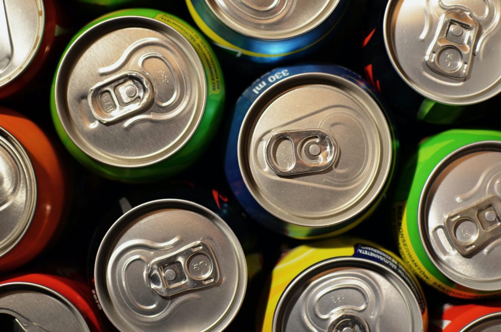
Sugar-sweetened beverages are the main source of added sugar in the diets of US adults – in 2017 and 2018, US adults consumed an average of 34.8g of added sugar each day from sugary beverages such as soda and sweetened fruit juice. Compared to added sugars in solid foods, added sugar in beverages “might be more easily absorbed, and they have a really high energy density because they’re just sugar and water,” says Qi.

Previous studies in Europe and China have shown that sugar-sweetened beverages alter gut microbiome composition, but this is the first study to investigate whether this microbial change impacts host metabolism and diabetes risk. It’s also the first study to investigate the issue in US-based Hispanic/Latino population — a group that experiences high rates of diabetes and is known to consume high volumes of sugar-sweetened beverages.

The team used data from the ongoing Hispanic Community Health Study/Study of Latinos (HCHS/SOL), a large-scale cohort study with data from over 16 000 participants living in San Diego, Chicago, Miami, and the Bronx. At an initial visit, participants were asked to recall their diet from the past 24 hours and had blood drawn to characterise their serum metabolites. The researchers collected faecal samples and characterized the gut microbiomes of a subset of the participants (n = 3035) at a follow-up visit and used these data to identify association between sugar-sweetened beverage intake, gut microbiome composition, and serum metabolites.

They found that high sugary beverage intake, defined as two or more sugary beverages per day, was associated with changes in the abundance of nine species of bacteria. Four of these species are known to produce short-chain fatty acids: molecules that are produced when bacteria digest fibre and that are known to positively impact glucose metabolism. In general, bacterial species that were positively associated with sugary beverage intake correlated with worse metabolic traits. Interestingly, these bacteria were not associated with sugar ingested from non-beverage sources.

The researchers also found associations between sugary beverage consumption and 56 serum metabolites, including several metabolites that are produced by gut microbiota or are derivatives of gut-microbiota-produced metabolites. These sugar-associated metabolites were associated with worse metabolic traits, including higher levels of fasting blood glucose and insulin, higher BMIs and waist-to-hip ratios, and lower levels of high-density lipoprotein cholesterol (“good” cholesterol). Notably, individuals with higher levels of these metabolites had a higher likelihood of developing diabetes in the 10 years following their initial visit.

“We found that several microbiota-related metabolites are associated with the risk of diabetes,” says Qi. “In other words, these metabolites may predict future diabetes.”

Because gut microbiome samples were only collected from a subset of the participants, the researchers had an insufficient sample size to determine whether any species of gut microbes were directly associated with diabetes risk, but this is something they plan to study further.

“In the future, we want to test whether the bacteria and metabolites can mediate or at least partially mediate the association between sugar-sweetened beverages and risk of diabetes,” says Qi.

The team plans to validate their findings in other populations and to extend their analysis to investigate whether microbial metabolites are involved in other chronic health issues linked to sugar consumption, such as cardiovascular disease.

Source: Science Direct

Amniotic Fluid Protects Both Baby and Birthing Parent, Research Finds

Image by Scientific Animations CC4.0

Researchers at Oregon Health & Science University have made new discoveries about amniotic fluid, which is historically not well understood in medical research due to the difficulty in obtaining it during pregnancy, especially across gestation in birthing parents.

In addition to providing much-needed cushion and protection for the foetus, amniotic fluid also aids in development of vital organs – especially the lungs, digestive tract and skin – and stabilises the temperature inside the womb.

The new study, published in the journal Research and Practice in Thrombosis and Haemostasis, found that the addition of amniotic fluid to plasma improves the blood’s ability to thicken and clot, which is a critical and likely a protective function throughout pregnancy and during delivery for both the birthing parent and the baby. It also appears to offer other unexpected functions, such as serving as a ‘pre-milk’ for foetuses.

The mechanism of amniotic fluid’s role in foetal development is not well understood and is understudied: The OHSU study is one of the first to identify how the features and properties of amniotic fluid change over time, especially those properties that play a role in thickening the blood, and how those changes can affect how maternal blood coagulates. If a pregnant person’s blood does not clot properly, it can create life-threatening complications for the foetus and birthing parent, including excessive bleeding during pregnancy and delivery.  

“We have always known that amniotic fluid is very important for foetal development and growth, but we don’t know much about it beyond that,” said the study’s corresponding author Jamie Lo, MD, MCR., associate professor of obstetrics and gynaecology (maternal-foetal medicine) in the OHSU School of Medicine, and Division of Reproductive & Developmental Sciences at the Oregon National Primate Research Center, or ONPRC. “We examined amniotic fluid across the pregnancy and found that indeed the composition and proteins in the amniotic fluid do change to match the growing needs of the developing baby.”

This discovery prompted Lo and her team to work with scientists in the Department of Biomedical Engineering at OHSU to take a deeper dive into the potential protective factors of amniotic fluid, and consider potential regenerative and therapeutic uses that could be developed down the road.

The research involved a multidisciplinary team including Lo, Chih Jen Yang, MD, Lyndsey Shorey-Kendrick, PhD, Joseph Shatzel, MD, MCR, Brian Scottoline, MD, PhD, and Owen McCarty, PhD.

Researchers analysed the properties of amniotic fluid obtained by amniocentesis, a prenatal test that involves sampling a small amount of amniotic fluid to examine the health of the pregnancy, from both human and non-human primates at gestational-age matched timepoints. The findings showed that amniotic fluid increases blood clotting through key fatty acids and proteins that change each trimester and help regulate coagulation.

With the untapped potential for amniotic fluid to aid in diagnosing and treating various prenatal conditions, researchers are now collaborating with Sanjay Malhotra, PhD, professor of cell, developmental and cancer biology in the OHSU School of Medicine, to target disorders of pregnancy – including disorders that affect the blood and blood-forming organs – that could benefit from the protective properties of proteins and other compounds within amniotic fluid.

Researchers are eager to learn more about the potential uses of amniotic fluid components and how they might be harnessed to improve prenatal and maternal health.

“Babies born prematurely miss out on critical weeks developing within amniotic fluid,” said the study’s co-senior author Brian Scottoline, MD, PhD, professor of paediatrics (neonatology), OHSU School of Medicine. “But if we have a better understanding of amniotic fluid, how it develops and what properties are valuable for what functions, that opens up many new possibilities for creating new therapies.”

“Through our research, our team is learning that amniotic fluid may be a critical precursor to breast milk – almost like ‘pre-term’ milk for a foetus in utero. With that analogy, could we eventually develop a formula that’s fit for preterm babies that mimics amniotic fluid, aiding in growth and development and protecting babies from complications of being born prematurely?” Lo added. “This is really the tip of the iceberg for what’s possible.”

Source: Oregon Health & Science University

Assessing Pain and Anxiety of Nursing Home Residents Unable to Speak

Photo by Pexels on Pixabay

As many as half of nursing home residents are cognitively impaired and may be unable to communicate symptoms such as pain or anxiety to the staff and clinicians caring for them. Therefore, information needed for the evaluation of symptoms and subsequent treatment decisions typically does not reliably exist in nursing home electronic health records (EHRs).

A new paper published in the International Journal of Geriatric Psychiatry reports on the novel adaptation of a commonly used symptom assessment instrument to more comprehensively acquire this difficult-to-obtain data with the ultimate goal of enabling knowledge-based expansion of palliative care services in nursing homes to address residents’ symptoms.

In the paper, part of the large, multi-state, multi-facility Utilizing Palliative Leaders in Facilities to Transform care for people with Alzheimer’s Disease (UPLIFT-AD) study researchers, including Regenstrief Institute, the Indiana University School of Medicine and the University of Maryland School of Social Work faculty, describe how they revamped and subsequently validated a symptom assessment tool used worldwide. The UPLIFT-AD researchers modified the instrument, originally designed for reporting by family members of individuals with dementia following their death, to enable reporting on the symptoms of current residents living with moderate to severe dementia by nursing home staff as well as family.

Led by Kathleen T. Unroe, MD, MHA, and John G. Cagle, PhD, the UPLIFT-AD team reports in the peer-reviewed paper that the tool they enhanced reliably addressed physical and emotional distress as well as well-being and symptoms that are precursors to end of life. This validation was critical as the researchers develop guidance for expansion of symptom recognition and management in any nursing home. Employing instruments used in other studies helps researchers to directly compare findings.

Dr. Unroe, Dr. Cagle and colleagues, including Wanzhu Tu, PhD, of the Regenstrief Institute and the IU School of Medicine, are in the late stages of the UPLIFT-AD clinical trial to enhance quality of care individuals with dementia by building capacity for palliative care within nursing homes.

“People receive care in nursing homes because they have significant needs – support for activities of daily living – as well as for complex, serious and multiple chronic conditions. But measuring symptoms of residents, especially those who are cognitively impaired, to address these needs is challenging,” said paper senior author Dr. Unroe, a Regenstrief Institute research scientist and an IU School of Medicine professor of medicine. “In my two decades of working as a clinician in nursing homes as well as a researcher, I have seen that often the information on symptoms that we want isn’t available consistently in the data that’s already collected or it isn’t collected at the frequency that we need to measure the impact of programs and approaches. And the gold standard for knowing if someone has a symptom, for example, if someone has pain or anxiety, to ask that person directly to assess the symptom, isn’t always possible for cognitively impaired residents. That’s why we took steps to validate a commonly used instrument in a wider population – individuals currently living with cognitive impairment – and added additional needed data points.

“While hospice care is typically available, there is widespread recognition that broader palliative care is needed in nursing homes. But there is no roadmap for how to provide it well. We hope that when we have our final results in 2026, UPLIFT-AD will prove to be a replicable model for implementing this much needed type of care.”

Source: Regenstrief Institute

New Antibodies Potentially Effective Against All SARS-CoV-2 Variants

Image by Fusion Medical on Unsplash

SARS-COV-2 has been very good at mutating to keep infecting people – so good that most antibody treatments developed during the pandemic are no longer effective. Now a team led by Stanford University researchers may have found a way to pin down the constantly evolving virus and develop longer-lasting treatments.

The researchers discovered a method to use two antibodies, one to serve as a type of anchor by attaching to an area of the virus that does not change very much and another to inhibit the virus’s ability to infect cells. This pairing of antibodies was shown to be effective against the initial SARS-CoV-2 virus that caused the pandemic and all its variants through omicron in laboratory testing. The findings are detailed in the journal Science Translational Medicine.

“In the face of an ever-changing virus, we engineered a new generation of therapeutics that have the ability to be resistant to viral evolution, which could be useful many years down the road for the treatment of people infected with SARS-CoV-2,” said Christopher O. Barnes, the study’s senior author, an assistant professor of biology.

An overlooked option

The team led by Barnes and first author Adonis Rubio, a doctoral candidate in the Stanford School of Medicine, conducted this investigation using donated antibodies from patients who had recovered from COVID-19. Analysing how these antibodies interacted with the virus, they found one that attaches to a region of the virus that does not mutate often.

This area, within the Spike N-terminal domain, or NTD, had been overlooked because it was not directly useful for treatment. However, when a specific antibody attaches to this area, it remains stuck to the virus. This is useful when designing new therapies that enable another type of antibody to get a foothold and attach to the receptor-binding domain, or RBD, of the virus, essentially blocking the virus from binding to receptors in human cells.

An illustration of the bispecific antibodies the Stanford-led research team developed to neutralise the virus that causes COVID-19. Named “CoV2-biRN,” these two antibodies work together by attaching to different areas of the virus.
The bispecific antibodies target two areas of the virus: One attaches to the “NTD,” or Spike N-terminal domain, an area on the virus that does not change very much. This allows the second antibody to attach to the “RBD,” or receptor-binding domain, essentially preventing the virus from infecting human cells. | Christopher O. Barnes and Adonis Rubio using Biorender stock images

The researchers designed a series of these dual or “bispecific” antibodies, called CoV2-biRN, and in laboratory tests they showed high neutralisation of all the variants of SARS-CoV-2 known to cause illness in humans. The antibodies also significantly reduced the viral load in the lungs of mice exposed to one version of the omicron variant.

More research, including clinical trials, would have to be done before this discovery could be used as a treatment in human patients, but the approach is promising – and not just for the virus that causes COVID-19.

Next, the researchers will work to design bispecific antibodies that would be effective against all coronaviruses, the virus family including the ones that cause the common cold, MERS, and COVID-19. This approach could potentially also be effective against influenza and HIV, the authors said.

“Viruses constantly evolve to maintain the ability to infect the population,” Barnes said. “To counter this, the antibodies we develop must continuously evolve as well to remain effective.”

Source: Stanford University

Twin Study Offers New Insights into Whether Exercise Extends life

Photo by Robert Ruggiero on Unsplash

Researchers from the University of Jyväskylä, Finland, investigated the links between long-term leisure-time physical activity and mortality, as well as whether physical activity can mitigate the increased risk of mortality due to genetic predisposition to diseases. Moreover, they examined the relationship between physical activity and later biological aging. 

The study included 22 750 Finnish twins born before 1958 whose leisure-time physical activity was assessed in 1975, 1981 and 1990. Mortality follow-up continued until the end of 2020.

Moderate activity yields maximum longevity benefits

Four distinct sub-groups were identified from the data, which was based on leisure-time physical activity over the 15-year follow-up: sedentary, moderately active, active and highly active groups. When the differences in mortality between the groups were examined at the 30-year follow-up, it was found that the greatest benefit – a 7% lower risk of mortality – was achieved between the sedentary and moderately active groups. A higher level of physical activity brought no additional benefit. 

When mortality was examined separately in the short and long term, a clear association was found in the short-term: the higher the level of physical activity, the lower the mortality risk. In the long term, however, those who were highly active did not differ from those who were sedentary in terms of mortality.

“An underlying pre-disease state can limit physical activity and ultimately lead to death, not the lack of exercise itself.”

“This can bias the association between physical activity and mortality in the short term”,  says Associate Professor Elina Sillanpää from the Faculty of Sports and Health Sciences. 

Meeting physical activity guidelines does not guarantee a lower mortality risk

The researchers also investigated whether following the World Health Organization’s physical activity guidelines affects mortality and genetic disease risk. The guidelines suggest 150 to 300 minutes of moderate or 75 to 150 minutes of vigorous activity weekly. The study found that meeting these guidelines did not lower mortality risk or alter genetic disease risk. Even for twins who met the recommended levels of PA over a 15-year period, no statistically significant difference in mortality rates was found compared to their less active twin pair.

“The widely observed favorable association between physical activity and mortality are based on observational studies that are prone to bias from different sources.” 

“In our studies, we aimed to account for various sources of biases, and combined with the long follow-up period, we could not confirm that adhering to physical activity guidelines mitigates genetic cardiovascular disease risk or causally reduces mortality”, says postdoctoral researcher Laura Joensuu from the Faculty of Sports and Health Sciences.  

Link between physical activity and biological aging is U-shaped

For the subsample of twins, biological aging was determined from blood samples using epigenetic clocks. Epigenetic clocks allow a person’s biological aging rate to be estimated based on methyl groups that regulate gene expression and are linked to aging process. 

“We found that the association between leisure-time physical activity and biological aging was U-shaped: Biological aging was accelerated in those who exercised the least and the most,” says Sillanpää.

Other lifestyles, such as smoking and alcohol consumption, largely explained the favourable associations of physical activity with biological aging. 

Genetic data were available for 4897 twins. The genetic susceptibility of twins to coronary artery disease, as well as systolic and diastolic blood pressure was assessed using new polygenic risk scores, which sum the genome-wide susceptibility to morbidity. In addition, all-cause and cardiovascular mortality was followed in 180 identical twin pairs. The biological aging rate of 1153 twins was assessed from a blood sample.

Source: University of Jyväskylä