Tag: 13/2/25

Cystic Fibrosis Damages the Immune System Early on
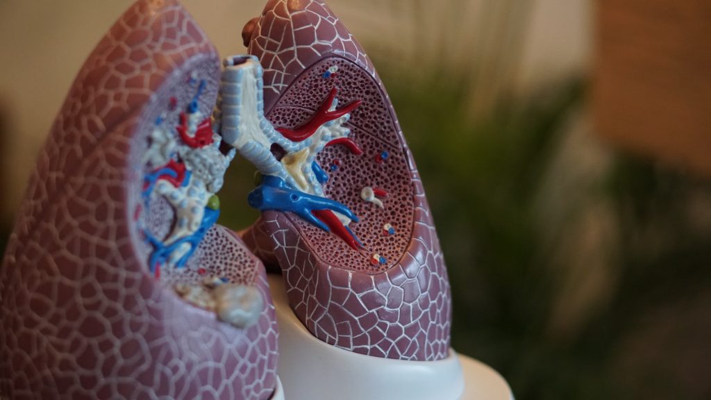
Photo by Robina Weermeijer on Unsplash

Despite new medication, cystic fibrosis often leads to permanent lung damage. Working with an international team, researchers from the Technical University of Munich (TUM) have discovered that the disease causes changes in the immune system early in life, presumably even in newborns. These changes lead to frequent inflammation and are not affected by drugs targeting the altered mucus production.

Cystic fibrosis is caused by hereditary genetic mutations that impair or halt the production of the CFTR protein. The respiratory tract is most severely affected. There, the mucus becomes so viscous that pathogens like bacteria cannot be removed by coughing. The result is often a deadly cycle of infection and inflammation.

In recent years, doctors have started using so-called CFTR modulator therapies to enhance the protein’s function. This reduces mucus formation and significantly improves the quality of life for those affected. However, clinical studies show that airway inflammation continues to occur frequently. In older patients, the decline in lung function seems unstoppable.

Current research aims to uncover additional processes in cystic fibrosis. “We specifically looked at how the immune system behaves in cystic fibrosis before the cycle of infection and inflammation begins,” said Prof. Nikolai Klymiuk from TUM. He is part of the international team that recently published a study on cystic fibrosis in Science Translational Medicine.

Immature immune cells in blood samples from children

The researchers found that in blood samples from children with cystic fibrosis and biological material from pigs with the same genetic defect, certain cells of the innate immune system are immature. This makes them less effective at fighting bacteria. Pigs with cystic fibrosis also showed an increased number and significantly altered composition of immune cells in the lungs at birth. The strong resemblance between the immune systems of pigs and humans suggests that this finding likely applies to human patients as well.

‘Emergency program’ responsible?

According to the authors, one possible explanation for the changes in the immune system could be a kind of “emergency program”. The program stimulates the body to produce a large number of immune cells particularly quickly or over a longer period of time. One consequence is the formation of immature immune cells, which could contribute to the fatal cycle of infections and inflammation in cystic fibrosis: Although immune cells are present in the lungs, they are ineffective and cause damage to the lung tissue without preventing infections in the long term.

Since immune cells generally produce only very small amounts of CFTR, the research team believes that the influence of cystic fibrosis on the immune system is indirect. This could explain why defective immune reactions cannot be treated well with novel CFTR modulator therapies.

Changes not a result of frequent infections

“We don’t yet know exactly why the immune cells in cystic fibrosis show such changes,” says Nikolai Klymiuk, Professor of Cardiovascular Translation in Large Animal Models. “However, we can show that these occur early in life. They then persist in the further course of life.” According to Klymiuk, although altered immune cells were known from blood samples of adults with cystic fibrosis, they were seen as a consequence of the numerous infections.

“To enable people with cystic fibrosis to live without symptoms, we probably need to tackle the disease on several levels,” said Klymiuk. “We hope our work will help us better understand the causes of the defective immune system and correct them in the future”


Source: Technical University of Munich (TUM)

Researchers Discover New Strength-boosting Mechanism in Androgens

Photo by Jonathan Borba on Unsplash

Researchers at Leipzig University’s Faculty of Medicine and Shandong University in China have discovered a new mechanism that is used by a key androgen essential for muscle and bone function. The findings could lead to the development of new drugs with fewer side effects, for use in applications such as strengthening the muscles of immobile patients. The researchers have published their findings in the prestigious journal Cell.

The most powerful of the androgens is called 5α-dihydrotestosterone (5α-DHT). Among other things, it is essential for bone and muscle function and for the development of secondary male sexual characteristics during puberty. As a driver of bone and muscle formation, 5α-DHT increases bone mineral density and promotes skeletal muscle growth to increase muscle strength. 

In this international study, the scientists were able to show that one of the adhesion G protein- coupled receptors – GPR133 – is activated by the androgenic steroid hormone 5α-DHT.

“This activation can, among other things, increase the contractile force of skeletal muscles, and our study also uses a newly developed, potent activator of this receptor to specifically trigger this effect,” says Professor Ines Liebscher, Professor of Signal Transduction at Leipzig University and co-leader of the study.

Increasing muscle strength with the chance of significantly fewer negative effects of androgens

Activation of GPR133 by the novel agonist AP503 increases muscle strength without triggering a specific negative effect that is otherwise observed when androgens are administered. For example, increased and prolonged exposure to testosterone can promote the development of prostate cancer, as evidenced by tissue changes in the prostate in mice after only two weeks of androgen administration. This side effect has not yet been observed with AP503.

In addition, the current study uses structural biology methods to elucidate the molecular basis of the interaction between the steroid hormone, the substance AP503 and GPR133. This will allow the activator to be specifically optimised and further developed into a new therapeutic agent. This could lead to the development of new muscle-strengthening drugs with a lower side-effect profile.

This publication is the result of a long-standing and successful collaboration between the Rudolf Schönheimer Institute of Biochemistry and the research group of Professor Jin-Peng Sun at Shandong University in China. The researchers are currently working on several follow-up studies to further investigate the use of AP503 in disease processes and the role of GPR133 in the organism. Here the data were analysed in animal models. Further studies are needed to investigate the applicability of the findings to humans.

Source: Universität Leipzig

Born to Heal: Why Babies Recover, but Adults scar, After Heart Damage

Photo by William Fortunato on Pexels

Newborns with heart complications can rely on their newly developed immune systems to regenerate cardiac tissues, but adults aren’t so lucky. After a heart attack, most adults struggle to regenerate healthy heart tissue, leading to scar-tissue buildup and, often, heart failure.

A new Northwestern Medicine study in experimental animals reveals a critical difference in how macrophages help repair the heart in newborns versus adults after a heart attack. The study highlights a fundamental difference in how the immune system drives healing based on age.

The study appears in the journal Immunity.

“Understanding why newborns can regenerate their hearts while adults cannot will open the door to developing treatments that could ‘reprogram’ adult macrophages,” said first and co-corresponding author Connor Lantz, lead scientist of the bioinformatics core at the Comprehensive Transplant Center at Northwestern University Feinberg School of Medicine.

In newborns, macrophages perform a process called efferocytosis, which recognizes and eats dying cells. This process triggers the production of a bioactive lipid called thromboxane, signaling nearby heart muscle cells to divide, and allowing the heart to regenerate damaged heart muscle, the study found. In adults, macrophages produce much less thromboxane, leading to a weaker repair signal.

“By mimicking the effects of thromboxane, we might one day improve tissue repair after a heart attack in adults,” Lantz said.

How the study worked

The study examined how the immune system responds to heart injury in mice of different ages, including newborn mice (one day old) and adult mice (eight weeks old). The researchers found the ability of macrophages to engulf dying cells was enhanced in newborn mice due to increased expression of MerTK, a receptor that recognizes dying cells. Therefore, when the scientists blocked this key receptor, newborn mice lost their ability to regenerate their hearts, resembling adult hearts after a heart attack.

Engulfment of dying cells by newborn macrophages triggered a chemical chain reaction that produced a molecule called thromboxane A2, which unexpectedly stimulated heart muscle cells to multiply and repair the damage, the study found. Additionally, nearby muscle heart cells in newborns are primed to respond to thromboxane A2, leading them to change their metabolism to support their growth and healing. But in adults, this process did not work the same way – after an injury, their macrophages did not produce enough thromboxane A2, limiting their ability to regenerate heart tissue.

Source: Northwestern University

Myth busted: Healthy Habits Take Longer than 21 Days to Set in

Photo by Ketut Subiyanto on Unsplash

Forming a healthy habit can take longer than you expect. In the first systematic review of its kind, University of South Australia researchers found that new habits can begin forming within about two months (median of 59-66 days) but can take up to 335 days to establish.

It’s an important finding that could inform health interventions to promote healthy behaviours and prevent chronic disease.

Many conditions, including type 2 diabetes, heart disease, lung diseases and stroke, can be prevented by changing unhealthy habits or lifestyle factors. University of South Australia researcher, Dr Ben Singh, says that contrary to popular belief, healthy habits take far longer than three weeks to lock down.

“Adopting healthy habits is essential for long-term well-being but forming these habits – and breaking unhealthy ones – can be challenging,” Dr Singh says.

“At the beginning of the year, many of us are setting goals and making plans for the months ahead –things like being more active, cutting back on sugar, or making healthier food choices – but while common wisdom suggests that it takes just 21 days to form such habits, these claims are not evidence-based.

“In our research, we’ve found that habit formation starts within around two months, but there is significant variability, with formation times ranging from four days to nearly a year.

“So, it’s important for people who are hoping to make healthier habits not to give up at that mythical three-week mark.”

The study of more than 2600 participants also found that certain factors can influence successful habit formation.

“When trying to establish a new healthy habit, success can be influenced by a range of things including how frequently we undertake the new activity, the timing of the practice, and whether we enjoy it or not,” Dr Singh says.

“If you add a new practice to your morning routine, the data shows that you’re more likely to achieve it. You’re also more likely to stick to a new habit if you enjoy it.

“Planning and intending to complete a new behaviour can also help solidify a new habit, so make sure you continue to make time to include your new healthy habits into your everyday activities. This could be as easy as laying out your gym clothes the night before a morning walk or having a healthy lunch ready to go in the fridge.

“Tailoring habit-building strategies into our day and making plans on how we can achieve them, will put you in a position for success.”

While more research is needed, researchers say that these findings can guide public health initiatives and personalised programs that support sustained and healthy behaviour change.

Source: University of South Australia