Tag: 12/3/24

Salt Substitute is an Effective Way of Cutting Hypertension in Older Adults

Credit: Pixabay CC0

Replacing table salt with a salt substitute can reduce incidence of hypertension in older adults without increasing their risk of hypotension episodes, according to a recent study in the Journal of the American College of Cardiology. Participants using a salt substitute had a 40% lower incidence and likelihood of experiencing hypertension compared to those who used regular salt.

One of the most effective ways to reduce hypertension risk, one of the world’s leading health risks, is to reduce sodium intake. This study looks at salt substitutes as a better solution to control and maintain healthy blood pressure than reducing salt alone.

“Adults frequently fall into the trap of consuming excess salt through easily accessible and budget-friendly processed foods,” said Yangfeng Wu, MD, PhD, lead author of the study and Executive Director of Peking University Clinical Research Institute in Beijing, China.

“It’s crucial to recognise the impact of our dietary choices on heart health and increase the public’s awareness of lower-sodium options.”

Researchers in this study evaluated the impact of sodium reduction strategies on blood pressure in elderly adults residing in care facilities in China.

While previous studies prove that reducing salt intake can prevent or delay new-onset hypertension, long-term salt reduction and avoidance can be challenging.

The DECIDE-Salt study included 611 participants 55 years or older from 48 care facilities split into two groups: 24 facilities (313 participants) replacing usual salt with the salt substitute and 24 facilities (298 participants) continuing the use of usual salt.

All participants had blood pressure <140/90mmHg and were not using anti-hypertension medications at baseline.

The primary outcome was participants who had incident hypertension, initiated anti-hypertension medications or developed major cardiovascular adverse events during follow-up.

At two years, the incidence of hypertension was 11.7 per 100 people-years in participants with salt substitute and 24.3 per 100 people-years in participants with regular salt.

People using the salt substitute were 40% less likely to develop hypertension compared to those using regular salt. Furthermore, the salt substitutes did not cause hypotension, which can be a common issue in older adults.

“Our results showcase an exciting breakthrough in maintaining blood pressure that offers a way for people to safeguard their health and minimise the potential for cardiovascular risks, all while being able to enjoy the perks of adding delicious flavour to their favourite meals,” Wu said.

“Considering its blood pressure – lowering effect, proven in previous studies, the salt substitute shows beneficial to all people, either hypertensive or normotensive, thus a desirable population strategy for prevention and control of hypertension and cardiovascular disease.”

Limitations of the study include that it is a post-hoc analysis, study outcomes were not pre-specified and there was a loss of follow-up visits in many patients.

Analyses indicated that these missing values were at random, and multiple sensitivity analyses supports the robustness of the results.

In an accompanying editorial comment, Rik Olde Engberink, MD, PhD, researcher, nephrologist and clinical pharmacologist at Amsterdam University Medical Center’s Department of Internal Medicine, said the study provides an attractive alternative to the failing strategy to reduce the intake of salt worldwide, but questions and effort remain.

“In the DECIDE-Salt trial, the salt substitute was given to the kitchen staff, and the facilities were not allowed to provide externally sourced food more than once per week,” Olde Engberink said. “This approach potentially has a greater impact on blood pressure outcomes, and for this reason, salt substitutes should be adopted early in the food chain by the food industry so that the sodium-potassium ratio of processed foods will improve.”

Source: American College of Cardiology

Microplastics Found in Every Human Placenta Tested in Study

Image by Scientific Animations CC4.0

A flurry of recent studies has found that microplastics are present in virtually everything we consume, from bottled water to meat and plant-based food. Now, University of New Mexico Health Sciences researchers have used a new analytical tool to measure the microplastics present in human placentas.

In a study published in the journal Toxicological Sciences, a team led by Matthew Campen, PhD, Regents’ Professor in the UNM Department of Pharmaceutical Sciences, reported finding microplastics in all 62 of the placenta samples tested, with concentrations ranging from 6.5 to 790 micrograms per gram of tissue.

Although those numbers may seem small, Campen is worried about the health effects of a steadily rising volume of microplastics in the environment.

For toxicologists, “dose makes the poison,” he said. “If the dose keeps going up, we start to worry. If we’re seeing effects on placentas, then all mammalian life on this plant could be impacted. That’s not good.”

In the study, Campen and his team, partnering with colleagues at the Baylor College of Medicine and Oklahoma State University, analyzed donated placenta tissue. In a process called saponification, they chemically treated the samples to “digest” the fat and proteins into a kind of soap.

Then, they spun each sample in an ultracentrifuge, which left a small nugget of plastic at the bottom of a tube. Next, using a technique called pyrolysis, they put the plastic pellet in a metal cup and heated it to 600 degrees Celsius, then captured gas emissions as different types of plastic combusted at specific temperatures.

“The gas emission goes into a mass spectrometer and gives you a specific fingerprint,” Campen said. “It’s really cool.”

The researchers found the most prevalent polymer in placental tissue was polyethylene, which is used to make plastic bags and bottles. It accounted for 54% of the total plastics. Polyvinyl chloride (better known as PVC) and nylon each represented about 10% of the total, with the remainder consisting of nine other polymers.

Marcus Garcia, PharmD, a postdoctoral fellow in Campen’s lab who performed many of the experiments, said that until now, it has been difficult to quantify how much microplastic was present in human tissue. Typically, researchers would simply count the number of particles visible under a microscope, even though some particles are too small to be seen.

With the new analytical method, he said, “We can take it to that next step to be able to adequately quantify it and say, ‘This is how many micrograms or milligrams,’ depending on the plastics that we have.”

Plastic use worldwide has grown exponentially since the early 1950s, producing a metric ton of plastic waste for every person on the planet. About a third of the plastic that has been produced is still in use, but most of the rest has been discarded or sent to landfills, where it starts to break down from exposure to ultraviolet radiation present in sunlight.

“That ends up in groundwater, and sometimes it aerosolizes and ends up in our environment,” Garcia said. “We’re not only getting it from ingestion but also through inhalation as well. It not only affects us as humans, but all off our animals — chickens, livestock — and all of our plants. We’re seeing it in everything.”

Campen points out that many plastics have a long half-life — the amount of time needed for half of a sample to degrade. “So, the half-life of some things is 300 years and the half-life of others is 50 years, but between now and 300 years some of that plastic gets degraded,” he said. “Those microplastics that we’re seeing in the environment are probably 40 or 50 years old.”

While microplastics are already present in our bodies, it is unclear what health effects they might have, if any. Traditionally, plastics have been assumed to be biologically inert, but some microplastics are nanometres in size and are capable of crossing cell membranes, he said.

Campen said the growing concentration of microplastics in human tissue might explain puzzling increases in some types of health problems, such as inflammatory bowel disease and colon cancer in people under 50, as well as declining sperm counts.

The concentration of microplastics in placentas is particularly troubling, he said, because the tissue has only been growing for eight months (it starts to form about a month into a pregnancy). “Other organs of your body are accumulating over much longer periods of time.”

Campen and his colleagues are planning further research to answer some of these questions, but in the meantime he is deeply concerned by the growing production of plastics worldwide.

“It’s only getting worse, and the trajectory is it will double every 10 to 15 years,” he said. “So, even if we were to stop it today, in 2050 there will be three times as much plastic in the background as there is now. And we’re not going to stop it today.”

Source: University of New Mexico Health Sciences Center

Artificial Intelligence Probes Link between Smoking in Pregnancy and Behavioural Disorders in Newborns

Source: Pixabay CC0

Although several studies have linked smoking during pregnancy with neurodevelopmental disorders, the results of behavioural experiments in mice prenatally exposed to nicotine have been inconsistent. In a recent study, scientists from Japan developed a deep learning-based framework to automatically observe and classify mice behaviour in such experiments, producing more accurate and unbiased results. In their results, published recently in Cells, they show that prenatal exposure to nicotine could increase the risk of autism spectrum- and attention deficit/hyperactivity disorders in newborns.

The fact that smoking is a risk factor for several diseases, including cancer, stroke, and diabetes, has been known for approximately half a century. However, over the past few decades, scientists have brought to light many of the detrimental effects of smoking during pregnancy, linking this habit to high infant mortality, failed delivery, and low body weight at birth. In addition, recent studies suggest that prenatal nicotine exposure (PNE) may be related to neurodevelopmental disorders, such as attention deficit/hyperactivity disorder (ADHD) and autism spectrum disorder (ASD).

For a long time now, scientists have used animal models (like rodents) to understand how PNE leads to neurodevelopmental disorders. By carefully analysing the behaviour of rodents, they can infer whether PNE is causing neurological changes and the brain regions affected by it; this can later be confirmed through histological analyses.

Unfortunately, thus far, studies on behavioural changes induced by PNE in mice have shown varied results, some of which are contradictory. Although there could be multiple reasons behind these discrepancies, human error and bias are prime suspects. In general, the assessment of complex animal behaviours, especially social interactions, relies on the efforts of human observers, which introduces a baseline level of subjectivity that is hard to dispel. But what if we can leverage artificial intelligence (AI) to produce more accurate and unbiased results from observations of PNE mice behaviour?

In this study , researchers from the Department of Molecular and Cellular Physiology at the Shinshu University School of Medicine, including graduate student Mengyun Zhou, Assistant Professor Takuma Mori, and Professor Katsuhiko Tabuchi, developed and trained a deep learning-based system to automatically analyse footage from behavioural experiments on mice. They used this tool to explore the behavioural changes induced by PNE in mice without observer biases, seeking to shed light on the link between nicotine and neurodevelopmental disorders.

The proposed AI-based framework relied on a combination of two well-established open-source toolkits, namely DeepLabCut and Simple Behavioral Analysis (SimBA). “AI tools can label the body parts of animals in a markerless video footage and precisely estimate their poses using supervised machine learning,” explains Prof Tabuchi. “Since animal behaviours are defined as a specific arrangement of body parts over a short period of time, deep-learning toolkits like SimBA can use the pose estimations obtained with DeepLabCut to classify different types of animal behaviours.”

After reaching an optimal training protocol for their framework using manually labelled data, the researchers conducted several experiments using PNE and control mice, looking for indicators of ADHD- and ASD-like behaviours. First, they carried out cliff avoidance reaction tests, which are used to test impulsivity. In these tests, they placed the subject mouse on top of a slightly elevated platform and took note, both manually and with the AI system, of how long the mouse waited before jumping down the platform. The test results suggested that PNE mice are more impulsive, a behavioural feature of ADHD in humans.

They also tested the working memory of mice using a Y-shaped maze and counted the number of times each mouse spontaneously switched from one arm of the maze to another. “We observed a decrease in the spontaneous alteration in PNE mice, suggesting that their working memory was altered, which is another behavioural feature of ADHD,” comments Mengyun Zhou. “These results suggest prenatal exposure to nicotine may cause ADHD in mice, which is consistent with clinical reports in humans.”

Finally, the researchers conducted open-field and social-interaction experiments, which represented the main challenge for their AI-based system. In these experiments, the researchers observed either one or two mice behaving freely in a large enclosure and looked for indicators of anxiety and social behaviours, such as grooming and following. Interestingly, PNE mice exhibited social behavioural deficits and increased anxiety which are features of ASD. Subsequent histological analysis of hippocampal brain tissue confirmed decreased neurogenesis, a hallmark of ASD. Thus, it appears that smoking may not only increase the risk of ADHD, but also ASD.

Worth noting, the results obtained using the AI-based system were highly reliable, as Prof Tabuchi highlights: “We validated the accuracy of our behavioural analysis framework by drawing a careful comparison between the results generated by the model and behaviour assessments made by multiple human annotators, which is considered the gold standard.” These analyses cement the potential of the proposed approach and showcase its capabilities for many types of behavioural studies.

With any luck, further efforts will pave the way to a solid understanding of mechanisms behind neurodevelopmental disorders like ASD and ADHD, ultimately leading to better diagnostic tools and therapeutic methods.

Source: Shinshu University

Revealing a Hidden Threat: Viral Infections Pose Early Heart Risks

Human heart. Credit: Scientific Animations CC4.0

In a potentially game-changing development, scientists at Virginia Tech have revealed a new understanding of sometimes fatal viral infections that affect the heart.

The focus has mostly been on myocarditis, which is often triggered by the body’s immune response to a viral infection. Now, a new study led by James Smyth, associate professor at the Fralin Biomedical Research Institute, sheds new light on this notion, revealing that the virus itself creates potentially dangerous conditions in the heart before inflammation sets in.

The discovery, now online and set to appear in the March 29 issue of Circulation Research, suggests completely new directions to diagnose and treat viral infections affecting the heart.

Given the high incidence of viral-related myocarditis leading to sudden cardiac death, the insight is crucial. Up to 42% of sudden cardiac deaths in young adults are attributed to myocarditis, and of these cases viral infection is the leading cause.

“From a clinical perspective, our understanding of viral infection of the heart has focused on inflammation, causing problems with the rate or rhythm of the heartbeat,” Smyth said. “But we have found an acute stage when the virus first infects the heart and before the body’s immune response causes inflammation. So even before the tissue is inflamed, the heart is being set up for arrhythmia.”

To make this discovery, researchers focused on adenovirus, a common culprit in cardiac infection and myocarditis, using Mouse Adenovirus Type-3 to replicate the human infection process.

They found that early in the infection, the virus disrupts critical components of the heart’s communication and electrical systems.

As a result, even before symptoms appear, the adenoviral infection creates conditions that disrupt the heart’s gap junctions and ion channels, according to virologist Rachel Padget, the study’s first author who worked in the Smyth lab while completing a doctoral degree from the Virginia Tech Translational Biology, Medicine, and Health graduate program.

Gap junctions are like tiny tunnels between heart cells that allow them to communicate, and ion channels are like gates in the cell membranes that help maintain the right balance of ions needed for the heart to generate normal patterns of electrical activity that allow it to beat properly.

When adenoviral infection disturbs these communication bridges and gatekeepers, it creates a situation where the heart might develop irregular patterns of electrical activity called arrhythmias affecting its mechanical beating and blood pumping capacity, and that can lead to sudden cardiac problems, especially in people with active infections.

Now, by targeting specific heart changes induced by viral infections at the molecular level, researchers aim to reduce the risk of cardiac issues in people grappling with viral illnesses.

“Individuals who have acute infections can look normal by MRI and echocardiography, but when we delved into the molecular level, we saw that something very dangerous could occur,” Smyth said. “In terms of diagnostics, we can now work with our colleagues here to start looking ways to analyse blood for a biomarker of the more serious problem. People get cardiac infections all the time and they recover. But can we identify what’s different about individuals that are at a higher risk to have the arrhythmia, possibly through a simple blood test in the doctor’s office.”

Source: Virginia Tech

Leakage of Mitochondrial DNA may Drive Many Inflammatory Disorders

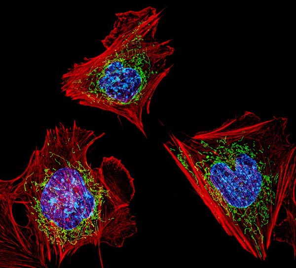
Cells with nuclei in blue, energy factories in green and the actin cytoskeleton in red. Credit: NIH

A new discovery, which was published in Nature Cell Biology, reveals how genetic material can escape mitochondria, prompting the body to launch a damaging immune response, setting off diseases such as lupus and rheumatoid arthritis. By developing therapies to target this process, doctors may one day be able to stop the harmful inflammation and prevent the toll it takes on our bodies.

“When mitochondria don’t correctly replicate their genetic material, they try to eliminate it. However, if this is happening too often and the cell can’t dispose of all of it, it can cause inflammation, and too much inflammation can lead to disease, including autoimmune and chronic diseases,” said researcher Laura E. Newman, PhD, of the University of Virginia School of Medicine. “Now that we are beginning to understand how this inflammation starts, we might be able to prevent this process, with the ultimate goal of limiting inflammation and treating disease.”

Powering inflammation

Mitochondria have their own set of genetic material, separate from the DNA that serves as the operating instructions for our cells. Scientists have known that this mitochondrial DNA, known as mtDNA, can escape into our cells and cause inflammation. But exactly what caused this has been a mystery until now.

“We knew that mtDNA was escaping mitochondria, but how was still unclear,” said Gerald Shadel, PhD, director of the San Diego-Nathan Shock Center of Excellence in the Basic Biology of Aging at the Salk Institute. “Using imaging and cell biology approaches, we’re able to trace the steps of the pathway for moving mtDNA out of the mitochondria, which we can now try to target with therapeutic interventions to hopefully prevent the resulting inflammation.”

Shadel and Newman, then a postdoctoral researcher in Shadel’s lab, and their collaborators used sophisticated imaging techniques to determine what was happening inside the leaky mitochondria. They found that the leak was triggered by a malfunction in mtDNA replication. This caused the accumulation of protein masses caused nucleoids.

To try to fix this problem, the cell containing the faulty mitochondrion begins to export the excess nucleoids to its cellular trash bins. But the trash bins, called endosomes, can become overwhelmed by the volume of debris, the scientists found. These overburdened endosomes respond by releasing mtDNA into the cell — in short, the trash can overflows.

“We had a huge breakthrough when we saw that mtDNA was inside of a mysterious membrane structure once it left mitochondria. After assembling all of the puzzle pieces, we realised that structure was an endosome,” Newman said. “That discovery eventually led us to the realisation that the mtDNA was being disposed of and, in the process, some of it was leaking out.”

The cell responds to this hazardous waste spill by flagging the nucleoids as foreign DNA, like a virus, and launches an immune response that results in harmful inflammation, the scientists determined.

“Using our cutting-edge imaging tools for probing mitochondria dynamics and mtDNA release, we have discovered an entirely novel release mechanism for mtDNA,” said researcher Uri Manor, PhD, former director of the Waitt Advanced Biophotonics Core at Salk and current assistant professor at UC San Diego. “There are so many follow-up questions we cannot wait to ask, like how other interactions between organelles control innate immune pathways, how different cell types release mtDNA, and how we can target this new pathway to reduce inflammation during disease and aging.”

Newman will continue to seek these answers in her new role at the UVA School of Medicine’s Department of Cell Biology. “We want to understand the physiological and disease contexts where this process can become activated,” she said. “For example, many viruses attack mitochondria during infection, so we will be testing whether mitochondria purposely use this pathway to sound the alarm against invading viruses, and whether over-reliance on this pathway to fight off infection can later trigger chronic diseases.”

Source: University of Virginia Health System