The anticoagulant drug heparin is widely given to patients with blood clotting disorders or after surgery to prevent complications. But it remains difficult to dose correctly, potentially leading to overdosing or underdosing. A team of Penn State researchers combined heparin with a protein fragment, peptide, to slow down the release of the drug and convey the medication directly to the site of a clot. They published their findings in the journal Small.
“We wanted to develop a material that can gradually deliver heparin over time rather than the current iteration that gets cleared from the body in a couple of hours,” said corresponding author Scott Medina, Penn State associate professor of biomedical engineering. “We also wanted to deliver the drug through the skin instead of through an IV.”
When mixed, positively charged peptides and negatively charged heparin bind to create a nanogranular paste that can be injected under the skin, forming a cache of material that is then diffused in the circulatory system and travels to blood clots when they appear. The turbulent flow of fluid near a blood clot triggers the two materials to separate, allowing heparin to begin its anticoagulating action.
“The peptide is ideal for pairing with heparin because it essentially blocks heparin’s action until it is needed in the body,” said Atip Lawanprasert, doctoral student in biomedical engineering and first author on the paper. “The peptide also has some anticoagulating properties on its own: It binds to platelets in the blood, enabling action at the clotting site.”
Without an added bonding agent, heparin applies its anti-clotting properties indiscriminately, not just at blood clot sites, and clears quickly, its half-life only 60 to 90 minutes. Using preclinical animal trials, researchers determined that the addition of peptide allows for a dramatic increase of heparin’s half-life, to up to nearly 24 hours.
“The peptide increases heparin’s effects by more than ten times longer than what is currently being used,” Medina said. “The increased half-life allows for sustained treatments for patients, less medication waste and more accurate dosing. Eventually, this could allow the medication to be injected under the skin just once a day, rather than an all-day IV drip.”
Next, researchers plan to replicate the study in a clinical setting, as well as study the effect of the medication’s toxicity with multi-day administration.
Vaccines that provide long-lasting protection against influenza, coronaviruses and respiratory syncytial virus (RSV) have proved exceptionally difficult to develop. In a new review article in Cell Host & Microbe, NIH researchers explore the challenges and outline approaches to improved vaccines and describe a promising path forward: mucosal vaccines.
Unlike the respiratory viruses that cause measles, mumps and rubella — for which vaccination or recovery from illness provides decades-long protection against future infection – flu, RSV, SARS-CoV-2 and “common cold” coronaviruses share several characteristics that enable them to cause repeated re-infections. These include very short incubation periods, rapid host-to-host transmission and replication in the nasal mucosa rather than throughout the body. This last feature – non-systemic replication – means these viruses do not stimulate the full force of the adaptive immune response, which typically takes a week or more to mount.
A next generation of improved vaccines for mucosa-replicating viruses will require advances in understanding on several fronts, the authors say. For instance, more must be learned about interactions between flu viruses, coronaviruses and RSV and the components of the immune response that operate largely or exclusively in the upper respiratory system. Over time, these interactions have evolved and led to “immune tolerance,” wherein the human host tolerates transient, limited infections by viruses that are generally non-lethal to avoid the destructive consequences of an all-out immune system attack.
The authors note that mucosal immunisation appears to be an optimal route of vaccination for the viruses of interest, when feasible. However, to develop useful mucosal vaccines, significant knowledge gaps must be filled including finding ideal vaccine formulations; determining dosage size, frequency and timing; and developing techniques for overcoming immune tolerance.
Cambridge scientists have successfully trialled an artificial pancreas for use by patients living with type 2 diabetes. They report in Nature Medicine that the device doubled the amount of time patients were in the target range for glucose compared to standard treatment and halved the time spent experiencing high glucose levels.
The artificial pancreas developed by University of Cambridge researchers combines an off-the-shelf glucose monitor and insulin pump with an app developed by the team, known as CamAPS HX. This app is run by an algorithm that predicts how much insulin is required to maintain glucose levels in the target range.
The researchers have previously shown that an artificial pancreas run by a similar algorithm is effective for patients living with type 1 diabetes, from adults through to very young children. They have also successfully trialled the device in patients with type 2 diabetes who require kidney dialysis.
Today, in Nature Medicine, the team report the first trial of the device in a wider population living with type 2 diabetes (not requiring kidney dialysis). Unlike the artificial pancreas used for type 1 diabetes, this new version is a fully closed loop system, whereas patients with type 1 diabetes need to tell their artificial pancreas that they are about to eat to allow adjustment of insulin, for example, with this version they can leave the device to function entirely automatically.
The researchers recruited 26 patients who were randomised to one of two groups – the first group would trial the artificial pancreas for eight weeks and then switch to the standard therapy of multiple daily insulin injections; the second group would take this control therapy first and then switch to the artificial pancreas after eight weeks.
The team used several measures to assess how effectively the artificial pancreas worked. The first was the proportion of time that patients spent with their glucose levels within a target range of between 3.9 and 10.0mmol/L. On average, patients using the artificial pancreas spent two-thirds (66%) of their time within the target range, compared to control (32%).
A second measure was the proportion of time spent with glucose levels above 10.0mmol/L. Over time, high glucose levels raise the risk of potentially serious complications. Patients taking the control therapy spent two-thirds (67%) of their time with high glucose levels — this was halved to 33% when using the artificial pancreas.
Average glucose levels fell from 12.6mmol/L when taking the control therapy to 9.2mmol/L while using the artificial pancreas.
The app also reduced levels of a molecule known as glycated haemoglobin, or HbA1c. Glycated haemoglobin develops when haemoglobin, a protein within red blood cells that carries oxygen throughout the body, joins with glucose in the blood, becoming ‘glycated’. By measuring HbA1c, clinicians are able to get an overall picture of what a person’s average blood sugar levels have been over a period of weeks or months. For people with diabetes, the higher the HbA1c, the greater the risk of developing diabetes-related complications. After the control therapy, average HbA1c levels were 8.7%, while after using the artificial pancreas they were 7.3%.
No patients experienced dangerously-low blood sugar levels (hypoglycaemia) during the study. One patient was admitted to hospital while using the artificial pancreas, due to an abscess at the site of the pump cannula.
Dr Charlotte Boughton from the Wellcome-MRC Institute of Metabolic Science at the University of Cambridge, who co-led the study, said: “Many people with type 2 diabetes struggle to manage their blood sugar levels using the currently available treatments, such as insulin injections. The artificial pancreas can provide a safe and effective approach to help them, and the technology is simple to use and can be implemented safely at home.”
Dr Aideen Daly, also from the Wellcome-MRC Institute of Metabolic Science, said: “One of the barriers to widespread use of insulin therapy has been concern over the risk of severe ‘hypos’ — dangerously low blood sugar levels. But we found that no patients on our trial experienced these and patients spent very little time with blood sugar levels lower than the target levels.”
Feedback from participants suggested that participants were happy to have their glucose levels controlled automatically by the system, and nine out of ten (89%) reported spending less time managing their diabetes overall. Users highlighted the elimination of the need for injections or fingerprick testing, and increased confidence in managing blood glucose as key benefits. Downsides included increased anxiety about the risk of hypoglycaemia, which the researchers say may reflect increased awareness and monitoring of glucose levels, and practical annoyances with wearing of devices.
The team now plan to carry out a much larger multicentre study to build on their findings and have submitted the device for regulatory approval with a view to making it commercially available for outpatients with type 2 diabetes.
Using new microscopic methods in combination with machine learning-based image analysis, researchers fromAlbert Ludwigs University of Freiburghave discovered new structures on the surface of living B cells that affect the distribution and possibly the function of their antigen receptors. The researchers’ study has been published in The EMBO Journal.
B cells recognise pathogens through specialised receptors on their surface. For the first time, scientists were able to observe how these receptors are distributed on the surface of living and moving cells. They found that the B cell surface is shaped into a characteristic landscape of interconnected ridges and protrusions. On this landscape, the IgM-class B cell antigen receptors (IgM-BCR) accumulate in specific areas. The precision of the receptors’ localisation and their clustering into larger units likely constitute a mechanism that controls receptor signalling and facilitates antigen sensing and thereby the activation of B cells.
In most immunological textbooks, lymphocytes are depicted as round, ball-like cells whose smooth surface carries randomly distributed receptors. The notion of a smooth unstructured B cell surface has already been challenged by electron micrographs of fixed and frozen lymphocytes, revealing thin membrane protrusions called microvilli on the cells’ surface. These tentacle-like structures help immune cells to search for molecular markers of pathogens, so-called antigens. B lymphocytes recognise such antigens through different classes of their B cell antigen receptors (BCR). These antigen receptors are complex molecular machines that, when activated, interact with other molecules to initiate a signalling cascade, leading to the differentiation of B cells into plasma cells and the production of protective antibodies.
The research group of Prof. Dr. Michael Reth collaborated with other imaging specialists to analyse how the IgM-BCR is distributed across the 3D surface of living B cells. For this, they used a technique called lattice light sheet microscopy, LLSM for short. “This method can capture volumetric images of living cells at a very high speed,” explains Dr. Deniz Saltukoglu from Freiburg University, the first author of the study. “In other types of high-resolution microscopy, cells need to be attached to a flat surface, which completely alters the B cells’ outer structures. LLSM allowed us to observe the cells in an environment that mimics biological tissues, meaning that the structures and movements that we saw were largely undisturbed,” she says.
The researchers then developed custom image analysis tools to quantify and objectively characterise the microscopic data. “We needed to segment the images and isolate morphological features,” says Saltukoglu. “So far this had only been done with two-dimensional data, so we had to develop new computational tools for volumetric, time course data.” For this, the researchers drew inspiration from algorithms that are used to map geographical data for archaeological surveys. With this approach, they found that the B cell surface carries a network of elevated ridges, with microvilli growing from the intersections of the network. Within this “cellular landscape”, the IgM-BCRs form clusters that concentrate along the ridges, in close proximity to the bases of the microvilli. The position of these clusters was linked with the dynamic movement of the ridges on the cells’ surface.
“We think that the 3-D location of the antigen receptors controls their activity,” says Reth. “Localisation at the microvilli base may prevent their unwanted activation. Once B cells receive a danger signal, they extent their microvilli and we assume that the IgM-BCR clusters then get recruited to the tip where they are localized in an optimal position for antigen sensing.”
This hypothesis is in line with other findings from Reth’s group, which suggest that the IgM-BCRs are regulated via lateral interactions with regulatory coreceptors. This means that the position and distribution of antigen receptors likely represent additional control mechanisms that affect signalling and activation of cells of the immune system.
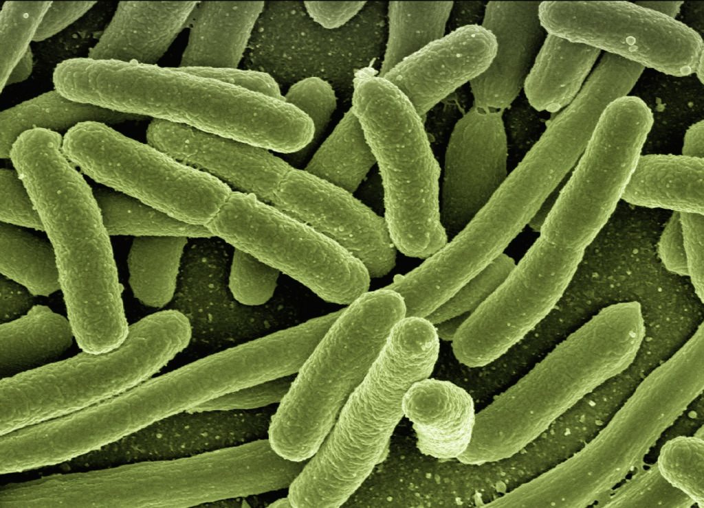
Despite stringent infection-control efforts around the world, hospital-acquired infections (HAIs)keep on popping up from new strains of bacteria. In Science Translational Medicine, researchers report evidence pointing to an unexpected source of such bacteria: the hospitalised patients themselves.
From experiments with mice, researchers at Washington University School of Medicine in St. Louis discovered that urinary tract infections (UTIs) can arise after sterile tubes, called catheters, are inserted into the urinary tract, even when no bacteria are detectable in the bladder beforehand. Such tubes are commonly used in hospitals to empty the bladders of people undergoing surgery. In the mice, inserting the tubes activated dormant Acinetobacter baumannii bacteria hidden in bladder cells, triggering them to emerge, multiply and cause UTIs, the researchers said.
The findings suggest that screening patients for hidden reservoirs of dangerous bacteria could supplement infection-control efforts and help prevent deadly HAIs.
“You could sterilise the whole hospital, and you would still have new strains of A. baumannii popping up,” said co-senior author Mario Feldman, PhD, a professor of molecular microbiology. “Cleaning is just not enough, and nobody really knows why. This study shows that patients may be unwittingly carrying the bacteria into the hospital themselves, and that has implications for infection control. If someone has a planned surgery and is going to be catheterised, we could try to determine whether the patient is carrying the bacteria and cure that person of it before the surgery. Ideally, that would reduce the chances of developing one of these life-threatening infections.”
The notoriously multidrug-resistant A. baumannii is a major threat to patients, causing many cases of UTIs in people with urinary catheters, pneumonia in people on ventilators, and bloodstream infections in people with central-line catheters into their veins.
The researchers set out to investigate why so many A. baumannii UTIs develop after people receive catheters.
Most UTIs among otherwise healthy people are caused by the bacterium Escherichia coli. Research has shown that E. coli can hide out in bladder cells for months after a UTI seems to have been cured, and then re-emerge to cause another infection.
The researchers investigated whether A. baumannii can hide inside cells like E. coli can. They studied mice with UTIs caused by A. baumannii. They used mice with weakened immune systems because, like people, healthy mice can fight off A. baumannii.
Once the infections had resolved and no bacteria were detected in the mice’s urine for two months, the researchers inserted catheters into the mice’s urinary tracts with a sterile technique. Within 24 hours, about half of the mice developed UTIs caused by the same strain of A. baumannii as the initial infection.
“The bacteria must have been there all along, hiding inside bladder cells until the catheter was introduced,” said co-senior author Scott J. Hultgren, PhD, a professor and expert on UTIs. “Catheterisation induces inflammation, and inflammation causes the reservoir to activate, and the infection blooms.”
Since A. baumannii rarely causes symptoms in otherwise healthy people, many people who carry the bacteria may never know they’re infected, the researchers said. According to the researchers’ literature search, 2% of healthy people carry A. baumannii in their urine.
“I wouldn’t put much weight on the precise percentage, but I think we can say with certainty that some percentage of the population is walking around with A. baumannii,” Feldman said. “As long as they’re basically healthy, it doesn’t cause any problems, but once they’re hospitalised, it’s a different matter. This changes how we think about infection control. We can start considering how to check if patients already have Acinetobacter before they receive certain types of treatment; how we can get rid of it; and if other bacteria that cause deadly outbreaks in hospitals, such as Klebsiella, hide in the body in the same way. That’s what we’re working on figuring out now.”