Tag: 1/12/23

Abnormally High Levels of HDL-C Linked to Dementia in Older Adults

Photo by Matteo Vistocco on Unsplash

Abnormally high levels of high density lipoprotein cholesterol (HDL-C), are associated with an increased risk of dementia in older adults, according to study led by Monash University. Researchers said very high levels of the ‘good cholesterol’ HDL-C linked to dementia risk in this study were uncommon and not diet related, but more likely to reflect a metabolic disorder. The findings may help doctors to recognise a group of older patients potentially at risk of dementia, particularly in those aged 75 and older.

Published in The Lancet Regional Health – Western Pacific, this is one of the largest studies of elevated HDL-C levels and dementia in initially healthy older people aged mostly over 70, enrolled in the ASPREE* study.

Over an average 6.3 years, participants with very high HDL-C (> 80mg/dL or > 2.07mmol/L) at study entry were observed to have a 27% higher risk of dementia compared to participants with optimal HDL-C levels, while those aged 75 years and older also showed a 42% increased risk compared to those with optimal levels.

Very high HDL-C levels were categorised as 80mg/dL (> 2.07mmol/L) or above.

The optimal level of HDL-C of 40 to 60mg/dL (1.03–1.55mmol/L) for men and 50 to 60mg/dL (1.55–2.07mmol/L) for women was generally beneficial for heart health.

Among 18 668 participants included in this analysis, 2709 had very high HDL-C at study entry, with 38 incidents of dementia in those aged less than 75 years with very high levels, and 101 in those aged 75 and more with very high levels.

First author and Monash University School of Public Health and Preventive Medicine senior research fellow Dr Monira Hussain said that further research was needed to explain why a very high HDL cholesterol level appeared to affect the risk of dementia.

Dr Hussain said these study findings could help improve our understanding of the mechanisms behind dementia, but more research was required.

“While we know HDL cholesterol is important for cardiovascular health, this study suggests that we need further research to understand the role of very high HDL cholesterol in the context of brain health,” she said.

“It may be beneficial to consider very high HDL cholesterol levels in prediction algorithms for dementia risk.”

*The Aspirin in Reducing Events in the Elderly (ASPREE) trial is a double-blind, randomised, placebo-controlled trial of daily aspirin in healthy older people. 

Source: Monash University

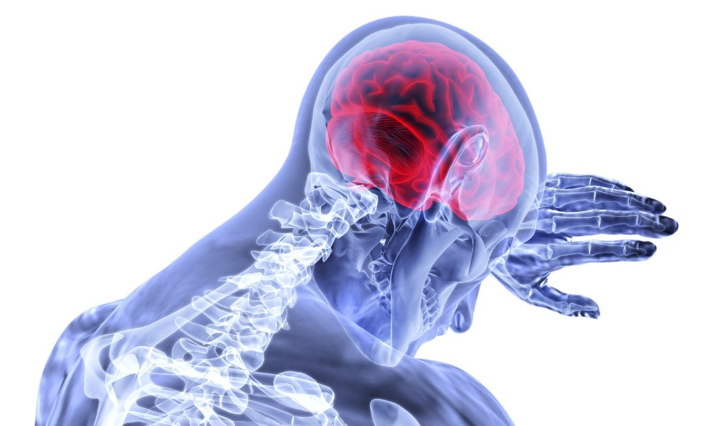
At Last, Objective Evidence for the Involvement of Neck Muscles in Headaches

Source: CC0

Researchers have identified objective evidence of how the neck muscles are involved in primary headaches. The study findings, being presented at the annual meeting of the Radiological Society of North America (RSNA), could lead to better treatments.

The distinct underlying causes of primary headaches, comprising tension-type headaches and migraines, are still not fully understood.

“Our imaging approach provides first objective evidence for the very frequent involvement of the neck muscles in primary headaches, such as neck pain in migraine or tension-type headache, using the ability to quantify subtle inflammation within muscles,” said Nico Sollmann, MD, PhD, resident at University Hospital Ulm and University Hospital Rechts der Isar in Munich, Germany.

In tension-type headaches there is often the perception of a tightening in the head and mild to moderate dull pain on both sides of the head. While these headaches are typically associated with stress and muscle tension, their exact origin is not fully understood.

Migraines are characterised by a severe throbbing pain and generally occur or are worse on one side of the head. Migraines may also cause nausea, weakness and light sensitivity.

Neck pain is commonly associated with primary headaches but there are no objective biomarkers for myofascial involvement. Myofascial pain is associated with inflammation or irritation of muscle or of the connective tissue, known as fascia, that surrounds the muscle.

For the study, Dr Sollmann and colleagues aimed to investigate the involvement of the trapezius muscles in primary headache disorders by quantitative magnetic resonance imaging (MRI) and to explore associations between muscle T2 values and headache and neck pain frequency.

The prospective study recruited 50 participants, mostly women, ranging in age from 20 to 31 years old. Of the participants, 16 had tension-type headache, and 12 had tension-type headache plus migraine episodes. The groups were matched with 22 healthy controls.

All participants underwent 3D turbo spin-echo MRI. The bilateral trapezius muscles were manually segmented, followed by muscle T2 extraction.

Associations between muscle T2 values and the presence of neck pain, number of days with headache, and number of myofascial trigger points as determined by manual palpation of the trapezius muscles were analysed (adjusting for age, sex and body mass index).

The tension-type headache plus migraine group demonstrated the highest muscle T2 values. Muscle T2 was significantly associated with the number of headache days and the presence of neck pain.

The increased muscle T2 values could be interpreted as a surrogate of inflammation arising from the nervous system and increased sensitivity of nerve fibres within myofascial tissues.

“The quantified inflammatory changes of neck muscles significantly correlate with the number of days lived with headache and the presence of subjectively perceived neck pain,” Dr Sollmann said.

“Those changes allow us to differentiate between healthy individuals and patients suffering from primary headaches.”

Muscle T2 mapping could be used to stratify patients with primary headaches and to track potential treatment effects for monitoring.

“Our findings support the role of neck muscles in the pathophysiology of primary headaches,” Dr Sollmann said. “Therefore, treatments that target the neck muscles could lead to a simultaneous relief of neck pain, as well as headache.”

Dr Sollmann pointed out that non-invasive treatment options that directly target the site of pain in the neck muscles could be highly effective and safer than systemic drugs.

“Our imaging approach with delivery of an objective biomarker could facilitate therapy monitoring and patient selection for certain treatments in the near future,” he added.

Source: Radiological Society of North America

New Device Uses an Eye-safe Laser to Detect Traumatic Brain Injury

Photo: Unsplash

Researchers from the University of Birmingham have designed and developed a novel diagnostic device to detect traumatic brain injury (TBI) by shining a safe laser into the eye.

The technique is radically different from other diagnostic methods and is expected to be developed into a hand-held device for use in the critical ‘golden hour’ after traumatic brain injury, when life critical decisions on treatment must be made.

The device, described in Science Advances, incorporates a class 1, CE marked, eye-safe laser and a unique Raman spectroscopy system, which uses light to reveal the biochemical and structural properties of molecules by detecting how they scatter light, to detect the presence and levels of known biomarkers for brain injury.

There is an urgent need for new technologies to improve the timeliness of TBI diagnosis. TBI is caused by sudden shock or impact to the head, which can cause mild to severe injury to the brain, and rapid intervention is necessary to prevent further irreversible damage.

Diagnosis at the point of injury is difficult. Moreover, radiological investigations such as X-ray or MRI are very expensive and slow to show results.

Birmingham researchers, led by Professor Pola Goldberg Oppenheimer from the School of Chemical Engineering, designed and developed the novel diagnostic hand-held device to assess patients as soon as injury occurs.

It is fast, precise and non-invasive for the patient, causing no additional discomfort, can provide information on the severity of the trauma, and will be suitable to be used on-site to assess TBI.

Professor Pola Goldberg Oppenheimer said: “Early diagnosis of TBI is crucial, as life-critical decisions on treatment must be made with the first ‘golden hour’ after injury. However current diagnostic procedure relies on observation by ambulance crews, and MRI or CT scans at a hospital – which may be some distance away.”

The device works by scanning the retina where the optic nerve sits. Since the optic nerve is so closely linked to the brain, it carries the same biological information in the form of protein and lipid biomarkers.

These biomarkers exist in a very tightly regulated balance, meaning even the slightest change may have serious effects on the ‘brain-health’. TBI causes these biomarkers to change, indicating that something is wrong.

Previous research has demonstrated the technology can accurately detect the changes in animal brain and eye tissues with different levels of brain injuries — picking up the slightest changes.1,2,3

The device detailed in the current paper detects and analyses the composition and balance of these biomarkers to create ‘molecular fingerprints’.

The current study details the development, manufacture, and optimisation of a proof-of-concept prototype, and its use in reading biochemical fingerprints of brain injury on the optic nerve, to see whether it is a viable and effective approach for initial ‘on the scene’ diagnosis of TBI.

The researchers constructed a phantom eye to test its alignment and ability to focus on the back of the eye, used animal tissue to test whether it could discern between TBI and non-TBI states, and also developed decision support tools for the device, using AI, to rapidly classify TBIs.

The device is now ready for further evaluation including clinical feasibility and efficacy studies, and patient acceptability.

The researchers expect the diagnostic device to be developed into a portable technology which is suitable for use in point-of-care conditions capable to rapidly determine whether TBI occurs as well as classify whether it is mild, moderate or severe, and therefore, direct triage appropriately and in timely manner.

Source: University of Birmingham

No-aspirin Regimen Benefits Heart Failure Patients with LVADs

Photo by cottonbro studio

A recent clinical trial published in JAMA found that excluding aspirin for advanced heart failure (HF) patients with a ventricular assist device saw a reduction in bleeding events while maintaining their survival rates.

The ARIES-HM3 Randomised Clinical Trial assessed the safety and efficacy of excluding aspirin from the antithrombotic regimen in patients with advanced HF who have undergone implantation of a fully magnetically levitated left ventricular assist device (LVAD).

“We can now safely say that not giving aspirin is not only safe from a thromboembolic risk profile but results in improved adverse event rate by a significant reduction in non-surgical bleeding which is a well-known complication related to LVAD therapy,” said Mirnela Byku, MD, P.D, MBA, co-author of the study and director of the UNC Durable Mechanical Circulatory Device Program at the UNC School of Medicine.

“Improving not only longevity but also reducing morbidity and improving quality of life is a big focus in the field of MCS.”

Until this study, there had been no consensus in the field about use of or dose of aspirin in the LVAD population.

The international clinical trial followed a randomised, double-blind, placebo-controlled design and involved 628 patients across 51 centres in 9 countries.

The patients were divided into two groups: one receiving aspirin (100mg/d) and the other receiving a placebo in addition to vitamin K antagonist (VKA) therapy.

A focus was to determine if the likelihood a patient experiences major nonsurgical haemocompatibility-related adverse events (such as stroke, pump thrombosis, major bleeding, or arterial peripheral thromboembolism) within 12 months differed between the two groups.

The results showed that not giving aspirin to patients with advanced HF, treated with a fully magnetically levitated LVAD who are receiving VKAs, did not make their survival worse. Furthermore, aspirin avoidance was associated with a significant reduction (34%) in major nonsurgical bleeding events.

Source: University of North Carolina Health Care