Category: Uncategorized

Study Shows How Present Fathers Support Adolescent Resilience

Photo by Kaysha on Unsplash

A new study published in Psychological Reports has shed light on the way present fathers play a positive role in adolescent psychological development. The researchers observed that when adolescents grow up with fathers present, they experience more ‘failure learning’, which supports the development of resilience.

Adolescence is a difficult time, with new social and academic pressures emerging. Resilience, or the capacity to cope with and recover from challenges, has been identified as a critical component to success in adolescence.

They explored the mediating effects of four subfactors of failure learning: failure cognition, reflection and analysis, experience transformation, and prudent attempt.

Failure cognition is the understanding and perception of a failure event. It includes recognising the occurrence of a failure and understanding the causes.

Reflective analysis involves evaluation of the events leading to the failure, and critically analysing one’s own actions and decisions. This helps identification of mistakes and areas for improvement.

Experience transformation involves taking the insights from reflective analysis and transforming the experience of failure into a learning opportunity.

Prudent attempt involves putting the lessons learned into practice, and taking calculated risks to improve subsequent performance. This component emphasises the importance of persistence and not being put off by prior failures.

Using questionnaires, the researchers assessed 626 Chinese middle school students, average age 14. They measured levels of father presence, resilience, and failure learning. The researchers found that: (1) there was a significant association between father presence, failure learning, and resilience; (2) failure learning played a mediating role between father presence and adolescents’ resilience; (3) the mediating effect of experience transformation and prudent attempt between father presence and adolescents’ resilience was significant, but not failure cognition and reflective analysis.

The researchers posited that the presence of a father helped to mediate resilience, especially in the two aspects of failure learning most linked to resilience. By supporting the decisions made around the failure, they help their adolescent children to recognise that a prudent attempt was made and to accept the failure.

Limitations included being the adolescents being exclusively Chinese, with cultural factors that may not be applicable to adolescents of other cultures. Additionally, the effect of mothers was not accounted for, and it was possible that the positive effects were only possible through the co-parenting support of a mother,

Source: PsyPost

Low Dietary Potassium can Cause Direct Kidney Injury

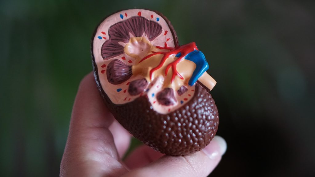
Anatomic model of a kidney
Photo by Robina Weermeijer on Unsplash

It is well known that diets with a high sodium-to-potassium ratio are linked to poor cardiovascular outcomes. To date, most attention has mostly focused on high sodium, but low potassium is also a culprit in cardiovascular disease. Now, research published in Cell Reports has revealed that low dietary potassium also causes direct kidney injury.

Using in vitro and in vivo approaches, Andrew Terker, MD, PhD, and colleagues demonstrated that the injury effects depend on the Kir4.2 potassium channel in kidney proximal tubule cells. First, they reduced dietary potassium levels to determine changes in kidney injury markers, and then lowered blood potassium levels to confirm that it indeed drove kidney injury.

Efflux of potassium from the cells caused intracellular acidosis and activated the enzyme glutaminase. This increased enzyme activity contributed to kidney injury, leading to hypertrophy, inflammation and fibrosis. They found that deleting Kir4.2 or glutaminase protected proximal tubule cells from injury in both cell culture and animal models. 

The findings identify Kir4.2 and glutaminase as mediators of low potassium-related kidney injury and potential therapeutic targets. The findings also suggest that the standard practice of recommending excessive restriction of dietary potassium for patients with chronic kidney disease could unintentionally contribute to disease progression in certain settings

Source: Vanderbilt University

Delivery Method Affects Babies’ Vaccine Responses

Photo by Christian Bowen on Unsplash

The method by which a baby was delivered is associated with how its immune system will respond to pneumococcal and meningococcal vaccinations, according to a study in Nature Communications. After vaccination, babies born naturally had higher antibody levels than those born via Caesarian section.

Experts say the findings could help to inform conversations about C-sections between expectant mothers and their doctors, and shape the design of more tailored vaccination programmes.

Researchers studied the relationship between gut microbes and antibody levels after vaccination in a cohort of 120 babies, who were vaccinated at 8 and 12 weeks against lung infections and meningitis.

The researchers tracked the development of the gut microbiome in the child’s first year of life and their immune response to the vaccines by testing saliva samples at 12 and 18 months.

Research was carried out by a team from the University of Edinburgh, Spaarne Hospital and University Medical Centre in Utrecht and the National Institute for Public Health and the Environment in The Netherlands.

In the 101 babies tested for antibodies as a result of the vaccine that protects against lung infections, the investigators found double the antibody levels in babies delivered naturally compared with those delivered by C-section.

Breastfeeding was linked with 3.5 times higher antibody levels compared with formula-fed children who had been delivered naturally.

Levels of antibodies as a result of the vaccine that protects against meningitis were tested in 66 babies. Experts found the levels of antibodies were 1.7 times higher for naturally delivered babies, regardless of breastfeeding, compared with those delivered via C-section.

The gut microbiome is seeded at birth, developing rapidly over the first few months of life, and is influenced mostly by delivery mode, breastfeeding, and antibiotic use.

The team found a clear relationship between microbes in the gut of those babies and levels of antibodies.

For example, among a host of bacteria in the gut, high levels of two in particular — Bifidobacterium and E. Coli — were associated with a high antibody response to the vaccine that protects against lung infections.

High levels of E. Coli were also linked with a high antibody response to the vaccine that protects against meningitis.

The baby acquires the Bifidobacterium and E.coli bacteria through natural birth and human milk is needed to provide the sugars for these bacteria to thrive on.

The team concludes that the babies’ microbiome in early life contributes the immune system’s response to the vaccines and sets the level of protection against certain infections in childhood.

Vaccination schedules could also be adjusted based on mode of delivery or an analysis of the baby’s microbiome in the future, experts say.

Dr Emma de Koff, first author and microbiology trainee at the Amsterdam University Medical Center, said: “We expected to find a link between the gut microbiome and the babies’ vaccine responses, however we never thought to find the strongest effects in the first weeks of life.”

Professor Debby Bogaert study lead and Chair of Paediatric Medicine at the University of Edinburgh said “I think it is especially interesting that we identified several beneficial microbes to be the link between mode of delivery and vaccine responses. In the future, we may be able to supplement those bacteria to children born by C-section shortly after birth through, for example, mother-to-baby ‘faecal transplants’ or the use of specifically designed probiotics.”

Probing why Vaccine Responses Vary among Individuals

Photo by Gustavo Fring at Pexels

Many factors dictate whether a vaccine provokes an immune response, including specific biomarkers within a person’s immune system, but until now there has been no evidence showing whether these factors were universal across a wide range of vaccines.

New findings from a meta-analysis published in Nature Immunology examine the biological mechanisms responsible for why some people’s immune systems respond differently to vaccinations, which could have global implications for the development and administration of vaccines.

As part of a series of studies for The Human Immunology Project Consortium (HIPC), a network of national research institutions studying the range of responses to different infections and vaccinations, Emory researchers analysed the molecular characteristics of 820 healthy young adults who were immunised with 13 different vaccines to identify specific biomarkers that generate antibody response to vaccines.

The participants were separated into three endotypes, or groups with a common gene expression, based on the level of inflammatory response prior to vaccination – a high inflammatory group, a low inflammatory group, and a mid-inflammatory group. After studying the immunological changes that occurred in participants following vaccination, researchers found the group that had the highest levels of inflammation prior to vaccine had the strongest antibody response.

“We were surprised because inflammation is usually depicted as something that is bad,” says Slim Fourati, PhD, bioinformatic research associate at Emory University and first author on the paper. “These data indicate that some types of inflammation can actually foster a stronger response from a vaccine.”

Fourati, Dr Rafick-Pierre Sekaly, professor and senior author of the paper, and the HIPC team identified specific biomarkers among this group and cellular features that characterised the pre-vaccination inflammatory signature, information that can be used to predict how well an individual will respond to a vaccine.

“With the knowledge we now have about what characteristics of the immune system enable a more robust response, vaccines can be tailored to induce this response and maximize their effectiveness,” says Fourati. “But we still have more questions to answer.”

More research is needed to determine the cause of this inflammation in otherwise healthy adults. Additionally, Fourati suggests future studies should look at how these biomarkers facilitate vaccine protection in older age groups and among populations who are immunocompromised.

These findings can serve to improve vaccine response across all individuals. Better understanding of how various pre-vaccine immune states impact antibody responses opens the possibility of altering these states in more vulnerable individuals. For example, scientists may give patients predicted to have a weaker immune response an adjuvant with the vaccine to trigger the inflammatory genes associated with greater protection.

This work will help enable improved, more efficient clinical trials for the development of new vaccines.

Source: Emory Health Sciences

Reawakening a Foetal Gene Promotes Diabetic Wound Healing

Photo by Diana Polekhina on Unsplash

In the journal Molecular Therapy, researchers report that it may be possible to heal wounds by using a healing protein that is active in foetuses, but largely inactive in adults and absent in diabetic adults.

“We already know from previous studies at other institutions that if a foetus is wounded, it can regenerate the tissue, or repair it to be like new,” said Chandan K. Sen, PhD, at Indiana University School of Medicine. “But after birth, such regenerative wound healing ability is lost. Healing in adults is relatively inefficient often associated with undesirable scar formation.”

In the study,  the team focused on a protein called nonselenocysteine-containing phospholipid hydroperoxide glutathione peroxidase, or NPGPx. NPGPx is active in foetal tissue but becomes mostly inactive in the skin after birth.

“Nature essentially hides this foetal regenerative repair pathway in the adult body,” Sen said. “We spotted its absence, and then activated it to improve healing of diabetic wounds.”

Researchers used tissue ‘nanotransfection’ technology to deliver the NPGPx gene to the wound site. Diabetic wounds, which are complicated skin injuries in people with diabetes, are particularly difficult to treat and often lead to amputations or other complications because of how easily they can become infected.

“This is an exciting new approach to harness foetal repair mechanisms to close diabetic wounds in adults,” Sen said. “The study results show that while NPGPx has been known to be abundant in the foetal skin, but not after birth, it can be reactivated in the skin after an injury. We look forward to continued study aiming to achieve a more complete regenerative repair by improving our understanding of how NPGPx functions.”

Source: Indiana University School of Medicine

Pregnancy Permanently Alters Skeletal Composition

pregnant woman holding her belly
Source: Anna Hecker on Unsplash

Reproduction permanently alters the skeletons of females in ways not previously known, a team of anthropologists has concluded from research findings published in PLOS ONE. This discovery, based on an analysis of primates, sheds new light on how giving birth can permanently change the body.

“Our findings provide additional evidence of the profound impact that reproduction has on the female organism, further demonstrating that the skeleton is not a static organ, but a dynamic one that changes with life events,” explains Paola Cerrito, who led the research as a doctoral student in NYU’s Department of Anthropology and College of Dentistry.

Specifically, the researchers found that calcium, magnesium, and phosphorus concentrations are lower in females who have experienced reproduction. These changes are linked to giving birth itself and to lactation.

However, they caution that while other clinical studies show calcium and phosphorus are necessary for optimal bone strength, the new findings do not address overall health implications for either primates or humans. Rather, they say, the work illuminates the dynamic nature of our bones.

“A bone is not a static and dead portion of the skeleton,” notes NYU anthropologist Shara Bailey, one of the study’s authors. “It continuously adjusts and responds to physiological processes.”

It’s been long-established that menopause can have an effect on females’ bones. Less clear is how preceding life-cycle events, such as reproduction, can influence skeletal composition. To address this, the researchers studied the primary lamellar bone, the main type of bone in a mature skeleton. This aspect of the skeleton is an ideal part of the body to examine because it changes over time and leaves biological markers of these changes, allowing scientists to monitor alterations during the life span.

The researchers examined the growth rate of lamellar bone in the femora, or thigh bones, of both female and male primates who had lived at the Sabana Seca Field Station in Puerto Rico and died of natural causes. Veterinarians at the field station had monitored and recorded information on these primates’ health and reproductive history, allowing the researchers to match bone-composition changes to life events with notable precision.

Cerrito and her colleagues used electron microscopy and energy-dispersive X-ray analysis to calculate changes in concentrations of calcium, phosphorus, oxygen, magnesium, and sodium in the primates’ bones.

Their results showed different concentrations of some of these elements in females who gave birth compared males as well as females who did not give birth. Specifically, in females who gave birth, calcium and phosphorus were lower in bone formed during reproductive events. Moreover, there was a significant decline in magnesium concentration during these primates’ breastfeeding of infants.

“Our research shows that even before the cessation of fertility the skeleton responds dynamically to changes in reproductive status,” says Cerrito, now a research fellow at ETH Zurich. “Moreover, these findings reaffirm the significant impact giving birth has on a female organism – quite simply, evidence of reproduction is ‘written in the bones’ for life.”

Source: New York University

Arthritis Link in Immune Response Against Gut Bacteria Protein

Photo by Cara Shelton on Unsplash

Rheumatoid arthritis (RA) is a chronic autoimmune disease that is associated with aberrant immune responses. In a recent analysis published in Arthritis & Rheumatology, people with RA and those at risk for the disease had higher blood levels of antibodies against a protein expressed by Prevotella copri, a common gut bacteria. 

The study compared 98 participants with established RA who were compared with 98 controls without the condition, as well as 67 participants at high risk for RA who were compared with 67 controls. The researchers measured levels of antibodies against Pc-p27, a protein expressed by P. copri.

Participants with RA had significantly higher levels of IgA anti-Pc-p27 antibodies and trends towards higher levels of IgG anti-Pc-p27 antibodies when compared to their matched controls. When stratified by early versus established RA, early RA participants had median values of IgG anti-Pc-p27 antibodies that were overall higher, whereas median values of IgA anti-Pc-p27 were statistically significantly higher in participants with established RA, compared with their matched controls.

The authors noted that additional research into the roles of this and other microorganisms in rheumatoid arthritis is warranted.

“Our hope is that these findings can help to further elucidate the complex etiologic role of bacterial commensals in people who are at-risk of developing RA and in those with RA so that targeted therapies can be developed with the goals of providing better treatment and ultimately, prevention of the disease,” said corresponding author Jennifer A. Seifert, MPH, of the University of Colorado Denver.

Source: Wiley

Making Friends with Active People Encourages Exercise in Sedentary People

Man and woman about to sprint
Source: Andrea Piacquadio on Pexels

A study published in PLOS ONE suggests that interacting with moderately active people is an important factor that could influence sedentary people into becoming more active. The researchers developed a mathematical model that takes into account the influence of social interactions on community exercise trends.

To help address shortfalls in people getting recommended levels of exercise, Ensela Mema of Kean University in Union, New Jersey, and colleagues drew on previous research showing that social interactions with peers can play a key role in boosting physical activity within a community. In line with that knowledge, they developed a mathematical model that simulates how social interactions can affect a population’s exercise trends over time. The model incorporates data from the US Military Academy.

The model simulations showed that, without social interactions, populations experienced a long-term decrease in physically active individuals, and sedentary behaviour began to dominate. However, when the simulations included social interactions between sedentary and moderately active people, sedentary populations became more physically active in the long term. Still, in simulations where moderately active people became more sedentary over time, overall physical activity trends plummeted.

While these simulations were not validated with real-world data, the researchers say they provide new insights that could inform public health efforts to boost community physical activity levels. The researchers outline a number of recommendations for such efforts, such as social activities designed to boost interactions between sedentary and moderately active people.

The researchers said that more research will be needed to better understand the balance between encouraging exercise among sedentary people while retaining activity levels in moderately active people.

The authors added: “We have traditionally directed physical activity interventions by engaging sedentary individuals to become more active. Our model suggests that focusing on the moderately active population to sustain their activity and increasing their interactions with sedentary people could stimulate higher levels of overall physical activity in the population.”

Source: ScienceDaily

Some Long COVID Cognitive Problems Stem from Immune Response

Source: Pixabay

Using brain ‘organoids’, researchers at Karolinska Institutet have found that COVID infection results in damage from immune cells and gene expressions similar to those found in neurodegenerative disorders. Their findings were published in Molecular Psychiatry.

The findings could help to identify new treatments against persistent cognitive symptoms after a COVID infection.

Neurological symptoms in ‘long COVID’ have been widely reported but the underlying mechanisms for this remains unknown. To find out, the study’s researchers created brain organoids from human induced pluripotent stem (iPS) cells. The model differs from previous organoid models as the researchers also included the brain immune cells – microglia – in the model. In the infected models, microglia excessively engulfed synaptic structures and displayed upregulation of factors involved in phagocytosis. The developed model and the findings in the study could help to guide future efforts to target cognitive symptoms in the aftermath of COVID and other neuroinvasive viral infections.

Post-infection cognitive deficits

 “Interestingly, our results to a large extent mimic what has recently been observed in mouse models infected with other neuroinvasive RNA viruses such as the West Nile virus. These viruses are also linked to residual cognitive deficits after the infection, and a persisting activation of microglia leading to an excessive engulfment of synapses, which has been suggested to drive these symptoms. Multiple studies have now also reported remaining cognitive symptoms after a COVID infection, as well as an increased risk of receiving a diagnosis of a disorder characterised by cognitive symptoms,” says co-first author of the study Samudyata, a postdoctoral fellow at Karolinska Institutet.

Connections to Parkinson’s and Alzheimer’s disease

Microglia also carry out important regulatory functions of the neuronal circuitries in the brain, one of which is engulfing unwanted synapses, a process that is believed to improve and maintain cognitive functions. However, excessive engulfment of synapses has been linked to both neurodevelopmental disorders, such as  schizophrenia, as well as to neurodegenerative disorders including Alzheimer’s disease.

By sequencing genes in single cells, the authors could also study how different cell types in the model responded to the virus.  

“Microglia displayed a distinct gene signature largely characterized by an upregulation of interferon-responsive genes, and included pathways previously linked to neurodegenerative disorders such as Parkinson’s and Alzheimer’s disease. This signature was also observed at a later time-point when the virus load was minimal,” says co-author of the study Susmita Malwade, a doctoral student at Karolinska Institutet.

The researchers will now study how different pharmacological approaches can reverse the observed changes in the infected models.

Source: Karolinska Institutet

Genes That Protected against Black Death now Linked to Autoimmune Disease

Plague doctor costume
Photo by Peter Kvetny on Unsplash

A team of researchers analysed centuries-old DNA samples from plague burial pits in London identified genes that protected some against the Black Death that swept through Europe, Asia and Africa nearly 700 years ago. Their study, which showed how those aspects of our immune systems have continued to evolve since that time, is published in the journal Nature.

According to the researchers’ findings, the same genes that once conferred protection against the Black Death are today linked to an increased susceptibility to autoimmune diseases such as Crohn’s and rheumatoid arthritis.

The team focused on a 100-year window before, during and after the Black Death, which reached London in the mid-1300s. It remains the single greatest human mortality event in recorded history, killing upwards of 50% of the people in what were then some of the most densely populated parts of the world.

More than 500 ancient DNA samples were extracted and screened from the remains of individuals who had died before the plague, died from it or survived the Black Death in London, including individuals buried in the East Smithfield plague pits used for mass burials in 1348–9. Additional samples were taken from remains in Denmark.

Scientists searched for signs of genetic adaptation related to the plague, which is caused by the bacterium Yersinia pestis.

They identified four genes that were under selection, all of which are involved in the production of proteins that defend our systems from invading pathogens and found that alleles of the genes either protected or rendered one susceptible to plague.

Individuals with two identical copies of a particular gene, known as ERAP2, survived the pandemic at a much higher rates than those with the opposing set of copies, because the ‘good’ copies allowed for more efficient neutralisation of Y. pestis by immune cells.

“When a pandemic of this nature – killing 30 to 50 per cent of the population – occurs, there is bound to be selection for protective alleles in humans, which is to say people susceptible to the circulating pathogen will succumb. Even a slight advantage means the difference between surviving or passing. Of course, those survivors who are of breeding age will pass on their genes,” explained evolutionary geneticist Hendrik Poinar, an author of the Nature paper.

Europeans living at the time of the Black Death had were extremely vulnerable at first as they had no recent exposure to Yersinia pestis. Mortality rates fell in subsequent waves of the pandemic over the following centuries.

Researchers estimate that people with the ERAP2 protective allele (the good copy of the gene, or trait), were 40 to 50 per cent more likely to survive than those who did not.

“The selective advantage associated with the selected loci are among the strongest ever reported in humans showing how a single pathogen can have such a strong impact to the evolution of the immune system,” says human geneticist Luis Barreiro, an author on the paper, and professor in Genetic Medicine at the University of Chicago.

Source: McMaster University