Category: Uncategorized

Sacubitril/Valsartan Shines in HF with Ejection Fraction > 40%

Sacubitril/valsartan leads to greater reduction in plasma NT-proBNP levels compared to valsartan alone after stabilisation for worsening heart failure (HF) in patients with an ejection fraction (EF) above 40%, according to late breaking science presented today at Heart Failure 2023, a scientific congress of the European Society of Cardiology (ESC), and published in the Journal of the American College of Cardiology.

Principal investigator Dr Robert Mentz of Duke University Medical Center, Durham, US said: “These data add to the evidence supporting a potential treatment benefit of sacubitril/valsartan in patients with EF over 40% and particularly in those with EF below normal (< 60%). The findings may influence future guidance for the use of the drug in this population, both in and out of hospital and for those with acute, chronic or de novo heart failure.”

Guidelines recommend consideration of sacubitril/valsartan to reduce hospitalisations in patients with HF with preserved EF (HFpEF; EF >50%) and/or mildly reduced EF (HFmrEF; EF 41–49%). Recommendations differ around the world, with some noting benefits are more evident in those with EF on the lower end of this spectrum (ie below normal).

The PARAGON-HF trial excluded patients with decompensated heart failure, but a post-hoc analysis suggested a larger benefit with sacubitril/valsartan in those recently hospitalised. Whether initiation of sacubitril/valsartan is safe and effective in patients with EF over 40% stabilised after a worsening HF event was unknown. In addition, further data were needed in populations excluded by PARAGON-HF (estimated glomerular filtration rate [eGFR] < 30 mL/min/1.73 m2, systolic blood pressure < 110mmHg, and body mass index [BMI] > 40kg/m2).

PARAGLIDE-HF evaluated the effect of sacubitril/valsartan versus valsartan on changes in NT-proBNP, safety and tolerability in HF patients with EF above 40% who had been stabilised after a worsening HF event. The primary endpoint was the time-averaged proportional change in NT-proBNP from baseline through Weeks 4 and 8. It was chosen to mirror the PIONEER-HF trial, which found that among patients with heart failure with reduced EF (< 40%) who were hospitalised for acute decompensated HF, sacubitril/valsartan led to a greater reduction in NT-proBNP concentration than enalapril.

Patients were recruited from 100 sites in the US and Canada. A total of 466 patients with EF above 40% were enrolled within 30 days of a worsening heart failure event (69% were enrolled while in hospital). The average age was 70 years, 52% were women and 22% were Black. Participants were randomly allocated in a 1:1 ratio to sacubitril/valsartan or valsartan. The time-averaged reduction in NT-proBNP was greater with sacubitril/valsartan compared with valsartan (ratio of change 0.85; p=0.049).

The secondary composite hierarchical outcome consisted of a) time to cardiovascular death, b)number and timing of HF hospitalisations, c) number and timing of urgent HF visits and d) time averaged proportional change in NT-proBNP from baseline to Weeks 4 and 8. This outcome was evaluated using a win ratio analysis, which considers the clinical hierarchy and timing of each component of the endpoint. More serious events are given a higher priority and are analysed first. The hierarchical outcome favoured sacubitril/valsartan (as did each of the components) but was not significant (unmatched win ratio 1.19; 95% CI 0.93–1.52; p=0.16).

Regarding other secondary endpoints, compared with valsartan, sacubitril/valsartan reduced worsening renal function (odds ratio [OR] 0.61). There was more symptomatic hypotension in the sacubitril/valsartan group (OR 1.73). Importantly, subgroup analyses showed evidence of a larger treatment effect in those with EF ≤60% for the change in NT-proBNP (0.78) and the hierarchical outcome (win ratio 1.46).

Dr Mentz said: “PARAGLIDE-HF complements PARAGON-HF by focusing on patients stabilised after a worsening heart failure event with EF above 40% similar to the manner in which PIONEER-HF complemented PARADIGM-HF in patients with reduced EF. PARAGLIDE-HF had no run-in period, allowed both newly diagnosed heart failure and improved EF, included those with acute heart failure without specific echocardiographic requirements and overall had a diverse study population (52% women, 22% Black individuals). The trial also permitted patients with eGFR down to 20 mL/min/1.73m2, systolic blood pressure as low as 100mmHg, and any BMI. The broad and diverse population included in PARAGLIDE-HF supports the generalisability of these data to similar patients seen in routine practice.”

Source: European Society of Cardiology

Monkeypox Virus can Linger for up to a Month on Surfaces

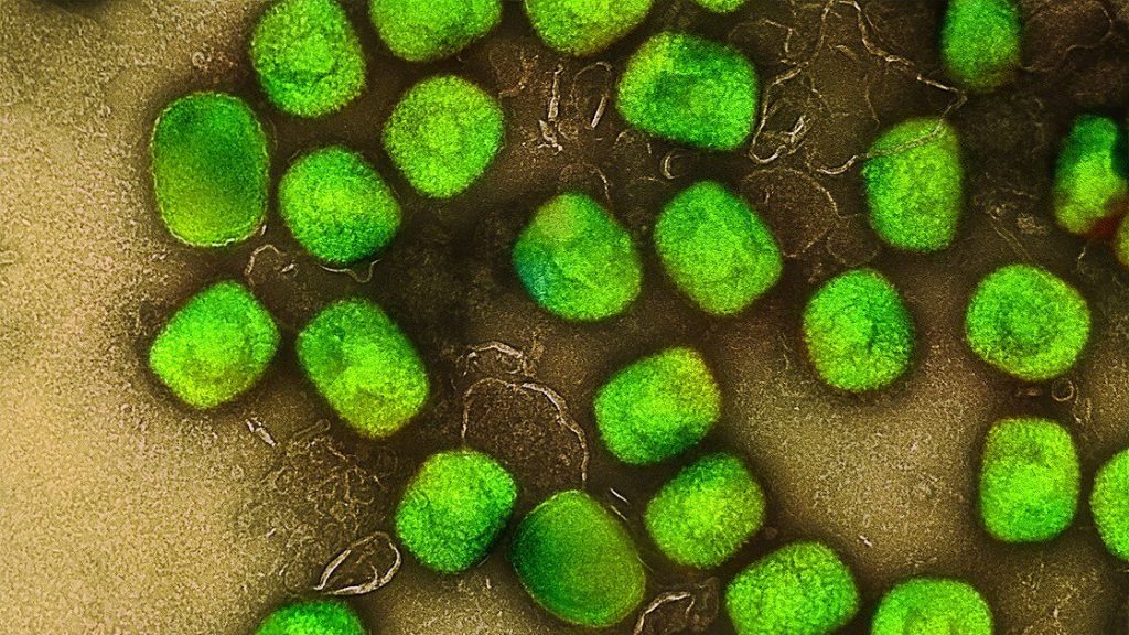
Monkeypox virus. Source: NIH

According to a study published in the Journal of Infectious Diseases, the monkeypox virus remains infectious on steel surfaces for up to 30 days, especially in cold conditions, but can be effectively inactivated by alcohol-based disinfectants.

Smallpox viruses are notorious for their ability to remain infectious in the environment for a very long time. A study conducted by the Department of Molecular and Medical Virology at Ruhr University Bochum, Germany, has shown that temperature is a major factor in this process: at room temperature, a monkeypox virus that is capable of replicating can survive on a stainless steel surface for up to 11 days, and at 4°C for up to a month. Consequently, it’s very important to disinfect surfaces. According to the study, alcohol-based disinfectants are very effective against monkeypox viruses, whereas hydrogen peroxide-based disinfectants have proved inadequate.

Weeks of monitoring

Since 2022, the monkeypox virus has been transmitted more and more frequently from one human host to another. Although infections primarily result from direct physical contact, it’s also possible to contract the virus through contaminated surfaces, for example in the household or in hospital rooms. “Smallpox viruses are notorious for their ability to remain infectious in the environment for a very long time,” explains Dr Toni Meister from the Department for Molecular and Medical Virology at Ruhr University Bochum. “For monkeypox, however, we didn’t know the exact time frames until now.”

The researchers therefore studied them by applying the virus to sanitised stainless steel plates and storing them at different temperatures: at 4°C, at 22°C, which roughly corresponds to room temperature, and at 37°C. They determined the amount of infectious virus after different periods of time, ranging from 15 minutes to several days to weeks.

Viruses remain infectious for a long time

Regardless of the temperature, there was little change in the amount of infectious virus during the first few days. At 22 and 37°C, the virus concentration dropped significantly only after five days. At 37°C, no virus capable of reproducing was detected after six to seven days, at 22°C it took 10–11 days until infection was no longer possible. At 4°C, the amount of virus only dropped sharply after 20 days, and after 30 days there was no longer any danger of infection. “This is consistent with our experience that people can still contract monkeypox from surfaces in the household after almost two weeks,” points out Professor Eike Steinmann, Head of the Department for Molecular and Medical Virology.

In order to reduce the risk of infection in the event of an outbreak, it is therefore extremely important to disinfect surfaces. This is why the researchers tested the effectiveness of five common disinfectants. They found that alcohol-based or aldehyde-based disinfectants reliably reduced the risk of infection. A hydrogen peroxide-based disinfectant, however, didn’t inactivate the virus effectively enough in the study. “Our results support the WHO’s recommendation to use alcohol-based surface disinfectants,” concludes Toni Meister.

Source: Ruhr-University Bochum

In Wounds, Fibroblasts also Clear Away Damaged Tissue

Photo by Diana Polekhina on Unsplash

Burn wounds are notoriously prone to bacterial infection and typically lead to a larger amount of scar tissue than laceration wounds, but new research shows that fibroblasts – normally considered as construction cells – clear away damaged tissue before depositing new material. The more damaged tissue there is, the longer it takes for the fibroblasts to remove the material and heal.

In APL Bioengineering, researchers from Boston University and Harvard University describe how they created a biomimetic model to study wound healing in burn and laceration wounds, which is slower in burn wound as more tissue damage is present.

Cell biologists identify four phases of wound healing: bleeding stoppage, inflammation, new tissue formation, and tissue strengthening. During the inflammation and formation stages, immune cells are thought to clear bacteria and dead cells from the wound. They also activate fibroblasts and blood vessels to begin repairs.

“Depending on the injury, the extent and duration of these four phases can wildly vary across different wound types,” said author Jeroen Eyckmans. “Given that laceration wounds are well perfused with blood, they tend to heal well. However, in burns, the blood vessels are cauterised, preventing blood from entering the wound bed and slowing down the healing process. Severe burn wounds also have large amounts of dead tissue that physically block new tissue formation.”

To study how the mode of injury impacts the healing rate of wounds, the team designed an in vitro model system made of fibroblasts embedded in a collagen hydrogel. Wounds were created in this microtissue using a microdissection knife to mimic laceration or a high-energy laser to simulate a burn.

Although both wound types were equal in size, laser ablation caused more cell death and tissue damage next to the wound margins compared to knife wounds.

“During healing, we found that the fibroblasts first cleared the damaged material from the wound before depositing new material,” said Eyckmans. “This was a surprising finding because removal of dead tissue has been attributed to specialised immune cells such as macrophages, and fibroblasts have been considered to be tissue-building cells, not tissue-removal cells.”

Given that there was more tissue damage in the laser ablation wounds, it took fibroblasts more time to remove the damage, ultimately delaying tissue healing.

Based on these findings, therapies that promote wound clearance could accelerate healing. Genetically engineered white blood cells, designed to remove dead tissue, could be particularly useful for reaching injured organs and tissues deep in the body.

Source: American Institute of Physics

New Review Finds Spinal Cord Stimulation Ineffective for Low Back Pain

Photo by Sasun Bughdaryan on Unsplash

Spinal cord stimulation for the treatment of chronic pain does not provide long-term relief and may cause harm, according to a new Cochrane Review. Spinal cord stimulation involves an implanted device that sends electrical pulses to the spinal cord to interrupt nerve signals before they get to the brain.

The study reviewed published clinical data on spinal cord stimulation, including randomised controlled trials, the ‘gold standard’ for medical research. The researchers analysed the results of 13 clinical trials, looking at data from 699 participants, comparing spinal cord stimulation treatment with placebo or no treatment for low back pain.

Cochrane reviews are trusted by researchers, medical professionals and policymakers because they use robust methodologies to combine evidence from multiple sources, reducing the impact of bias and random error that can make individual studies less reliable.

The review concluded that spinal cord stimulation is no better than a placebo for treating low back pain, with probably little to no benefit for people with low back pain or improvement in their quality of life.

There was little to no clinical data regarding the long-term effectiveness of spinal cord stimulation.

The researchers also found that adverse side effects to the surgery were poorly documented overall, preventing them from concluding the level of risk involved. Harms from spinal cord stimulation could include nerve damage, infection, and the electrical leads moving, all of which may need repeated surgeries.

The review findings have been submitted to the Federal Department of Health and Aged Care prosthesis list review taskforce. The taskforce is reviewing the eligibility of current prostheses subsidised by Medicare.

In Australia, the devices’ long-term safety and performance are also being re-assessed by The Therapeutic Goods Administration (TGA), the country’s regulatory authority for therapeutic goods.

“Spinal cord stimulation is invasive and has a great financial cost to people who choose surgery as a last resort to alleviate their pain. Our review found that the long-term benefits and harms are essentially unknown,” said lead researcher Dr Adrian Traeger from Sydney Musculoskeletal Health, an initiative of the University of Sydney, Sydney Local Health District and Northern Sydney Local Health District.

“Our review of the clinical data suggests no sustained benefits to the surgery outweigh the costs and risks.

“Low back pain is one of the leading causes of disability worldwide. Our findings further emphasise the urgent need to review funding arrangements for chronic pain care to help patients in their search for relief. There are evidence-based physical and psychological therapies for back pain; ensuring access to these is essential.”

The review team found multiple gaps in clinical data.

There were no studies that investigated the long-term (more than 12 months) impact of spinal cord stimulation on low back pain. The longest was a single six-month trial.

The majority of clinical trials only looked at the immediate impact of the device, which is a time frame of less than a month.

The review team provided a list of recommendations, including that future spinal cord stimulation clinical trials be at least 12 months, clearly document the number of people who experience adverse events and make comparisons with other pain treatment options.

Professor Chris Maher,Co-Director of Sydney Musculoskeletal Health, said:

“Our review found that the clinical benefit of adding spinal cord stimulation to treat low back pain remains unknown. When coupled with the reality that these devices are very expensive and often break down there is clearly a problem here that should be of concern to regulators.”

A separate Cochrane review, in which the researchers were not involved, examined the effect of spinal cord stimulation versus placebo in people with chronic pain. Similar to this review, it concluded there was a lack of evidence to suggest long-term benefits in treating chronic pain.

Source: University of Sydney

Study Shows How Present Fathers Support Adolescent Resilience

Photo by Kaysha on Unsplash

A new study published in Psychological Reports has shed light on the way present fathers play a positive role in adolescent psychological development. The researchers observed that when adolescents grow up with fathers present, they experience more ‘failure learning’, which supports the development of resilience.

Adolescence is a difficult time, with new social and academic pressures emerging. Resilience, or the capacity to cope with and recover from challenges, has been identified as a critical component to success in adolescence.

They explored the mediating effects of four subfactors of failure learning: failure cognition, reflection and analysis, experience transformation, and prudent attempt.

Failure cognition is the understanding and perception of a failure event. It includes recognising the occurrence of a failure and understanding the causes.

Reflective analysis involves evaluation of the events leading to the failure, and critically analysing one’s own actions and decisions. This helps identification of mistakes and areas for improvement.

Experience transformation involves taking the insights from reflective analysis and transforming the experience of failure into a learning opportunity.

Prudent attempt involves putting the lessons learned into practice, and taking calculated risks to improve subsequent performance. This component emphasises the importance of persistence and not being put off by prior failures.

Using questionnaires, the researchers assessed 626 Chinese middle school students, average age 14. They measured levels of father presence, resilience, and failure learning. The researchers found that: (1) there was a significant association between father presence, failure learning, and resilience; (2) failure learning played a mediating role between father presence and adolescents’ resilience; (3) the mediating effect of experience transformation and prudent attempt between father presence and adolescents’ resilience was significant, but not failure cognition and reflective analysis.

The researchers posited that the presence of a father helped to mediate resilience, especially in the two aspects of failure learning most linked to resilience. By supporting the decisions made around the failure, they help their adolescent children to recognise that a prudent attempt was made and to accept the failure.

Limitations included being the adolescents being exclusively Chinese, with cultural factors that may not be applicable to adolescents of other cultures. Additionally, the effect of mothers was not accounted for, and it was possible that the positive effects were only possible through the co-parenting support of a mother,

Source: PsyPost

Low Dietary Potassium can Cause Direct Kidney Injury

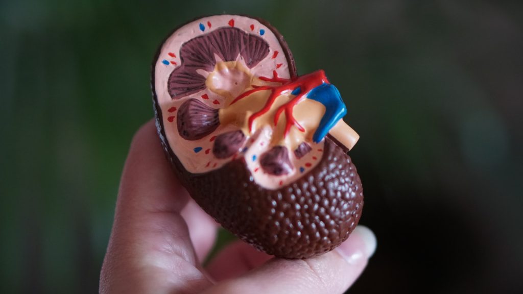
Anatomic model of a kidney
Photo by Robina Weermeijer on Unsplash

It is well known that diets with a high sodium-to-potassium ratio are linked to poor cardiovascular outcomes. To date, most attention has mostly focused on high sodium, but low potassium is also a culprit in cardiovascular disease. Now, research published in Cell Reports has revealed that low dietary potassium also causes direct kidney injury.

Using in vitro and in vivo approaches, Andrew Terker, MD, PhD, and colleagues demonstrated that the injury effects depend on the Kir4.2 potassium channel in kidney proximal tubule cells. First, they reduced dietary potassium levels to determine changes in kidney injury markers, and then lowered blood potassium levels to confirm that it indeed drove kidney injury.

Efflux of potassium from the cells caused intracellular acidosis and activated the enzyme glutaminase. This increased enzyme activity contributed to kidney injury, leading to hypertrophy, inflammation and fibrosis. They found that deleting Kir4.2 or glutaminase protected proximal tubule cells from injury in both cell culture and animal models. 

The findings identify Kir4.2 and glutaminase as mediators of low potassium-related kidney injury and potential therapeutic targets. The findings also suggest that the standard practice of recommending excessive restriction of dietary potassium for patients with chronic kidney disease could unintentionally contribute to disease progression in certain settings

Source: Vanderbilt University

Delivery Method Affects Babies’ Vaccine Responses

Photo by Christian Bowen on Unsplash

The method by which a baby was delivered is associated with how its immune system will respond to pneumococcal and meningococcal vaccinations, according to a study in Nature Communications. After vaccination, babies born naturally had higher antibody levels than those born via Caesarian section.

Experts say the findings could help to inform conversations about C-sections between expectant mothers and their doctors, and shape the design of more tailored vaccination programmes.

Researchers studied the relationship between gut microbes and antibody levels after vaccination in a cohort of 120 babies, who were vaccinated at 8 and 12 weeks against lung infections and meningitis.

The researchers tracked the development of the gut microbiome in the child’s first year of life and their immune response to the vaccines by testing saliva samples at 12 and 18 months.

Research was carried out by a team from the University of Edinburgh, Spaarne Hospital and University Medical Centre in Utrecht and the National Institute for Public Health and the Environment in The Netherlands.

In the 101 babies tested for antibodies as a result of the vaccine that protects against lung infections, the investigators found double the antibody levels in babies delivered naturally compared with those delivered by C-section.

Breastfeeding was linked with 3.5 times higher antibody levels compared with formula-fed children who had been delivered naturally.

Levels of antibodies as a result of the vaccine that protects against meningitis were tested in 66 babies. Experts found the levels of antibodies were 1.7 times higher for naturally delivered babies, regardless of breastfeeding, compared with those delivered via C-section.

The gut microbiome is seeded at birth, developing rapidly over the first few months of life, and is influenced mostly by delivery mode, breastfeeding, and antibiotic use.

The team found a clear relationship between microbes in the gut of those babies and levels of antibodies.

For example, among a host of bacteria in the gut, high levels of two in particular — Bifidobacterium and E. Coli — were associated with a high antibody response to the vaccine that protects against lung infections.

High levels of E. Coli were also linked with a high antibody response to the vaccine that protects against meningitis.

The baby acquires the Bifidobacterium and E.coli bacteria through natural birth and human milk is needed to provide the sugars for these bacteria to thrive on.

The team concludes that the babies’ microbiome in early life contributes the immune system’s response to the vaccines and sets the level of protection against certain infections in childhood.

Vaccination schedules could also be adjusted based on mode of delivery or an analysis of the baby’s microbiome in the future, experts say.

Dr Emma de Koff, first author and microbiology trainee at the Amsterdam University Medical Center, said: “We expected to find a link between the gut microbiome and the babies’ vaccine responses, however we never thought to find the strongest effects in the first weeks of life.”

Professor Debby Bogaert study lead and Chair of Paediatric Medicine at the University of Edinburgh said “I think it is especially interesting that we identified several beneficial microbes to be the link between mode of delivery and vaccine responses. In the future, we may be able to supplement those bacteria to children born by C-section shortly after birth through, for example, mother-to-baby ‘faecal transplants’ or the use of specifically designed probiotics.”

Probing why Vaccine Responses Vary among Individuals

Photo by Gustavo Fring at Pexels

Many factors dictate whether a vaccine provokes an immune response, including specific biomarkers within a person’s immune system, but until now there has been no evidence showing whether these factors were universal across a wide range of vaccines.

New findings from a meta-analysis published in Nature Immunology examine the biological mechanisms responsible for why some people’s immune systems respond differently to vaccinations, which could have global implications for the development and administration of vaccines.

As part of a series of studies for The Human Immunology Project Consortium (HIPC), a network of national research institutions studying the range of responses to different infections and vaccinations, Emory researchers analysed the molecular characteristics of 820 healthy young adults who were immunised with 13 different vaccines to identify specific biomarkers that generate antibody response to vaccines.

The participants were separated into three endotypes, or groups with a common gene expression, based on the level of inflammatory response prior to vaccination – a high inflammatory group, a low inflammatory group, and a mid-inflammatory group. After studying the immunological changes that occurred in participants following vaccination, researchers found the group that had the highest levels of inflammation prior to vaccine had the strongest antibody response.

“We were surprised because inflammation is usually depicted as something that is bad,” says Slim Fourati, PhD, bioinformatic research associate at Emory University and first author on the paper. “These data indicate that some types of inflammation can actually foster a stronger response from a vaccine.”

Fourati, Dr Rafick-Pierre Sekaly, professor and senior author of the paper, and the HIPC team identified specific biomarkers among this group and cellular features that characterised the pre-vaccination inflammatory signature, information that can be used to predict how well an individual will respond to a vaccine.

“With the knowledge we now have about what characteristics of the immune system enable a more robust response, vaccines can be tailored to induce this response and maximize their effectiveness,” says Fourati. “But we still have more questions to answer.”

More research is needed to determine the cause of this inflammation in otherwise healthy adults. Additionally, Fourati suggests future studies should look at how these biomarkers facilitate vaccine protection in older age groups and among populations who are immunocompromised.

These findings can serve to improve vaccine response across all individuals. Better understanding of how various pre-vaccine immune states impact antibody responses opens the possibility of altering these states in more vulnerable individuals. For example, scientists may give patients predicted to have a weaker immune response an adjuvant with the vaccine to trigger the inflammatory genes associated with greater protection.

This work will help enable improved, more efficient clinical trials for the development of new vaccines.

Source: Emory Health Sciences

Reawakening a Foetal Gene Promotes Diabetic Wound Healing

Photo by Diana Polekhina on Unsplash

In the journal Molecular Therapy, researchers report that it may be possible to heal wounds by using a healing protein that is active in foetuses, but largely inactive in adults and absent in diabetic adults.

“We already know from previous studies at other institutions that if a foetus is wounded, it can regenerate the tissue, or repair it to be like new,” said Chandan K. Sen, PhD, at Indiana University School of Medicine. “But after birth, such regenerative wound healing ability is lost. Healing in adults is relatively inefficient often associated with undesirable scar formation.”

In the study,  the team focused on a protein called nonselenocysteine-containing phospholipid hydroperoxide glutathione peroxidase, or NPGPx. NPGPx is active in foetal tissue but becomes mostly inactive in the skin after birth.

“Nature essentially hides this foetal regenerative repair pathway in the adult body,” Sen said. “We spotted its absence, and then activated it to improve healing of diabetic wounds.”

Researchers used tissue ‘nanotransfection’ technology to deliver the NPGPx gene to the wound site. Diabetic wounds, which are complicated skin injuries in people with diabetes, are particularly difficult to treat and often lead to amputations or other complications because of how easily they can become infected.

“This is an exciting new approach to harness foetal repair mechanisms to close diabetic wounds in adults,” Sen said. “The study results show that while NPGPx has been known to be abundant in the foetal skin, but not after birth, it can be reactivated in the skin after an injury. We look forward to continued study aiming to achieve a more complete regenerative repair by improving our understanding of how NPGPx functions.”

Source: Indiana University School of Medicine

Pregnancy Permanently Alters Skeletal Composition

pregnant woman holding her belly
Source: Anna Hecker on Unsplash

Reproduction permanently alters the skeletons of females in ways not previously known, a team of anthropologists has concluded from research findings published in PLOS ONE. This discovery, based on an analysis of primates, sheds new light on how giving birth can permanently change the body.

“Our findings provide additional evidence of the profound impact that reproduction has on the female organism, further demonstrating that the skeleton is not a static organ, but a dynamic one that changes with life events,” explains Paola Cerrito, who led the research as a doctoral student in NYU’s Department of Anthropology and College of Dentistry.

Specifically, the researchers found that calcium, magnesium, and phosphorus concentrations are lower in females who have experienced reproduction. These changes are linked to giving birth itself and to lactation.

However, they caution that while other clinical studies show calcium and phosphorus are necessary for optimal bone strength, the new findings do not address overall health implications for either primates or humans. Rather, they say, the work illuminates the dynamic nature of our bones.

“A bone is not a static and dead portion of the skeleton,” notes NYU anthropologist Shara Bailey, one of the study’s authors. “It continuously adjusts and responds to physiological processes.”

It’s been long-established that menopause can have an effect on females’ bones. Less clear is how preceding life-cycle events, such as reproduction, can influence skeletal composition. To address this, the researchers studied the primary lamellar bone, the main type of bone in a mature skeleton. This aspect of the skeleton is an ideal part of the body to examine because it changes over time and leaves biological markers of these changes, allowing scientists to monitor alterations during the life span.

The researchers examined the growth rate of lamellar bone in the femora, or thigh bones, of both female and male primates who had lived at the Sabana Seca Field Station in Puerto Rico and died of natural causes. Veterinarians at the field station had monitored and recorded information on these primates’ health and reproductive history, allowing the researchers to match bone-composition changes to life events with notable precision.

Cerrito and her colleagues used electron microscopy and energy-dispersive X-ray analysis to calculate changes in concentrations of calcium, phosphorus, oxygen, magnesium, and sodium in the primates’ bones.

Their results showed different concentrations of some of these elements in females who gave birth compared males as well as females who did not give birth. Specifically, in females who gave birth, calcium and phosphorus were lower in bone formed during reproductive events. Moreover, there was a significant decline in magnesium concentration during these primates’ breastfeeding of infants.

“Our research shows that even before the cessation of fertility the skeleton responds dynamically to changes in reproductive status,” says Cerrito, now a research fellow at ETH Zurich. “Moreover, these findings reaffirm the significant impact giving birth has on a female organism – quite simply, evidence of reproduction is ‘written in the bones’ for life.”

Source: New York University