Category: Respiratory Diseases

Scientists Discover Macrophages on Standby in the Pleural Cavity

Credit: Scientific Animations CC4.0

Scientists have long thought of the pleural cavity merely as a cushion from external damage. Turns out, it also houses macrophages that rush into the lungs during flu infections.

“We were surprised to find them in the lungs because nobody has seen this before, that these cells go into the lung when there’s an infection,” said UC Riverside virologist Juliet Morrison, who led the discovery team.

A paper published in the Proceedings of the National Academy of Sciences details how during an influenza infection, macrophages leave the exterior cavity and cross into the lungs where they decrease inflammation and reduce levels of disease.

“This study shows it’s not just what happens in the lung that matters, but also what’s outside of the lung. Cell types not normally connected to the lung can have outsized impacts on lung disease and health,” Morrison said.

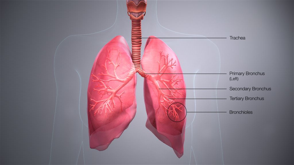
There are three main cavities in the body: one around the heart, the abdominal cavity, and the pleural cavity surrounding the lungs.

“Because it contains fluid, it prevents the lungs from collapsing. However, people have not thought much about the pleural cavity being a whole organ within itself. This research may change that perception,” Morrison said.

Initially, the researchers set out to understand the more general question of what types of cells are present in the lungs during flu infections. They took existing data on lung-related genes from studies of mice that either died from the flu or survived. They then mined the data using an algorithm to predict cell types that change in the lungs during infections.

“We took big data and broke it down to assign which potential immune cells are in the lung tissues. That’s where I got a hint that maybe we had a previously unknown external source of cells in the lung,” Morrison said.

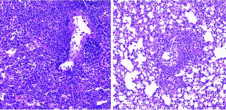
Next, using a laser-based technique, the team tracked macrophages going into the lungs of mice, and observed what happened if they took these cells out of the equation.

“When you take them out of the mouse you see more disease and more lung inflammation,” Morrison said.

Morrison says she hopes this study will encourage other scientists to reevaluate data sets from older studies.

“Our approach was to take information already out there and put it to new use, and we were able to see something new,” she said.

Moving forward, the research team is hoping to determine which proteins “tell” the macrophages to move into the lungs. Once the protein signals have been identified, it may be possible to create drugs that boost either the number of macrophages, or their activity.

The strategy of boosting human defences to infection, rather than developing another antiviral to which viruses could become resistant, could offer people a flu treatment that would be more effective for much longer.

“If we can boost what resolves infection in us, we probably have a better shot. We’re less likely to have resistance. The immune system is so complicated, but it’s our best bet in the long run to work with what we have rather than chase viruses that continue to escape our therapeutics,” Morrison said.

Understanding How T Cells Target Tuberculosis will Enhance Vaccines and Therapies

Tuberculosis bacteria. Credit: CDC

La Jolla Institute for Immunology (LJI) is working to guide the development of new tuberculosis vaccines and drug therapies. Now a team of LJI scientists has uncovered important clues to how human T cells combat Mycobacterium tuberculosis, the bacterium that causes TB. Their findings were published recently in Nature Communications.

“This research gives us a better understanding of T cell responses to different stages in tuberculosis infection and helps us figure out is there are additional diagnostic targets, vaccine targets, or drug candidates to help people with the disease,” says LJI Research Assistant Professor Cecilia Lindestam Arlehamn, PhD, who led the new research in collaboration with LJI Professors Bjoern Peters, PhD, and Alessandro Sette, Dr.Biol.Sci.

The urgent need for TB research

According to the World Health Organization, more than 1.3 million people died of TB in 2022, making it the second-leading infectious cause-of-death after COVID. “TB is a huge problem in many countries,” says Lindestam Arlehamn.

Currently, a vaccine called bacille Calmette-Guerin (BCG) protects against some severe cases of TB. Unfortunately, BCG doesn’t consistently prevent cases of pulmonary TB, which can also be deadly.

Although there are drug treatments for TB, more and more cases around the world have proven drug resistant.

To help stop TB, Lindestam Arlehamn and her colleagues are learning from T cells. Instead of targeting an entire pathogen, T cells look for specific markers, called peptides sequences, that belong to the pathogen.

When a T cell recognises a certain part of a pathogen’s peptide sequence, that area is termed an “epitope.”

Uncovering T cell epitopes gives scientists vital information on how vaccines and drug treatments might take aim at the same epitopes to stop a pathogen.

T cells take aim at a range of TB epitopes

For the new study, the researchers worked with samples from patients who were mid-treatment for active TB. These samples came from study participants in Peru, Sri Lanka, and Moldova.

By looking at T cells in patients from three different continents, the researchers hoped to capture a wide diversity of genetics and environmental factors that can affect immune system activity.

In their analysis, the LJI team uncovered 137 unique T cell epitopes. They found that 16% of these epitopes were targeted by T cells found in two or more patients. The immune system appeared to be working hard to zoom in on these epitopes.

Going forward, Lindestam Arlehamn’s laboratory will investigate which of these epitopes may be promising targets for future TB vaccines and drug therapies.

A step toward better diagnostics

The new study is also a step toward catching TB cases before they turn deadly.

Because Mycobacterium tuberculosis is an airborne bacteria, a person can be exposed without ever realizing it. Once exposed, many people go months or years without any symptoms.

This inactive, or “latent,” TB can turn into active TB if a person’s immune system weakens, for example, during pregnancy or due to an infection such as HIV.

For the new study, the researchers also compared samples from active TB patients with samples from healthy individuals.

The scientists uncovered key differences in T cell reactivity between the two groups.

“For the first time, we could distinguish people with active TB versus those that have been exposed to TB – or unexposed individuals,” says Lindestam Arlehamn.

Lindestam Arlehamn says it may be possible to develop diagnostics that detect this tell-tale T cell reactivity that marks a person’s shift from latent to active TB. “Can we use this peptide pool to look for high-risk individuals and try and follow them over time?” she says.

Source: La Jolla Institute for Immunology

New T Cell ‘Rescue’ Therapy Promising for ARDS

Credit: Scientific Animations CC4.0

Promising trial results indicate that a new type of cell therapy could improve the prognosis of those who are critically ill with acute respiratory distress syndrome (ARDS) resulting from severe COVID.

Published in the journal Nature Communications, Professor Justin Stebbing of Anglia Ruskin University (ARU) is the joint senior author of the new study investigating the use of agenT-797, MiNK Therapeutic’s allogeneic, unmodified invariant natural killer T (iNKT) cell therapy.

The iNKT cell therapy has the effect of rescuing exhausted T cells and prompting an anti-inflammatory cytokine response, potentially activating anti-viral immunity to help these patients fight infection as well as to reduce severe, pathogenic inflammation of the lung.

The new research was carried out at three medical centres and found that agenT-797, which is also under investigation in cancer trials, could be manufactured rapidly, had a tolerable safety profile, and appeared to have a positive effect on mortality among critically unwell Covid-19 ARDS patients receiving intensive care.

The exploratory trial included 20 mechanically ventilated patients with severe ARDS secondary to Covid-19. Of the 20 patients in the trial, 14 survived (70%) at 30 days (compared to a control group of 10%), and there was an 80% lower occurrence of bacterial pneumonia amongst those who received the highest dosage of agenT-797, compared to those who received fewer cells.

Twenty-one patients were treated overall (the main trial, plus one under compassionate use), which included five who were also receiving veno-venous extracorporeal membrane oxygenation (VV-ECMO), known as ‘the most aggressive salvage therapy’ for critically ill patients with ARDS.

In VV-ECMO, deoxygenated blood is pumped through a membrane lung and returned to the body via a cannula. This trial is believed to be the first immune cell therapy of any type to be used in critically unwell patients undergoing VV-ECMO.

Survival of the VV-ECMO cohort was 80% after 30 and 90 days, and 60% after 120 days. This compares favourably to overall survival of 51% for patients with Covid-19 who were treated with just VV-ECMO at the same institution, during the same timeframe.

Joint senior author Justin Stebbing, Professor of Biomedical Sciences at Anglia Ruskin University (ARU) in Cambridge, England, said: “During this small, exploratory study we observed that MiNK’s iNKT cell treatment, which is also being advanced for people with cancer, triggered an anti-inflammatory response in ARDS patients.

“Despite a poor prognosis, critically ill patients treated with this therapy showed favourable mortality rates and those treated at the highest dose also had reduced rates of pneumonia, underscoring the potential application of iNKT cells, and agenT-797 in particular, in treating viral diseases and infections more broadly.

Source: Anglia Ruskin University

Appeasing the Wheezing: Determinants and Outcomes of Respiratory Disease in Childhood

Credit: Pixabay CC0

Asthma and cystic fibrosis are diseases which affect the lungs of children and adults. Previous research has shown that genetic and environmental factors during pregnancy and early childhood can contribute to the way children and young adults are affected by these lung diseases.

In her thesis, Emma Caffrey Osvald, PhD student at Karolinska Institutet looked for new factors that may influence the development and outcomes of asthma and cystic fibrosis. In the four included studies, Emma used data from a clinical cohort and national health and demographic registers and a quality register on individuals born in Sweden to shed light on potential factors which impact the course of asthma and cystic fibrosis. Her findings should be useful when creating clinical guidelines and policies for the prevention and management of respiratory disease in children and young adults.

What are the most important results in your thesis?

“In my first study, we show that mothers with asthma have an increased likelihood of having a child with asthma and that higher lung function in pregnancy is associated with a decreased likelihood of having a child with asthma. However, asthma or lung function in the mother does not impact childhood growth. In the second study, we see that parental social standing (socioeconomic status, measured as parents’ education and income) is associated with the onset of asthma in childhood. By comparing the social standing and onset of asthma among first cousins we see that parental education may be directly linked to the onset of asthma. In the third study, we also show that there is a connection between having asthma in childhood or young adulthood and death between 1 to 25 years of age. The likelihood of death between 1 to 25 years of age is higher if the person also has a life-limiting disease but not altered by the parents social standing at the child’s birth. In the final study, we see some association between low parental social standing and severe disease and lung function decline among persons with cystic fibrosis, however low parental social standing does not impact growth. So we found that there are factors in the parents (including during the pregnancy and social standing) which impacts the onset of asthma. Asthma increases the risk of mortality between 1 to 25 years and low parental social standing is shown to be associated with severe disease and lung function decline in persons with cystic fibrosis.”

Why did you become interested in this topic?

“I have wanted to learn more about epidemiology ever since my ex-job project as a medical student and these PhD projects have allowed me, as a paediatric pulmonologist, to explore the factors which influence onset and outcomes for children and young adults with respiratory disease. Asthma and CF are two chronic diseases which we meet as part of our routine clinical practice and for me it has been really interesting to avail of both clinical data and national register data and a variety of statistical methods to further our understanding of these diseases.

What do you think should be done in future research?

“Areas which will interest me in my future research continues to be the determinants and outcomes of respiratory disease in childhood. For me, the future of register-based research lies in the combining of clinical data with register data. There is more to explore in regards to risk factors for acute respiratory disease such as severe pneumonia and empyema, but also the outcomes for persons with asthma and CF, such as presence of comorbidity or educational attainment.”

Doctoral thesis: Appeasing the wheezing: determinants and outcomes of respiratory disease in childhood.

Source: Karolinska Institutet

Gut Microbiome Composition Affects Sensitivity to Respiratory Viruses

Gut Microbiome. Credit Darryl Leja National Human Genome Research Institute National Institutes Of Health

The composition of microbiota found in the gut influences how susceptible mice are to respiratory virus infections and the severity of these infections, according to Georgia State University researchers. The findings, published in the journal Cell Host & Microbe, report that segmented filamentous bacteria, a bacterial species found in the intestines, protected mice against influenza virus infection when these bacteria were either naturally acquired or administered.

This protection against infection also applied to respiratory syncytial virus (RSV) and severe acute SARS-CoV-2. To maintain this protection, the study noted that segmented filamentous bacteria required immune cells in the lungs called basally resident alveolar macrophages.

In this study, the researchers investigated how differences in specific microbial species can impact outcomes of respiratory virus infections and how they might do so, which hasn’t been well defined previously.

They studied mice with discrete microbiome differences and mice differing in only the presence or absence of segmented filamentous bacteria.

Viral titers in the lung were measured several days after infection and varied significantly depending on the nature of the microbiome of the different animal groups.

“These findings uncover complex interactions that mechanistically link the intestinal microbiota with the functionality of basally resident alveolar macrophages and severity of respiratory virus infection,” said Dr. Andrew Gewirtz, co-senior author of the study and Regents’ Professor in the Institute for Biomedical Sciences at Georgia State.

The study found that in segmented filamentous bacteria-negative mice, basally resident alveolar macrophages were quickly depleted as respiratory virus infection progressed.

However, in segmented filamentous bacteria-colonised mice, basally resident alveolar macrophages were altered to resist influenza virus infection depletion and inflammatory signaling.

The basally resident alveolar macrophages disabled influenza virus, in large part by activating a component of the immune system referred to as the complement system.

“We find it remarkable that the presence of a single common commensal bacterial species, amidst the thousands of different microbial species that inhabit the mouse gut, had such strong impacts in respiratory virus infection models and that such impacts were largely attributable to reprogramming of basally resident alveolar macrophages,” said D. Richard Plemper, co-senior author of the study, Regents’ Professor and director of the Center for Translational Antiviral Research at Georgia State.

“If applicable to human infections, these findings will have major implications for the future risk assessment of a patient to advance to severe disease.”

“We find it highly unlikely that segmented filamentous bacteria is the only gut microbe capable of impacting the phenotype of alveolar macrophages, and consequently, proneness to respiratory virus infection,” Gewirtz said.

“Rather, we hypothesize that gut microbiota composition broadly influences proneness to respiratory virus infection. Microbiota mediated programming of basally resident alveolar macrophages may not only influence the severity of acute respiratory virus infection, but may also be a long-term post-respiratory virus infection health determinant.”

Source: Georgia State University

Can Toothbrushing Reduce Rates of Hospital-acquired Pneumonia?

Photo by Towfiqu barbhuiya: https://www.pexels.com/photo/a-toothbrush-with-toothpaste-on-a-white-surface-12065623/

A new study by investigators from Brigham and Women’s Hospital examined whether daily toothbrushing among hospitalised patients is associated with lower rates of hospital-acquired pneumonia and other outcomes. Their analysis of 15 randomised clinical trials found that hospital-acquired pneumonia rates were lower among patients who received daily toothbrushing compared to those who did not. The results were especially compelling among patients on mechanical ventilation. Their results are published in JAMA Internal Medicine.

“The signal that we see here towards lower mortality is striking – it suggests that regular toothbrushing in the hospital may save lives,” said corresponding author Michael Klompas, MD, MPH, hospital epidemiologist and an infectious disease physician in the Department of Medicine at BWH and Professor of Population Medicine at Harvard Pilgrim Health Care Institute.

“It’s rare in the world of hospital preventative medicine to find something like this that is both effective and cheap. Instead of a new device or drug, our study indicates that something as simple as brushing teeth can make a big difference.”

Hospital-acquired pneumonia occurs when bacteria in the mouth enter a patient’s airways and infect their lungs.

Patients experiencing frailty or patients with a weakened immune system are particularly susceptible to developing hospital-acquired pneumonia during their hospital stay.

However, adopting a daily toothbrushing regimen can decrease the amount of bacteria in the mouth, potentially lowering the risk of hospital-acquired pneumonia from occurring.

The team conducted a systematic review and meta-analysis to determine the association between daily toothbrushing and hospital-acquired pneumonia.

Using a variety of databases, the researchers collected and analysed randomised clinical trials from around the world that compared the effect of regular oral care with toothbrushing versus oral care without toothbrushing on the occurrence of hospital-acquired pneumonia and other outcomes.

The team’s analysis found that daily toothbrushing was associated with a significantly lower risk for hospital-acquired pneumonia and ICU mortality.

In addition, the investigators identified that toothbrushing for patients in the ICU was associated with fewer days of mechanical ventilation and a shorter length of stay in the ICU.

Most of the studies in the team’s review explored the role of a teeth-cleaning regimen in adults in the ICU.

Only two of the 15 studies included in the authors’ analysis evaluated the impact of toothbrushing in non-ventilated patients.

The researchers are hopeful that the protective effect of toothbrushing will extend to non-ICU patients but additional studies focusing on this population are needed to clarify if in fact this is the case.

“The findings from our study emphasise the importance of implementing an oral health routine that includes toothbrushing for hospitalised patients. Our hope is that our study will help catalyse policies and programs to assure that hospitalised patients regularly brush their teeth. If a patient cannot perform the task themselves, we recommend a member of the patient’s care team assist,” said Klompas.

Source: Brigham and Women’s Hospital

How Measles Spreads to the Brain in Rare Cases

Mayo Clinic researchers mapped how the measles virus mutated and spread in the brain of a person who succumbed to a rare, lethal brain disease. New cases of this disease, which is a complication of the measles virus, may occur as measles re-emerges among the unvaccinated, say researchers.

Using the latest tools in genetic sequencing, researchers at Mayo Clinic reconstructed how a collective of viral genomes colonised a human brain.

The virus acquired distinct mutations that drove the spread of the virus from the frontal cortex outward.

The highly contagious measles virus infects the upper respiratory tract where it uses the trachea as a trampoline to launch and spread through droplets dispersed when an infected person coughs or sneezes.

Dr Cattaneo pioneered studies on how the measles virus spreads throughout the body. He first began to study the measles virus about 40 years ago and was fascinated by the rare, lethal brain disease called subacute sclerosing panencephalitis (SSPE), which occurs in about 1 in every 10 000 measles cases.

It can take about five to 10 years after the initial infection for the measles virus to mutate and spread throughout the brain.

Symptoms of this progressive neurological disease include memory loss, seizures and immobility.

Dr. Cattaneo studied SSPE for several years until the lethal disease nearly disappeared as more people were vaccinated against measles. But now, measles is resurging due to vaccine hesitancy and missed vaccinations.

During the COVID pandemic, millions of children missed receiving their measles vaccinations, which has resulted in an estimated 18% increase in measles cases and 43% increase in death from measles in 2021 compared to 2022 worldwide, according to a recent Centers for Disease Control and Prevention (CDC) report.

“We suspect SSPE cases will rise again as well. This is sad because this horrible disease can be prevented by vaccination. But now we are in the position to study SSPE with modern, genetic sequencing technology and learn more about it,” says Iris Yousaf, co-lead author of the study and a fifth-year Ph.D. candidate at Mayo Clinic Graduate School of Biomedical Sciences.

Dr Cattaneo and Yousaf had a unique research opportunity through a collaboration with the CDC. They studied the brain of a person who had contracted measles as a child and had succumbed to SSPE years later as an adult.

They investigated 15 specimens from different regions of the brain and conducted genetic sequencing on each region to piece together the puzzle of how the measles virus mutated and spread.

The researchers discovered that, after the measles virus entered the brain, its genome began to mutate in harmful ways over successive generations, creating a population of varied genomes.

“In this population, two specific genomes had a combination of characteristics that worked together to promote virus spread from the initial location of the infection – the frontal cortex of the brain – out to colonise the entire organ,” says Dr Cattaneo.

The next steps in this research are to understand how specific mutations favour virus spread in the brain. These studies will be done in cultivated brain cells brain organoids. This knowledge may help in creating effective antiviral drugs to combat virus spread in the brain. However, pharmacological intervention in advanced disease stages is challenging, and preventing SSPE through measles vaccination remains the best method.

Source: Mayo Clinic

Why People with Diabetes are More Vulnerable to Respiratory Infection

Credit: Scientific Animations CC4.0

It has long been known that people with diabetes are at a substantially increased risk of developing severe lung disease if they become infected with viruses such as influenza, as well as other pathogens. When the COVID-19 pandemic started in early 2020, it became even more important to understand this mysterious phenomenon. It became clear that people with diabetes were at a significantly higher risk of coming down with severe, even fatal, lung disease after developing severe COVID, but no one understood why. In fact, some 35% of the pandemic’s COVID mortalities had diabetes.

Now, research conducted at the Weizmann Institute of Science and published in Nature has revealed how, in diabetics, high levels of blood sugar disrupt the function of key cell subsets in the lungs that regulate the immune response. It also identifies a potential strategy for reversing this susceptibility and saving lives.

Prof. Eran Elinav‘s team in his lab at Weizmann, headed by Drs. Samuel Nobs, Aleksandra Kolodziejczyk and Suhaib K. Abdeen, subjected multiple mouse models of types 1 and 2 diabetes to a variety of viral lung infections. Just as in diabetic humans, in all these models the diabetic mice developed a severe, fatal lung infection following exposure to lung pathogens such as influenza. The immune reaction, which in nondiabetics eliminates the infection and drives tissue healing, was severely impaired in the diabetic mice, leading to uncontrolled infection, lung damage and eventual death.

Next, to decode the basis of this heightened risk, the team performed an evaluation of gene expression on the level of individual cells, in more than 150 000 single lung cells of infected diabetic and nondiabetic mice. The researchers also performed an extensive array of experiments involving immune and metabolic mechanisms, as well as an in-depth assessment of immune cell gene expression in infected diabetic mice. In the diabetic mice they identified a dysfunction of certain lung dendritic cells, the immune cells that orchestrate a targeted immune response against pathogenic infection. “High blood sugar levels severely disrupt certain subsets of dendritic cells in the lung, preventing these gatekeepers from sending the molecular messages that activate the critically important immune response,” says Nobs, postdoctoral fellow and study first author. “As a result, the infection rages on, uncontrolled.”

Next, they explored ways to prevent the harmful effects of hyperglycaemia in lung dendritic cells, as a means of lowering the infection’s risk in diabetic animals. Indeed, tight control of glycaemic levels by insulin supplementation prompted the dendritic cells to regain their capacity to generate a protective immune response that could prevent the cascade of events leading to a severe, life-threatening viral lung infection. Alternatively, administration of small molecules reversing the sugar-induced regulatory impairment corrected the dendritic cells’ dysfunction and enabled them to generate a protective immune response despite the presence of hyperglycaemia.

“Correcting blood sugar levels, or using drugs to reverse the gene regulatory impairment induced by high sugar, enabled our team to get the dendritic cells’ function back to normal,” says Abdeen, a senior intern who co-supervised the study. “This was very exciting because it means that it might be possible to block diabetes-induced susceptibility to viral lung infections and their devastating consequences.”

Lung tissue of a diabetic mouse (right) contains fewer immune cells (small purple dots) than that of a non-diabetic animal (left)

With over 500 million people around the world affected by diabetes, and with diabetes incidence expected to rise over the next decades, the new research has significant, promising clinical implications.

“Our findings provide, for the first time, an explanation as to why diabetics are more susceptible to respiratory infection,” Elinav says. “Controlling sugar levels may make it possible to reduce this pronounced diabetes-associated risk. In diabetic patients whose sugar levels are not easily normalized, small molecule drugs may correct the gene alterations caused by high sugar levels, potentially alleviating or even preventing severe lung infection. Local administration of such treatments by inhalation may minimize adverse effects while enhancing effectiveness, and merits future human clinical testing.”

Source: Weizmann Institute of Science

Biologics Alone may be Able to Control Severe Asthma

Credit: Pixabay CC0

A European study showed that 92% of patients using the biologic therapy benralizumab could safely reduce inhaled steroid dose and more than 60% could cease entirely. The results, published in The Lancet, could be transformative for severe asthma patients by minimising or eliminating the unpleasant, and often serious, side effects of inhaled steroids.

These include osteoporosis which leads to increased risk of fractures, diabetes and cataracts.

Around 3 to 5% of the 300 million people with asthma worldwide have severe asthma. This leads to daily symptoms of breathlessness, chest tightness and cough, along with repeated asthma attacks which require frequent hospitalisation.

The SHAMAL study was led by Professor David Jackson, head of the Severe Asthma Centre at Guy’s and St Thomas’ and Professor of Respiratory Medicine at King’s College London.

Professor Jackson said: “Biological therapies such as benralizumab have revolutionised severe asthma care in many ways, and the results of this study show for the first time that steroid related harm can be avoided for the majority of patients using this therapy.”

The monoclonal antibody benralizumab targets interleukin-5, reducing eosinophil count, which is elevated in the airway of patients with severe asthma and is critically involved in the development of asthma attacks.

Benralizumab is injected every four to eight weeks.

The SHAMAL study took place across 22 sites in the UK, France, Italy and Germany.

The 208 patients were randomly assigned to taper their high dose inhaled steroid by varying amounts over 32 weeks, followed by a 16 week maintenance period.

Approximately 90% of patients experienced no worsening of asthma symptoms and remained free of any exacerbations throughout the 48 week study.

Similar studies to SHAMAL will be necessary before firm recommendations can be made regarding the safety and efficacy of reducing or eliminating high dose steroid use with other biologic therapies.

Source: King’s College London

WHO Requests Information on Respiratory Illness Cluster in Northern China

The World Health Organization (WHO) noted an upsurge of unidentified pneumonia-like respiratory illnesses among children in Northern China, and asked China for more information. This is significant as previous outbreaks of severe respiratory illnesses have started out in this fashion, but such WHO requests for more information on disease clusters are routine as part of its monitoring. No “unusual or novel pathogens” have been found, according to China, which attributed it to an increase in multiple pathogens and the lifting of COVID restrictions.

Earlier this month, China’s National Health Commission reported a nationwide increase in respiratory disease incidence, mostly among children. This increase was attributed to lifting of COVID restrictions and the arrival of the cold season, and due to circulating known pathogens including Mycoplasma pneumonia and RSV, which are known to affect children more than adults.

On 22 November 2023, the WHO identified media and ProMED reports about clusters of undiagnosed pneumonia in children’s hospitals in Beijing, Liaoning and other places in China. The WHO requested from China additional epidemiologic and clinical information, as well as lab results from these cases and data about recent trends in circulating respiratory pathogens.

The WHO held a teleconference with Chinese health authorities and received data indicating an increase in outpatient consultations and hospital admissions of children due to Mycoplasma pneumoniae pneumonia since May, and RSV, adenovirus and influenza virus since October. Some of these increases are earlier in the season than usual, but not unexpected given the lifting of COVID restrictions, as similarly experienced in other countries. No changes in the disease presentation were reported by the Chinese health authorities, who said no unusual or novel pathogens or unusual clinical presentations had been detected, but only the general increase in respiratory illnesses by known pathogens. Local hospitals had not been overloaded by new cases.

Risk assessment

In the current outbreak of respiratory illness, the reported symptoms are common to several respiratory diseases and, as of now, at the present time, Chinese surveillance and hospital systems report that the clinical manifestations are caused by known pathogens in circulation. M. pneumoniae is a common respiratory pathogen and a common cause of paediatric pneumonia, and is readily treated with antibiotics.

China has stepped up its influenza-like illness (ILI) and severe acute respiratory infections (SARI) sentinel surveillance system since mid-October, including for M. pneumoniae.

There is limited detailed information available to fully characterize the overall risk of these reported cases of respiratory illness in children. However, due to the arrival of the winter season, the increasing trend in respiratory illnesses is expected; co-circulation of respiratory viruses may increase burden on health care facilities.

According to surveillance data reported to WHO’s FluNet and published by the National Influenza Centre in China, ILI was above usual levels for this time of year and increasing in the northern provinces. Influenza detections were predominantly A(H3N2) and B/Victoria lineage viruses.

WHO advice

The WHO advice was for people in China to take measures against respiratory illnesses, including vaccines, masking and social distancing. It also does not recommend any specific measures for travellers to China.

Source: WHO