Category: Obstetrics & Gynaecology

Researcher Discovers Ancient Egyptian Mugs Contained Hallucinogens

(a) Drinking vessel in shape of Bes head; El-Fayūm Oasis, Egypt; Ptolemaic-Roman period (4th century BCE − 3rd century CE), (courtesy of the Tampa Museum of Art, Florida). (b) Bes mug from the Ghalioungui collection, 10.7 × 7.9 cm (Ghalioungui, G. Wagner 1974, Kaiser 2003, cat. no. 342). (c) Bes mug inv. no. 14.415 from the Allard Pierson Museum, 11.5 × 9.3 cm (courtesy of the Allard Pierson Museum, Amsterdam; photo by Stephan van der Linden). (d) Bes mug from El-Fayum, dimensions unknown (Kaufmann 1913; Kaiser 2003, cat. no. 343). Credit: Scientific Reports, 2024

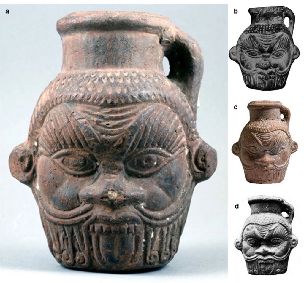
The first-ever physical evidence of hallucinogens in an Egyptian mug has been found, validating written records and centuries-old myths of ancient Egyptian rituals and practices. Through advanced chemical analyses, University of South Florida professor Davide Tanasi examined one of the world’s few remaining Egyptian Bes mugs.

Such mugs, including the one donated to the Tampa Museum of Art in 1984, are decorated with the head of Bes, an ancient Egyptian god or guardian demon worshiped for protection, fertility, medicinal healing and magical purification. Published in Nature’s Scientific Reports, the study sheds light on an ancient Egyptian mystery: The secret of how Bes mugs were used about 2000 years ago. 

“There’s no research out there that has ever found what we found in this study,” Tanasi said. “For the first time, we were able to identify all the chemical signatures of the components of the liquid concoction contained in the Tampa Museum of Art’s Bes mug, including the plants used by Egyptians, all of which have psychotropic and medicinal properties.”

The presence of Bes mugs in different contexts over a long period of time made it extremely difficult to speculate on their contents or roles in ancient Egyptian culture.

“For a very long time now, Egyptologists have been speculating what mugs with the head of Bes could have been used for, and for what kind of beverage, like sacred water, milk, wine or beer,” said Branko van Oppen, curator of Greek and Roman art at the Tampa Museum of Art. “Experts did not know if these mugs were used in daily life, for religious purposes or in magic rituals.”

Several theories about the mugs and vases were formulated on myths, but few of them were ever tested to reveal their exact ingredients until the truth was extracted layer by layer.

Tanasi, who developed this study as part of the Mediterranean Diet Archaeology project promoted by the USF Institute for the Advanced Study of Culture and the Environment, collaborated with several USF researchers and partners in Italy at the University of Trieste and the University of Milan to perform chemical and DNA analyses. With a pulverised sample from scraping the inner walls of the vase, the team combined numerous analytical techniques for the first time to uncover what the mug last held.

The new tactic was successful and revealed the vase had a cocktail of psychedelic drugs, bodily fluids and alcohol – a combination that Tanasi believes was used in a magical ritual re-enacting an Egyptian myth, likely for fertility. The concoction was flavoured with honey, sesame seeds, pine nuts, liquorice and grapes, which were commonly used to make the beverage look like blood.

“This research teaches us about magic rituals in the Greco-Roman period in Egypt,” Van Oppen said. “Egyptologists believe that people visited the so-called Bes Chambers at Saqqara when they wished to confirm a successful pregnancy because pregnancies in the ancient world were fraught with dangers. So, this combination of ingredients may have been used in a dream-vision inducing magic ritual within the context of this dangerous period of childbirth.”

“Religion is one of the most fascinating and puzzling aspects of ancient civilizations,” Tanasi said. “With this study, we’ve found scientific proof that the Egyptian myths have some kind of truth and it helps us shed light on the poorly understood rituals that were likely carried out in the Bes Chambers in Saqqara, near the Great Pyramids at Giza.”

The Bes mug is on display now at the Tampa Museum of Art and can be viewed in the exhibition, “Prelude: An Introduction to the Permanent Collection.” View a 3D model of the Bes mug produced by the USF Institute for Digital Exploration.

Many not Getting Enough Nutrients in Their Pregnancy, Study Finds

Source: Pixabay CC0

It’s generally estimated that around 10% of pregnant people struggle to meet their nutritional needs – but the real number could be far higher, according to new research in The Journal of Nutrition.

Over 90% of pregnant individuals are potentially failing to get enough iron, vitamin D, or vitamin E from the food they eat, while over one-third could be short of calcium, vitamin C, and vitamin A. Troublingly, almost two-thirds of pregnant people were also found to be getting insufficient dietary folate – a critical nutrient that helps prevent birth defects in the baby’s brain and spine.

“It’s important to remember that many pregnant people take prenatal vitamin supplements, which might help prevent nutritional deficiencies,” says lead author Dr Samantha Kleinberg, professor at Stevens Institute of Technology. “Nonetheless, this is a startling finding that suggests we need to be looking much more closely at whether pregnant individuals are getting the nutrients they need.”

Where most previous studies of nutrition during pregnancy relied on a few days of food diaries, or on simply asking people what they remembered eating, the Stevens team asked pregnant people to take before-and-after photos of everything they ate over two 14-day periods. Experts then reviewed the photos to assess the amount of food actually eaten and determine the nutrients consumed during each meal.

That’s a far more accurate approach, because people are notoriously bad at estimating portion size or accurately reporting what they’ve eaten, Dr Kleinberg explains. A photo-based approach is also much less laborious for pregnant people, making it easy to collect data over a period of weeks instead of just a few days.

“Most surveys only track diet over a day or two – but if you feel off one day and don’t eat much, or have a big celebratory meal over the weekend, that can skew the data,” Dr Kleinberg says. “By looking at a longer time period, and using photos to track diet and nutrition, we’re able to get a much richer and more precise picture of what people actually ate.”

The study found significant dietary variations between individuals, but also among the same individuals from one day to the next, suggesting that shorter studies and population-based reports might be failing to spot important nutritional deficits. “Some people eat really well, and others don’t – so if you just take an average, it looks like everything’s fine,” Dr Kleinberg explains. “This study suggests that in reality, an alarming number of pregnant people may not be getting the nutrients they need from their food.”

Using food photos also recorded the exact timing of meals and snacks, and to explore the way that patterns of eating behaviour correlated with total energy and nutrient intake. When pregnant people ate later in the day, the data shows, they were likely to consume significantly more total calories – potentially an important finding as researchers explore connections between eating behaviours and health problems such as gestational diabetes.

The current research didn’t directly study health outcomes, so it’s too early to say whether insufficient nutrition or excessive energy consumption is adversely impacting pregnant individuals or their babies. “We’ll be digging into that in future studies, and looking at possible connections with eating patterns and changes in glucose tolerance,” Dr Kleinberg says.

Source: Stevens Institute of Technology

How Human Brain Functional Networks Emerge and Develop during the Birth Transition

Shedding light on the growth trajectory of global functional neural networks before and after birth

Photo by Christian Bowen on Unsplash

Brain-imaging data collected from foetuses and infants has revealed a rapid surge in functional connectivity between brain regions on a global scale at birth, possibly reflecting neural processes that support the brain’s ability to adapt to the external world, according to a study published November 19th, in the open-access journal PLOS Biology led by Lanxin Ji and Moriah Thomason from the New York University School of Medicine, USA.

Understanding the sequence and timing of brain functional network development at the beginning of human life is critical. Yet many questions remain regarding how human brain functional networks emerge and develop during the birth transition. To fill this knowledge gap, Thomason and colleagues leveraged a large functional magnetic resonance imaging dataset to model developmental trajectories of brain functional networks spanning 25 to 55 weeks of post-conceptual gestational age. The final sample included 126 foetal scans and 58 infant scans from 140 subjects.

The researchers observed distinct growth patterns in different regions, showing that neural changes accompanying the birth transition are not uniform across the brain. Some areas exhibited minimal changes in resting-state functional connectivity (RSFC) – correlations between blood oxygen level-dependent signals between brain regions when no explicit task is being performed. But other areas showed dramatic changes in RSFC at birth. The subcortical network, sensorimotor network, and superior frontal network stand out as regions that undergo rapid reorganisation during this developmental stage.

Additional analysis highlighted the subcortical network as the only region that exhibited a significant increase in communication efficiency within neighbouring nodes. The subcortical network represents a central hub, relaying nearly all incoming and outgoing information to and from the cortex and mediating communication between cortical areas. On the other hand, there was a gradual increase in global efficiency in sensorimotor and parietal-frontal regions throughout the foetal to neonatal period, possibly reflecting the establishment or strengthening of connections as well as the elimination of redundant connections.

According to the authors, this work unveils fundamental aspects of early brain development and lays the foundation for future research on the influence of environmental factors on this process. In particular, further studies could reveal how factors such as sex, prematurity, and prenatal adversity interact with the timing and growth patterns of children’s brain network development.

The authors add, “This study for the first time documents the significant change of brain functional networks over the birth transition. We observe that growth patterns are regionally specific, with some areas of the functional connectome showing minimal changes, while others exhibit a dramatic increase at birth.”

Provided by PLOS

Antiseizure Drugs during Pregnancy may Affect Neurodevelopment

Photo by SHVETS production

Children whose mothers have taken antiseizure drugs during pregnancy are more likely than others to receive a neuropsychiatric diagnosis. This is according to a comprehensive study by researchers at Karolinska Institutet and elsewhere, published in Nature Communications. However, the researchers emphasise that the absolute risk is low.

Antiseizure drugs are used to treat epilepsy and to stabilise mood in certain psychiatric conditions. However, some of these drugs, such as valproate, are known to affect the foetus if used during pregnancy. 

The current study included data from over three million children in the UK and Sweden, 17 495 of whom had been exposed to antiseizure drugs during pregnancy. 

As expected, children exposed to valproate were more likely to be diagnosed with autism, intellectual disability or ADHD compared to children not exposed to antiseizure drugs. Children exposed to topiramate had a 2.5-fold increased risk of intellectual disability, while those exposed to carbamazepine had a 25 per cent increased risk of being diagnosed with autism and a 30 per cent increased risk of intellectual disability. 

No increased risk with lamotrigine 

However, the researchers found no evidence that taking the antiseizure drug lamotrigine during pregnancy increases the risk of neuropsychiatric diagnoses in the child. 

“Our findings suggest that while certain medications may pose some risk, lamotrigine may be a less risky option, but active monitoring of any antiseizure medication is critical to ensure safety and effectiveness, particularly during pregnancy,” says Brian K. Lee, Professor at Drexel University Dornsife School of Public Health, USA, and affiliated researcher at the Department of Global Public Health, Karolinska Institutet, Sweden. 

The researchers emphasise that the absolute risk of the child receiving a neuropsychiatric diagnosis is low and that there may also be risks associated with not taking antiseizure medication during pregnancy. 

“If you’re pregnant or trying to become pregnant, and taking one of these medications, it may be worth talking with your physician to make sure you’re taking the best medicine for your needs, while minimising risk to future children,” says Viktor H. Ahlqvist, researcher at the Institute of Environmental Medicine, Karolinska Institutet, and joint first author with Paul Madley-Dowd at the University of Bristol, UK. 

The results support previous findings from smaller studies that found links between antiseizure drugs during pregnancy and the risk of neuropsychiatric diagnoses in the child. One difference is that the new study found no statistically significant association between topiramate or levetiracetam and ADHD in the child. 

Source: Karolinska Institutet

Having the Choice of Birth Position is Key in Satisfaction of Expectant Mothers

Photo by Duda Oliveira

Whether supine, all-fours position, sitting upright or squatting – women adopt different birthing positions during childbirth. But until now, how the final birthing position affects the satisfaction of the woman giving birth had not been known. Researchers in Germany investigated the relationship. In particular, they also took into account whether the choice of birthing position was voluntary. The results, which appear in Archives of Gynecology and Obstetrics, showed that having the choice counted the most for mothers’ satisfaction.

Around three quarters of those surveyed were lying during the birth and were particularly dissatisfied if they felt that they had not made this choice themselves. But if the expectant mothers had chosen the supine or lateral supine position themselves, the position actually tended to make them more satisfied.

For a long time, the supine position was the most common birth position in Western countries, giving obstetricians unhindered access to the woman and child.  In various cultures, however, upright birthing positions, such as sitting or squatting, are also widespread. Which position is best for expectant mothers and the unborn child is controversial in the literature.

“Until today, international guidelines usually only recommend that women should adopt their preferred birthing position,” explains Prof Dr Nadine Scholten, professor in psychosomatic and psycho-oncological health services research at the University of Bonn. With regard to birthing positions, the German guideline also states that women should adopt the position that seems most comfortable to them. However, they should also be encouraged to adopt an upright position in the final phase of birth.

“In reality, whether they ultimately lie, sit or squat depends on the wishes of the expectant mothers themselves, but also on the suggestions of the midwives, obstetricians and sometimes necessary medical measures,” explains Prof Dr Brigitte Strizek, Director of the Clinic for Obstetrics and Prenatal Medicine at the University Hospital Bonn (UKB).

Focus on women’s satisfaction

A team led by Prof Scholten, who carried out the study at the Institute for Medical Sociology, Health Services Research and Rehabilitation Research (IMVR) at the University of Cologne and the UKB, wanted to find out which birthing position women were most satisfied with afterwards. Almost 800 mothers were asked about their final birthing position and how satisfied they were with the birth overall using an anonymous questionnaire. All of the data analysed here was from women who had given birth vaginally in a hospital without the use of a vacuum extraction or forceps, and who had given birth eight to twelve months previously at the time of the survey. In their study, the researchers also asked about the satisfaction of the mothers – depending on whether the birth position was freely chosen or not. The reasons for not choosing a free position were also asked.

It was found that over three quarters of mothers gave birth to their child lying on their side or on their back. Of these women, up to 40% stated that they had not chosen the birth position voluntarily. “The most common reason given by respondents was instructions from medical staff,” explains Prof Scholten, first and corresponding author. The most common position assigned by obstetricians was the supine position. It was striking that women were more satisfied with their birth if they were allowed to choose the position voluntarily – especially if they chose the supine position themselves. Women who were not free to choose their birthing position were particularly dissatisfied if the medical staff specified this and not the CTG to record the heart rate of the unborn child and labour activity or the epidural, the anaesthetic to relieve pain, prevented the desired position.

Self-determination not always given in the delivery room

“The number of women who did not choose the birth position themselves is particularly striking, as is the associated lower level of satisfaction with the birth,” summarises co-author Prof Strizek. However, the team cannot confirm whether an increased voluntary choice of birth position in the future would result in fewer women giving birth in the supine position. “In order to increase women’s subjective satisfaction with their birth experience, they should be given the opportunity to adopt their preferred position,” appeals lead author Prof Scholten. “The first step is to increase the awareness of medical staff and empower women to understand and better communicate their preferences.”

Prof Strizek adds: “If a certain position would be advantageous for the woman giving birth from a medical point of view, we as obstetric teams need to explain this better to the women so that they rarely have the feeling that they have not determined the birth position themselves.”

Source: Universitatsklinikum Bonn

Boy or Girl? This Genetic Mutation Increases Odds of Having a Daughter

Source: Pixabay CC0

Each year, roughly the same numbers of boys and girls are born. But in individual families, some couples have four or more daughters and no sons, and some have all male children and no female children, points out University of Michigan evolutionary geneticist Jianzhi Zhang. This has led some scientists to question whether this skewed sex ratio is a result of the genes of the parents.

Now, Zhang and U-M doctoral student Siliang Song have detected a human genetic variant that influences the sex ratio of children. Additionally, they found that many hidden genetic variants of sex ratio may exist in human populations. Their results are published in the Proceedings of the Royal Society B: Biological Sciences.

“Scientists have been pondering and researching a genetic basis for sex ratio for decades, yet no unambiguous evidence for a genetic variation that alters the human sex ratio from an approximately 50:50 ratio has been found,” said Zhang, professor of ecology and evolutionary biology.

Zhang says this has led some scientists to think that the human sex ratio is not subject to mutation.

“But this scenario seems unlikely, because almost all human characteristics are subject to mutation and genetic variation,” he said. “Instead, we think genetic variation of sex ratio is too difficult to detect because sex ratio is not measured precisely.”

That is, each person typically has a very small number of children, which can lead to large errors in the estimation of the true sex ratio of a person’s children. For example, if a person only has one child, the estimated sex ratio would be either zero (if it’s a girl) or 1 (if it’s a boy) even if the true sex ratio is 0.5.

To detect genetic influence on sex ratio, the researchers realised they needed a much larger sample than in all previous studies. They turned to the UK Biobank, a biomedical database that contains the genetic and phenotypic information of about 500 000 British participants.

Analysing this data, the researchers identified a single nucleotide change named rs144724107 that is associated with a 10% increase in the probability of giving birth to a girl as opposed to a boy. But this nucleotide change is rare among the UK Biobank participants: About 0.5% of the participants carry this change. The nucleotide change is located near a gene named ADAMTS14, which is a member of the ADAMTS gene family known to be involved in spermatogenesis and fertilisation. The researchers also note that their discovery has not yet been confirmed in other samples.

The researchers also identified two genes, called RLF and KIF20B, that may also influence the sex ratio.

The study’s findings align with a theory in evolutionary biology called Fisher’s principle, which states that natural selection favours the genetic variant that increases the births of the rare sex. That is, if fewer males than females are born in a population, natural selection favours genetic variants that increase the number of males born, and vice versa. As a result, this selection yields a more or less even sex ratio in the population

“For Fisher’s principle to work, there must be mutations that influence the sex ratio,” Zhang said. “The fact that no genetic variation on human sex ratio had been identified has led some scientists to question the applicability of Fisher’s principle in humans.

“Our study shows that in fact, human data are consistent with Fisher’s principle and the reason no genetic variants of sex ratio had been discovered was the imprecision of the measure of a person’s offspring sex ratio.”

Next, the researchers hope to verify their findings in other samples – not an easy task, Zhang says, because of the large sample size requirement and the rareness of the identified genetic variant.

Source: University of Michigan

A Potential Stool Test for Endometriosis also Suggests an IBD Link

Photo by Sora Shimazaki on Pexels

Promising findings by researchers at Baylor College of Medicine and collaborating institutions could lead to the development of a non-invasive stool test and a new therapy for endometriosis, a painful condition that affects nearly 200 million women worldwide. The study appeared in the journal Med.

“Endometriosis develops when lining inside the womb grows outside its normal location, for instance attached to surrounding intestine or the membrane lining the abdominal cavity. This typically causes bleeding, pain, inflammation and infertility,” said corresponding author Dr Rama Kommagani, associate professor in the Department of Pathology and Immunology at Baylor. “Generally, it takes approximately seven years to detect endometriosis and is often diagnosed incorrectly as a bowel condition. Thus, delayed diagnosis, together with the current use of invasive diagnostic procedures and ineffective treatments underscore the need for improvements in the management of endometriosis.”

“Our previous studies in mice have shown that the microbiome, the communities of bacteria living in the body, or their metabolites, the products they produce, can contribute to endometriosis progression,” Kommagani said. “In the current study, we took a closer look at the role of the microbiome in endometriosis by comparing the bacteria and metabolites present in stools of women with the condition with those of healthy women. We discovered significant differences between them.”

The findings suggested that stool metabolites found in women with endometriosis could be the basis for a non-invasive diagnostic test as well as a potential strategy to reduce disease progression.

The researchers discovered a combination of bacterial metabolites that is unique to endometriosis. Among them is the metabolite called 4-hydroxyindole. “This compound is produced by ‘good bacteria,’ but there is less of it in women with endometriosis than in women without the condition,” said first author Dr Chandni Talwar, postdoctoral associate in Kommagani’s lab.

“These findings are very exciting,” Talwar said. “There are studies in animal models of the disease that have shown specific bacterial metabolite signatures associated with endometriosis. Our study is the first to discover a unique metabolite profile linked to human endometriosis, which brings us closer to better understanding the human condition and potentially identifying better ways to manage it.”

Furthermore, extensive studies also showed that administering 4-hydroxyindole to animal models of the disease prevented the initiation and progression of endometriosis-associated inflammation and pain. 

“Interestingly, our findings also may have implications for another condition. The metabolite profile we identified in endometriosis is similar to that observed in inflammatory bowel disease (IBD), revealing intriguing connections between these two conditions,” Kommagani said. “Our findings support a role for the microbiome in endometriosis and IBD.”

The researchers are continuing their work toward the development of a non-invasive stool test for endometriosis. They are also conducting the necessary studies to evaluate the safety and efficacy of 4-hydroxyindole as a potential treatment for this condition.

Source: Baylor College of Medicine

Defective Sperm in IVF Doubles the Risk of Preeclampsia

Intracytoplasmic Sperm Injection (ICSI) procedure. Credit: Scientific Animations CC4.0

For the first time, researchers have linked specific frequent defects in sperm to risk of pregnancy complications and negative impacts on the health of the baby. The study from Lund University in Sweden shows that a high proportion of father’s spermatozoa possessing DNA strand breaks is associated with a doubled risk of preeclampsia in women who have become pregnant by IVF. It also increases the risk of the baby being born prematurely.

Infertility is a growing problem and the number of in vitro fertilisation procedures is increasing rapidly. It is already known that women who become pregnant by assisted reproduction techniques have an increased risk of preeclampsia, repeated miscarriages and the baby being born prematurely and with a lower birth weight. Yet, the reasons behind this have not been fully understood. 

“Before a planned in vitro fertilisation, the man’s sperm sample is analysed for concentration, motility and morphology. But there are men who, according to this analysis, have normal sperm, but still have reduced fertility,” says Amelie Stenqvist, lecturer at Lund University and first author of the study published in Fertility and Sterility. She received her PhD from Lund and now works as a specialist in gynaecology and obstetrics at Skåne University Hospital in Malmö.

Around 20-30% of babies born through IVF have fathers with damaged DNA in their sperm, as shown by elevated levels of DNA fragmentation. The DNA fragmentation index (DFI) is a measure of the amount of strand breaks in the DNA and is used to provide important new information about male fertility. Sperm with DNA damage may still be fertile, but the chances of fertilisation are lower and if the percentage of DFI exceeds 30%, the chances of natural conception are close to zero.

Although current in vitro techniques mean that men with a high DFI can become fathers, until now very little has been known about the impact of DNA fragmentation on pregnancy and the health of the baby. It has been difficult to research the topic because the DFI value is not included in the standard measurements currently taken by Sweden’s fertility clinics. It also requires a large study population and access to national medical registries.

“Since half of the placenta’s DNA comes from the father and placental development and function play a central role in preeclampsia, we wanted to investigate whether a high percentage of DNA damage in the sperm affected the risk of preeclampsia,” says Aleksander Giwercman.

He is a professor of reproductive medicine at Lund University, a consultant at Skåne University Hospital in Malmö. Aleksander Giwercman also led a research study that included 1660 children conceived through IVF and ICSI at the Reproductive Medicine Centre in Malmö over the period 2007-2018. 

The results showed that in the 841 couples who underwent IVF, a DFI of over 20% doubled the risk of the woman developing preeclampsia (10.5%) and also increased the risk of premature birth. In the IVF group with a DFI below 20%, there was a 4.8% risk of preeclampsia, which is comparable to pregnancies that occur naturally. For couples undergoing ICSI, there was no association with preeclampsia.

“Today, DFI analysis is only performed at some fertility clinics in Sweden, but we think that it should be introduced as standard at all clinics. It can give couples answers as to why they are not getting pregnant and can influence the chosen method of assisted fertilisation. Not only that, our latest results show that a DFI analysis could be used to identify high-risk pregnancies,” says Aleksander Giwercman.

 What makes this finding even more interesting is that high DNA fragmentation in sperm is linked to the overall health of the father and is potentially treatable. Most DNA damage is caused by oxidative stress, which is an imbalance between harmful molecules and the antioxidants that protect cells. Other factors that increase DNA fragmentation include the man’s age, smoking, obesity and infections. 

“The next step is to identify which group of men respond best to methods to prevent and treat sperm DNA damage, and to test these methods to prevent pregnancy complications,” concludes Amelie Stenqvist.

Source: Lund University

Sharp Spike Seen in Emergency Visits for Life Threatening Pregnancy Complication

Findings suggest significant increase in emergency department utilisation for hypertensive disorders of pregnancy over 14 year span

Photo by Camilo Jimenez on Unsplash

Hypertensive disorders of pregnancy, the second leading cause of maternal deaths worldwide, may be sending a significantly higher number of pregnant people to the emergency department. Between 2006 and 2020, researchers found a surge in emergency visits and admissions for the condition that causes serious maternal and neonatal complications and accounts for 6.3% of all pregnancy-related deaths in the United States.

The study, which appears in JAMA Network Open, also suggests greater emergency utilisation for the disease among underrepresented racial and ethnic groups. 

“Hypertensive disorders of pregnancy often develop suddenly, even in healthy women, and symptoms may appear without warning and progress rapidly,” said senior author Erica Marsh, MD, professor of obstetrics and gynaecology at the University of Michigan Medical School and chief of the division of reproductive endocrinology and infertility at U-M Health Von Voigtlander Women’s Hospital, of Michigan Medicine.

“Ideally, this risk would be detected during prenatal care and lead to early intervention. Our study indicates more people turning to the emergency department, which may reflect a higher prevalence of the condition or an increased awareness for prompt assessment and treatment.”

Hypertensive disorders of pregnancy, which could include preeclampsia, gestational hypertension, and eclampsia, are serious complications that involve elevated blood pressure. 

The American College of Obstetricians and Gynecologists recommends management of severe blood pressure in pregnancy within 30 to 60 minutes of diagnosis to prevent complications such as stroke, myocardial ischaemia, seizure, placental abruption, and maternal and neonatal mortality.

Disparities in ED reliance, disease severity

Researchers analysed nationally representative data, finding a 76% increase in emergency encounters related to the condition over the 14-year span, up from 31  623 to 55  893, and nearly 1.5 times as many ED admissions – up from 17 338 to 43 563.

Concerns about costs, time constraints, misconceptions about the necessity of early care or barriers to accessing prenatal care may be possible factors for the increase, authors say.

“The disparities in reliance on emergency rooms for this disease may imply limited access to timely outpatient care or other health system barriers,” said lead author Courtney Townsel, MD, MSc, who was at Michigan Medicine at the time of the study and is now at the University of Maryland.

Black, Hispanic, and Asian or Pacific Islander groups were also more likely to both utilise emergency care and be admitted to the hospital for hypertensive disorders of pregnancy.

“The disproportionate rate of admissions among certain racial and ethnic groups suggests worse disease severity by the time people seek care,” Townsel said.

“Racial differences in emergency care utilisation for hypertensive disorders of pregnancy underscore the ongoing racial disparities in US maternal morbidity and mortality and highlight a critical need for accessible, culturally competent community-level interventions for all.”

Original written by Beata Mostafavi. Republished under a Creative Commons Licence.

Source: Michigan Medicine – University of Michigan


New Treatment for Pregnancy Loss Caused by Specific Autoantibody

Photo by SHVETS production

Amongst women who experience recurrent pregnancy loss, around 20% test positive for a specific autoantibody. A Kobe University-led research team now found a treatment using either of two common drugs that drastically increases these women’s chances of carrying to full-term without complications, reporting their findings in Frontiers in Immunology.

Recurrent pregnancy loss is a condition of women who have lost two or more pregnancies for non-obvious reasons. Kobe University obstetrician Tanimura Kenji and his team have previously found that in 20% of these women, they can detect a specific antibody in their blood that targets their own bodies: anti-β2-glycoprotein I/HLA-DR autoantibodies.

Tanimura explains: “There is no known treatment for this particular condition, but the antibodies have a similar target to those that play a role in a different condition that has an established treatment.” Therefore, he wanted to test whether that treatment also works in the cases with the newly discovered antibody.

Tanimura enlisted the help of obstetricians across five hospitals in Japan and over the course of two years analysed the blood of consenting women suffering from recurrent pregnancy loss for the antibodies. If any of these women got pregnant during this time frame, their doctors would offer treatment options also containing those drugs that are effective against the chemically similar condition, specifically, low-dose aspirin or heparin. The research team then observed how many of the women who included these drugs in their treatment had full-term live births or pregnancy complications and compared that to the pregnancy outcomes in women who did not take either of the two drugs.

They report that women who received the treatment were much more likely to have live births (87% did) compared to the ones without treatment (of which only 50% had live births). In addition, amongst the live births, the treatment reduced the likelihood of complications from 50% to 6%. “The sample size was rather small (39 women received the treatment and 8 did not), but the results still clearly show that a treatment with low-dose aspirin or heparin is very effective in preventing pregnancy loss or complications also in women who have these newly discovered self-targeting antibodies,” summarises Tanimura.

Many women who tested positive for the newly discovered self-targeting antibodies also tested positive for the previously known ones. However, the Kobe University-led team found that women who only had the newly discovered antibodies and who received the treatment were even more likely to have a live birth (93%) and, amongst these, none had pregnancy complications.

Looking ahead, Tanimura says: “The newly discovered self-targeting antibody has been demonstrated to be involved also in infertility and recurrent implantation failure, as well as a risk factor for arterial thrombosis in women with systemic rheumatic diseases. I therefore expect that studies about the effectivity of the treatment against a broader range of conditions might produce encouraging results.”

Source: Kobe University