Category: Obstetrics & Gynaecology

A Short Course of Radiation Therapy for Endometrial Cancer Patients is Effective

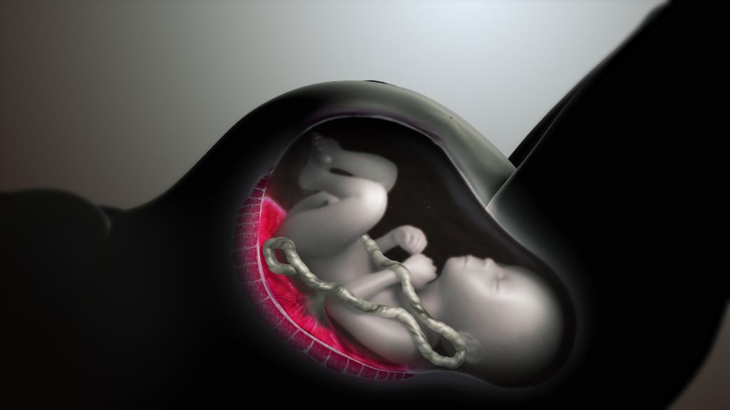
Female reproductive system. Credit: Scientific Animations CC4.0 BY-SA

In a randomised clinical trial, researchers from Huntsman Cancer Institute at the University of Utah (the U) have found that short-course, higher dose vaginal brachytherapy for endometrial cancer had similar effectiveness to more frequent, lower dose sessions.

Gita Suneja, MD, MS, physician-scientist at Huntsman Cancer Institute and professor of radiation oncology at the U, is the first author of the SAVE trial report – which stands for, Short-Course Adjuvant Vaginal Cuff Brachytherapy in Early Endometrial Cancer Compared with Standard of Care.

“There isn’t high quality-data on optimal dose and schedule for brachytherapy treatments. Because of this, practice patterns really vary,” says Suneja. “The SAVE trial sought to try to lower the number of treatments that patients were receiving but maintain short-term quality of life and disease control.”

Endometrial cancer is a disease that begins in the lining of the uterus. The primary treatment for endometrial cancer is surgery, including the removal of the uterus, cervix, and upper vagina. Brachytherapy, a form of internal radiation, is used as a secondary treatment to prevent the cancer’s return. Patients receiving vaginal cuff brachytherapy are treated with internal radiation by way of an applicator in the vaginal cavity.

The SAVE trial compared two groups who received different treatment doses over a varying number of sessions. The control group received the standard treatment – between three to five appointments with lower doses. The experimental group received higher doses of radiation in just two sessions.

“The study outcomes will help improve cancer care for Huntsman Cancer Institute patients across the five states of the Mountain West.”

Gita Suneja, MD, MS

The researchers found similarly effective short-term outcomes and few acute toxicities for the patients in the experimental group.

David Gaffney, MD, PhD, FACR, FABS, FASTRO, physician-scientist at Huntsman Cancer Institute and professor of radiation oncology at the U, developed the idea for the SAVE study after seeing patient need. According to the American Cancer Society, endometrial cancer is the most common cancer of the female reproductive organs. Incidence is on the rise, as is the mortality rate.

“It is a big win when we can preserve good outcomes and make cancer care easier,” says Gaffney.

The results of the SAVE trial were published in JCO Oncology Advances.

Source: Huntsman Cancer Institute

Study Probes How to Predict Complications from Preeclampsia

Data from 8843 women diagnosed with preeclampsia during pregnancy showed that existing risk prediction models are most accurate only in the days after diagnosis

Photo by Shvets Production on Pexels

The existing prediction models for severe complications of preeclampsia are most accurate only in the two days after hospital admission, with deteriorating performance over time, according to a new study published February 4th in the open-access journal PLOS Medicine by Henk Groen of University of Groningen, the Netherlands, and colleagues.

Preeclampsia is a potentially life-threatening condition that can occur during pregnancy; of women diagnosed with preeclampsia, 5-20% will develop severe complications. Two existing PIERS (Pre-eclampsia Integrated Estimate of RiSk) models, PIERS Machine Learning (PIERS-ML) and the logistic-regression-based fullPIERS, are designed to identify individuals at greatest or least risk of adverse maternal outcomes in the 48 hours following hospital admission for preeclampsia. However, both models are regularly used for ongoing assessment beyond the first 48 hours.

In the new study, researchers used data from 8843 women diagnosed with preeclampsia at a median gestational age of 36 weeks between 2003 and 2016. Data included PIERS-ML and fullPIERS assessments as well as health outcomes.

The study found that neither the PIERS-ML nor fullPIERS model maintained good performance over time for repeated risk stratification in women with preeclampsia. The PIERS-ML remained generally good at identifying the very high-risk and very-low risk groups over time, but performance of the larger high-risk and low-risk groups deteriorated significantly after 48 hours. The fullPIERS model underperformed compared to the PIERS-ML model.

“Since there are no better options, clinicians may still use these two models for ongoing assessments after the first admission with pre-eclampsia, but the predictions should be treated with increasing caution as the pregnancy progresses,” the authors say. More prediction models are needed that perform well over time, they add.

The authors add, “Pregnancy hypertension outcome prediction models were designed and validated for initial assessment of risks for mothers; this study shows that such ‘static’ models if used repeatedly over days yield increasingly inaccurate predictions.”

Provided by PLOS

Could the Contraceptive Pill Reduce the Risk of Ovarian Cancer?

Photo by Reproductive Health Supplies Coalition on Unsplash

It’s a little pill with big responsibilities. But despite its primary role to prevent pregnancy, the contraceptive pill (or ‘the Pill’) could also help reduce the risk of ovarian cancer, according to new research from the University of South Australia.

Screening for risk factors of ovarian cancer using artificial intelligence, UniSA researchers found that the oral contraceptive pill reduced the risk of ovarian cancer by 26% among women who had ever used the Pill, and by 43% for women who had used the Pill after the age of 45.

The study, published in the International Journal of Gynecologic Cancer, also identified some biomarkers associated with ovarian cancer risk, including several characteristics of red blood cells and certain liver enzymes in the blood, with lower body weight and shorter stature associating with a lower risk of ovarian cancer.

Researchers also found that women who had given birth to two or more children had a 39% reduced risk of developing ovarian cancer compared to those who had not had children.

UniSA researcher Dr Amanda Lumsden says understanding risks and preventative factors for ovarian cancer is key for improved treatment and outcomes.

“Ovarian cancer is notoriously diagnosed at a late stage, with about 70% of cases only identified when they are significantly advanced,” Dr Lumsden says.

“Late detection contributes to a survival rate of less than 30% over five years, in comparison to more than 90% for ovarian cancers that are caught early. That’s why it’s so important to identify risk factors.

“In this research, we found that women who had used the oral contraceptive pill had a lower risk of ovarian cancer. And those who had last used the Pill in their mid-40s, had an even lower level of risk.

“This poses the question as to whether interventions that reduce the number of ovulations could be used as a potential target for prevention strategies for ovarian cancer.”

Supported by the MRFF, the study used artificial intelligence to assess the data of 221 732 females (aged 37-73 at baseline) in the UK Biobank.

Machine learning specialist, UniSA’s Dr Iqbal Madakkatel, says the study shows how AI can help to identify risk factors that may otherwise have gone undetected.

“We included information from almost 3000 diverse characteristics related to health, medication use, diet and lifestyle, physical measures, metabolic, and hormonal factors, each measured at the start of the study,” Dr Madakkatel says.

Source: University of South Australia

Music Can Influence Foetal Heart Rate in the Womb

Photo by Sergio Capuzzimati on Unsplash

Playing music has long been a way for expectant parents to connect with their children in the womb, but a group of researchers has found evidence it can calm foetal heart rates, potentially providing developmental benefits.

In the interdisciplinary journal Chaos, researchers studied the effect of classical music on a foetal heartbeat. The team used mathematical analysis tools to identify patterns in heart rate variability.

Typical measures of heart rate are an average of several beats across multiple seconds. In contrast, heart rate variability measures the time between individual beats. This measure can provide insight into the maturation of the foetal autonomic nervous system, with greater variability often indicating healthy development.

To test the effects music can have on foetal heart rate, the group of researchers recruited 36 pregnant women and played a pair of classical pieces for their foetuses. For the experiment, they chose “The Swan,” by French composer Camille Saint-Saëns, and “Arpa de Oro,” by Mexican composer Abundio Martínez.

By attaching external heart rate monitors, the researchers could measure the foetal heart rate response to both songs. And by employing nonlinear recurrence quantification analysis, they could identify changes in heart rate variability during and after the music was played.

“Overall, we discovered that exposure to music resulted in more stable and predictable foetal heart rate patterns,” said author Claudia Lerma. “We speculate that this momentary effect could stimulate the development of the foetal autonomic nervous system.”

In addition to the overall effects of playing music, the researchers looked at the differences between the two classical pieces. While both were effective, they found that the Mexican guitar melody had a stronger effect.

“When contrasting ‘The Swan’ with ‘Arpa de Oro,’ we did notice some significant differences,” said author Eric Alonso Abarca-Castro. “In particular, the second piece appeared to have a stronger impact on some measures, indicating that it produced heart rate patterns that were more predictable and regular. Factors like rhythmic characteristics, melodic structure, or cultural familiarity may be linked to this differentiation.”

For expectant parents at home, the researchers suggest that classical music could help promote fetal development.

“Our results suggest that these changes in foetal heart rate dynamics occur instantly in short-term fluctuations, so parents might want to consider exposing their foetuses to quiet music,” said Abarca-Castro. “Parents who play soothing music may stimulate and benefit the foetal autonomic system.”

The authors plan to continue to explore this effect, looking at different genres and types of music to further their understanding.

“To ascertain whether rhythmic or cultural variations elicit distinct foetal cardiac responses, we intend to increase the size of our sample and expand our investigation to include a variety of musical styles beyond classical pieces,” said author José Javier Reyes-Lagos.

Source: American Institute of Physics

The Importance of Vitamin D during First Trimester

Photo by Michele Blackwell on Unsplash

Low vitamin D levels in the first trimester of pregnancy are associated with higher rates of preterm birth and decreased foetal length, according to a new study led by researchers in the Penn State Department of Nutritional Sciences. This research provides evidence that early pregnancy or even preconception may represent critical time points for intervening with women who have low vitamin D status, to optimise pregnancy outcomes.

Celeste Beck, who earned her doctorate in nutritional sciences from Penn State in 2023, and Alison Gernand, Beck’s doctoral adviser and associate professor of nutritional sciences at Penn State, led the study. Their results were recently published in The American Journal of Clinical Nutrition.

“More than 25% of women who are pregnant or lactating have lower than recommended levels of vitamin D,” Gernand said, explaining that prior research has demonstrated the effect of vitamin D on foetal skeletal growth, maternal immune function at the foetal interface, and the development of the placenta in pregnant women. “A lot of the development early in pregnancy requires vitamin D, so we conducted this study to better understand how early-pregnancy vitamin D status is related to pregnancy outcomes.”

Most prior studies on vitamin D status in pregnant women have measured vitamin D concentrations starting in the second trimester or later, the researchers said. The researchers said this study, to their knowledge, is the first to examine both first and second trimester maternal vitamin D status in relation to longitudinal foetal growth and pregnancy outcomes.

The researchers at Penn State partnered with colleagues at the University of Utah to test blood samples from 351 women collected as part of the Nulliparous Pregnancy Outcomes Study: Monitoring Mothers-to-Be, which was funded by the National Institute of Child Health and Human Development and recruited pregnant women across the United States between 2010 and 2013.

According to the Institute of Medicine, less than 50nmol/L represents an insufficiency of vitamin D. When the researchers compared outcomes for women with vitamin D insufficiency (less than 50nmol/L) to women with sufficient vitamin D (more than or equal to 50nmol/L), they found no statistical differences in pregnancy outcomes. However, when the researchers compared pregnancy outcomes across a wider range of vitamin D concentrations, they found that pregnant women with first trimester vitamin D concentrations lower than 40 nmol/L were four times more likely to experience a preterm birth compared to women with vitamin D concentrations more than or equal to 80nmol/L.

Despite the higher risk of preterm birth in women with low vitamin D status, the researchers cautioned that these results were based on a very low number of preterm births in this study and recommend that additional, larger studies be conducted.

The researchers also observed an association between first-trimester vitamin D concentrations and certain foetal growth patterns. Women with higher levels of vitamin D experienced a small but statistically significant increase in foetal length.

Source: Penn State

Removing Fallopian Tubes during Other Abdominal Surgeries may Lower Ovarian Cancer Risk

Mathematical modelling study suggests this approach could also reduce total healthcare costs in Germany

Female reproductive system. Credit: Scientific Animations CC4.0 BY-SA

A mathematical modelling study conducted in Germany suggests that ovarian cancer incidence could be reduced and healthcare savings boosted if women who have already completed their families were offered fallopian tube removal during any other suitable abdominal surgeries. Angela Kather and Ingo Runnebaum of Jena University Hospital, Germany, and colleagues present these findings on January 30th in the open-access journal PLOS Medicine.

Some of the most widespread and serious forms of ovarian cancer begin in the fallopian tubes, and removing them may reduce ovarian cancer risk. While women at average risk of ovarian cancer are not recommended to have surgery solely to remove their fallopian tubes, many surgeons offer “opportunistic” tube removal during other gynaecologic surgeries such as hysterectomy or tubal sterilisation. Opportunistic removal may also be feasible during other abdominal surgeries, such as gallbladder removal.

However, the overall potential benefits of opportunistic fallopian tube removal have been unclear. To help clarify, Kather and colleagues developed a mathematical model that incorporates real-world patient statistics to predict population-level risks of ovarian cancer after opportunistic fallopian tube removal, as well as the potential healthcare cost savings.

By applying the model to statistics from Germany, the researchers predicted that opportunistic fallopian tube removal during every hysterectomy and tubal sterilisation could reduce ovarian cancer cases by 5% across the female population of Germany. Removal during every suitable abdominal surgery for women who are done having children could reduce nationwide cancer cases by 15%, the analysis suggests, and it could save more than €10 million in healthcare costs annually.

Ovarian cancer is the third most common gynaecologic cancer in the world and has a mortality rate of 66%. Overall, these findings suggest that opportunistic fallopian tube removal during appropriate abdominal surgeries could not only lower population-level ovarian cancer risks and prevent ovarian cancer deaths, but also provide economic benefits. This study could help inform health policy and insurance costs for the procedure.

The authors add, “We developed a mathematical model to estimate the likelihood of women undergoing surgeries that offer an opportunity for fallopian tube removal and the potential for reducing their ovarian cancer risk. Applying this model to the entire female population of Germany revealed that 15% of ovarian cancer cases could be prevented if fallopian tubes were removed during every suitable abdominal surgery in women who have completed their families. This approach has the potential to extend healthy years of life and significantly save healthcare costs.”

Provided by PLOS

PTSD and Anxiety may Affect Reproductive Health in Female Firefighters

Source: CC0

A new study led by University of Arizona researchers in collaboration with fire service partners and other researchers around the country through the Fire Fighter Cancer Cohort Study showed that post-traumatic stress disorder and anxiety are associated with lower levels of anti-Müllerian hormone, a marker of ovarian reserve, among women firefighters.

The ovarian reserve, a measure of fertility, is the number of healthy eggs in a woman’s ovaries that could potentially be fertilised.

“These findings highlight the negative effect that mental health conditions can have on health – specifically, reproductive health,” said first author Michelle Valenti, MPH, a doctoral student in epidemiology at the Zuckerman College of Public Health and program coordinator of the Fire Fighter Cancer Cohort Study’s Women Firefighter Studies.

The paper was published in the Journal of Women’s Health.

Firefighters are exposed to high stress and traumatic situations in addition to chemical exposures and have a higher prevalence of PTSD compared with the general population. Previous research showed that women firefighters have lower levels of anti-Müllerian hormone compared with women who are not firefighters; however, the reason why was unknown.

The research team, which included personnel at the Zuckerman College of Public Health’s Center for Firefighter Health Collaborative Research, led this analysis to determine whether anxiety, depression or PTSD were associated with anti-Müllerian hormone levels. They found that clinical diagnoses of PTSD and anxiety among women firefighters were associated with reductions in anti-Müllerian hormone levels of 66% and 33%, respectively.

These findings highlight a potential mechanism through which adverse mental health conditions could lead to adverse reproductive outcomes. Further research is needed to identify potential areas for intervention.

“The work of this AMH study within the broader context of the FFCCS is imperative to taking care of all of our firefighters,” said Captain Caitlin St. Clair of the Puget Sound Regional Fire Authority. “These findings provide scientific leverage to fire departments to implement programs to reduce stress and improve the lives of our firefighter women.”

The Women Firefighter Study, a subgroup of the Fire Fighter Cancer Cohort Study, aims to identify causes of stress, cancer and adverse reproductive health effects in women firefighters that would inform effective interventions to mitigate these conditions.

“This study demonstrates the power of the Fire Fighter Cancer Cohort Study’s Women Firefighter Study to evaluate exposures that lead to adverse gynaecologic conditions,” Valenti said. “The Women Firefighter Study would not be possible without our amazing fire service partners who have championed women firefighter research.”

Source: University of Arizona Health Sciences

Human Papillomavirus Infection Kinetics Revealed in New Longitudinal Study

Improved understanding of why some infections persist could improve treatment, screening, and vaccination strategies for HPV infection

Non-persistent human papillomavirus (HPV) infections are characterized by a sharp increase in viral load followed by a long plateau, according to a study published January 21st in the open-access journal PLOS Biology by Samuel Alizon of the National Centre for Scientific Research (CNRS), France, and colleagues.

Chronic HPV infection is responsible for more than 600 000 new cancers each year, including nearly all cervical cancers. Infection among young women is common, impacting nearly 20% of women 25 years of age. Fortunately, the vast majority of these infections clear within two years without symptoms. Still, a better understanding of why some infections clear while others persist could improve treatment, screening, and vaccination strategies for HPV-associated diseases.

To monitor the dynamics of HPV infection, the PAPCLEAR cohort study in France followed 189 women aged 18 to 25 years for up to 24 months. Researchers followed participants every two months, collecting information on viral kinetics and immune markers.

By frequently monitoring participants, the researchers achieved unprecedented temporal resolution on viral and immune kinetics. They found that non-persistent infections are characterised by a plateau in HPV viral load that starts around 2 months after infection and lasts for 13 to 20 months before rapidly declining. Additionally, they found a strong correlation between a population of immune cells in between innate and adaptive immunity, namely TCRγδ cells, and the total amount of viruses produced.

The study is limited by the fact that many of the infection follow-ups were truncated, meaning that the participants were already infected when enrolled or were lost to follow-up before infection clearance. Furthermore, only 4 out of 76 infected participants were followed for 18 months or more. Therefore, differences between chronic and acute infections will have to wait for longer studies.

The authors add, “Asymptomatic genital infections by human papillomaviruses (HPVs) display striking dynamics at the frontier between acute and chronic infections. HPV infections appear to be associated with specific patterns of the innate and adaptative local immune response.”

Provided by PLOS

Point-of-care Ultrasound Enhances Early Pregnancy Care, Cuts Emergency Visits by 81%

Photo by Mart Production on Pexels

Published in Annals of Family Medicine, a University of Minnesota Medical School research team found that implementing point-of-care ultrasounds (POCUS) to assess the viability and gestational age of pregnancies in the first trimester enhanced care for pregnant patients and cut emergency visits by 81% for non-miscarrying patients.

Previously, early pregnancy care was provided through separate appointments for ultrasound, risk assessment and patient education. This new integrated approach allows patients who are under 14 weeks pregnant to receive comprehensive care during a single visit. This includes ultrasound-based pregnancy dating, immediate assessment of pregnancy viability, risk evaluation and on-site counselling – all based on real-time ultrasound results.

“Our study demonstrates that the use of point-of-care ultrasound provides meaningful benefit to the patients we serve by addressing early pregnancy problems at the time they are identified,” said Allison Newman, MD, an assistant professor at the U of M Medical School and family medicine physician at M Health Fairview Clinic. “POCUS in early pregnancy helps clinicians more efficiently and accurately diagnose problems without compromising the quality of needed first trimester assessments – saving time, money and stress for patients.”

The research team introduced this integrated approach at M Health Fairview Clinic – Bethesda in autumn 2022, allowing the clinic to quickly identify high-risk cases and offer timely intervention for issues such as miscarriage or abnormal pregnancies. They found:

  • The clinic saw an 81% reduction in emergency visits, urgent clinic appointments and first-trimester phone inquiries for non-miscarrying patients. 
  • Clinic implementation led to more timely diagnosis of abnormal pregnancies and improved education and support for all patients, including those who experience miscarriage. 
  • For miscarriage cases, the time from initial concern to diagnosis decreased from an average of 5.8 days to 1.7 days.

Suggested next steps include rolling out the process more widely within other family medicine practices and performing a wider study across multiple sites.

Source: University of Michigan

The Massive Changes to Mothers’ Intestines in Pregnancy and Breastfeeding

Source: Pixabay CC0

When women are pregnant and nurse their babies, their bodies change and various organs, such as the breasts or the immune system, are adapted to ensure the health of both mother and child. This happens throughout evolution in all mammals. An international research team led by Josef Penninger and Masahiro Onji, Medical University of Vienna, now reports the surprising finding that the intestine also changes completely in pregnant and nursing females, resulting in a doubling of the intestinal surface area and a striking structural reorganisation.

Reporting their findings in Nature, the researchers also provide the first genetic and mechanistic evidence of how this intestinal epithelial expansion occurs in mothers, with direct implications for the transgenerational health of the babies.

A multinational team led by Josef Penninger observed that the intestinal villi reorganise during pregnancy and breastfeeding and significantly enlarge, doubling their surface area. The studies were carried out in genetically modified mice and intestinal organoids from mice and humans – self-organised three-dimensional tissues derived from stem cells in the intestine. Mechanistically, the researchers identified the RANK receptor/RANK ligand (RANK/RANKL) system as the key to the villous enlargement of the small intestine during reproduction, which is regulated by sex and lactation hormones. When mice were engineered to lack the RANK/RANKL system in the intestine, the villous expansion during pregnancy and breastfeeding was significantly impaired.

For decades, researchers have studied the RANK/RANKL system as a key facilitator of essential, evolutionarily conserved processes. The Penninger group has already identified key functions of the RANK/RANKL system in bone turnover, in the biology of the mammary gland, in breast cancer, and in immune tolerance in pregnancy, contributing to the development of drugs against bone loss used by millions of people and clinical trials for breast cancer prevention and cancer immunotherapies are underway. The researchers now discovered that these intestinal changes, which appear to be completely reversible when nursing is stopped, are important for proper feeding and nourishment of the babies.

“Our study shows that the impairment of this intestinal expansion by the lack of the RANK/RANKL system during pregnancy changes the milk of the nursing mothers. This results in lower weights of the babies and transgenerational long-term metabolic consequences,” states lead author Masahiro Onji. “Mothers need to eat for themselves and their babies. These new studies provide for the first time a molecular and structural explanation of how and why the intestine changes to adapt to enhanced nutrient demand of mothers, which is probably the case in all pregnant and nursing mammals,” adds study leader Josef Penninger.

How mothers adapt to the demands of pregnancy and breastfeeding remains a central question of evolution and human health. During this phase, female hormones influence multiple organs to control and change their structure and functions, which is crucial for the health of the mother and the development of the offspring. It was known that pregnant women have enhanced nutrient demands. However, this fundamental aspect has not been well studied until now:

“By identifying the RANK/RANKL system as the driving force behind intestinal adaptation during pregnancy and lactation, our study contributes to a deeper understanding of biological processes that are of fundamental importance for evolution and human health”, says Josef Penninger, summarising the impact of the findings.

This massive expansion is controlled by sex and pregnancy hormones, which change the stem cells in the gut via the RANK/RANKL system and then give the intestinal cell a survival signal to grow much larger. This growth then leads to a near doubling of the intestinal surface area, which also increases the molecular machinery for the uptake of sugar, protein, and fat, and even leads to a profound architectural change in the intestinal villi, which probably slows down the flow of food, again maximising the uptake of nutrients.

Josef Penninger said: “Our team has discovered an amazing new way how mother’s bodies change to keep babies healthy. Hardly anybody knew about this, apart from a few old studies that have largely been forgotten. We have also found that this system, via stem cells, can directly affect tumours in the intestine; maybe we can learn from pregnant and nursing mothers to reversibly rewire this system to develop new treatments and a better understanding of intestinal cancer or gut regeneration.”

Source: Medical University of Vienna