Category: Ophthalmology

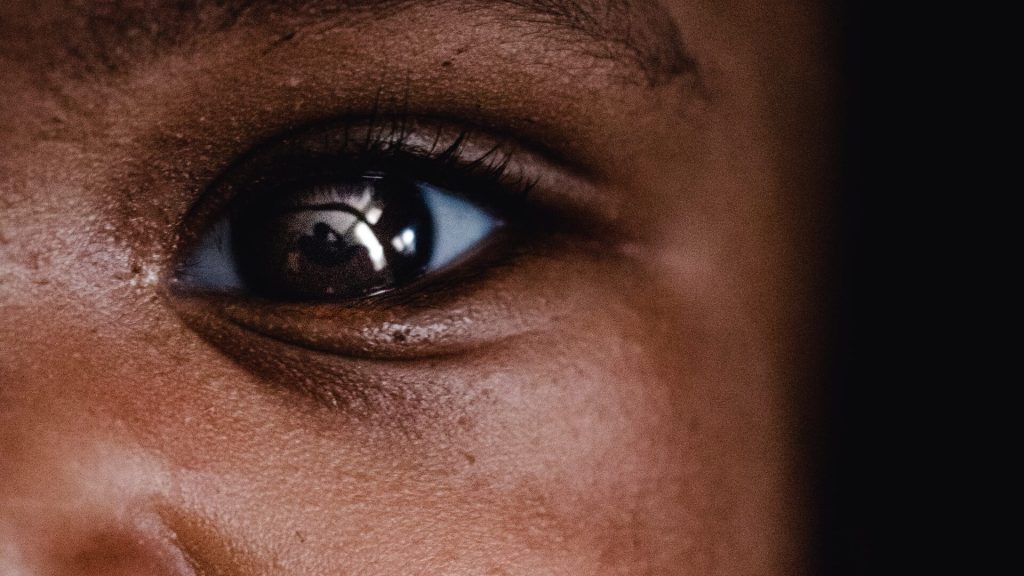
SARS-CoV-2 can Cross the Blood–Retinal Barrier, Infecting the Eyes

Photoreceptor cells in the retina. Credit: Scientific Animations

The blood-retinal barrier is designed to protect vision from infections by preventing microbial pathogens from reaching the retina where they could trigger an inflammatory response with potential vision loss. But researchers at the University of Missouri School of Medicine have discovered that SARS-CoV-2 can breach this protective retinal barrier with potential long-term consequences in the eye. Their findings are reported in PLOS Pathogens.

Pawan Kumar Singh, PhD, an assistant professor of ophthalmology, leads a team researching new ways to prevent and treat ocular infectious diseases. Using a humanised ACE2 mice model, the team found that SARS-CoV-2, can infect the inside of the eyes even when the virus doesn’t enter the body through the surface of the eyes. Instead, they found that when viruses enter the body through inhalation, it not only infects organs like lungs, but also reaches highly protected organs like eyes through the blood-retinal barrier by infecting the cells lining this barrier.

This finding is important as we increase our understanding of the long-term effects of SARS-CoV-2 infection,” said Singh. “Earlier, researchers were primarily focused on the ocular surface exposure of the virus. However, our findings reveal that SARS-CoV-2 not only reaches the eye during systemic infection but induces a hyperinflammatory response in the retina and causes cell death in the blood-retinal barrier. The longer viral remnants remain in the eye, the risk of damage to the retina and visual function increases.”

Singh also discovered that extended presence of SARS-CoV-2 spike antigen can cause retinal microaneurysm, retinal artery and vein occlusion, and vascular leakage.

“For those who have been diagnosed with COVID-19, we recommend you ask your ophthalmologist to check for signs of pathological changes to the retina,” Singh said. “Even those who were asymptomatic could suffer from damage in the eyes over time because of COVID-19 associated complications.”

While viruses and bacteria have been found to breach the blood-retinal-barrier in immunocompromised people, this research is the first to suggest that the virus that causes COVID-19 could breach the barrier even in otherwise healthy individuals, leading to an infection that manifests inside the eye itself. Immunocompromised patients or those with hypertension or diabetes may experience worse outcomes if they remain undiagnosed for COVID-19 associated ocular symptoms.

“Now that we know the risk of COVID-19 to the retina, our goal is to better understand the cellular and molecular mechanisms of how this virus breaches the blood-retinal barrier and associated pathological consequences in hopes of informing development of therapies to prevent and treat COVID-19 induced eye complications before a patient’s vision is compromised,” Singh said.

Source: University of Missouri-Columbia

Newly Found Retinal Cells may Paint a Complete Picture of Colour Vision

Photo by Jeffrey Riley on Unsplash

Scientists have long studied just how the eye’s three cone photoreceptor types work together to allow humans to perceive colour. In a new study in the Journal of Neuroscience, researchers at the University of Rochester used adaptive optics to identify rare retinal ganglion cells (RGCs) that could help fill in the gaps in existing theories of colour perception.

The retina has three types of cones to detect colour that are sensitive to either short, medium, or long wavelengths of light. Retinal ganglion cells transmit input from these cones to the central nervous system.

In the 1980s, David Williams, the William G. Allyn Professor of Medical Optics, helped map the “cardinal directions” that explain colour detection. However, there are differences in the way the eye detects colour and how colour appears to humans. Scientists suspected that while most RGCs follow the cardinal directions, they may work in tandem with small numbers of non-cardinal RGCs to create more complex perceptions.

Recently, a team of researchers from Rochester’s Center for Visual Science, the Institute of Optics, and the Flaum Eye Institute identified some of these elusive non-cardinal RGCs in the fovea that could explain how humans see red, green, blue, and yellow.

“We don’t really know anything for certain yet about these cells other than that they exist,” says Sara Patterson, a postdoctoral researcher at the Center for Visual Science who led the study. “There’s so much more that we have to learn about how their response properties operate, but they’re a compelling option as a missing link in how our retina processes colour.”

Adaptive optics peer past the eye’s natural distorations

The team took advantage of adaptive optics, which uses a deformable mirror to overcome light distortion and was first developed by astronomers to reduce atmospheric blurring in ground-based telescopes. In the 1990s, Williams and his colleagues began applying adaptive optics to study the human eye. They created a camera that compensated for distortions caused by the eye’s natural aberrations, producing a clear image of individual photoreceptor cells.

“The optics of the eye’s lens are imperfect and really reduce the amount of resolution you can get with an ophthalmoscope,” says Patterson. “Adaptive optics detects and corrects for these aberrations and gives us a crystal-clear look into the eye. This gives us unprecedented access to the retinal ganglion cells, which are the sole source of visual information to the brain.”

Patterson says improving our understanding of the retina’s complex processes could ultimately help lead to better methods for restoring vision for people who have lost it.

“Humans have more than 20 ganglion cells and our models of human vision are only based on three,” says Patterson. “There’s so much going on in the retina that we don’t know about. This is one of the rare areas where engineering has totally outpaced visual basic science. People are out there with retinal prosthetics in their eyes right now, but if we knew what all those cells do, we could actually have retinal prosthetics drive ganglion cells in accordance with their actual functional roles.”

Source: University of Rochester

A New Treatment for Retinal Detachment – Based on Seaweed

Photoreceptor cells in the retina. Credit: Scientific Animations

In Korea, it is taboo to consume seaweed soup before exams since it there is a belief that the slippery nature of seaweed can lead to slipping up in the exam and failing. The slick surface of seaweeds is attributed to alginate, a mucilaginous carbohydrate substance. Now, Korean researchers have made use of this substance in the treatment of retinal detachment.

The result is an artificial vitreous body for treating retinal detachment, based on alginate from seaweed.

The research findings were recently published in Biomaterials, an international journal of biomaterials published by Elsevier. The work was a collaborative effort between Professor Hyung Joon Cha from the Department of Chemical Engineering and the School of Convergence Science and Technology and Dr. Geunho Choi from the Department of Chemical Engineering at Pohang University of Science and Technology (POSTECH), and Professor Woo Jin Jeong, Professor Woo Chan Park, and Professor Seoung Hyun An from the Dong-A University Hospital’s Department of Ophthalmology.

The vitreous body is a gel-like substance that occupies the space between the lens and retina, contributing to the eye’s structural integrity. Retinal detachment occurs when the retina separates from the inner wall of the eye and moves into the vitreous cavity, leading to detachment and potentially resulting in blindness in severe cases.

While a common approach involves removing the vitreous body and substituting it with medical intraocular fillers like expandable gas or silicone oil, these fillers have been associated with various side effects. To address these concerns, the research team employed a modified form of alginate, a natural carbohydrate sourced from algae.

Alginate, also known as alginic acid, is widely utilised in various industries, including food and medicine, for its ability to create viscous products. In this research, the team crafted a medical composite hydrogel based on alginate, offering a potential alternative for vitreous replacement.

The hydrogel has high biocompatibility and optical properties akin to authentic vitreous body, preserving the vision of patients post-surgery. Its distinctive viscoelasticity effectively regulates fluid dynamics within the eye, contributing to retinal stabilisation and the elimination of air bubbles.

To validate the hydrogel’s stability and effectiveness, the team conducted experiments using rabbit eyes, which closely resemble human eyes in structure, size, and physiological response.

Implanting the hydrogel into rabbit eyes demonstrated its success in preventing the recurrence of retinal detachment, maintaining stability, and functioning well over an extended period without any adverse effects.

Professor Hyung Joon Cha of the POSTECH who led the study remarked, “There is a correlation between retinal detachment and severe myopia and the prevalence of retinal detachment is increasing, particularly in young people. The incidence of retinal detachment cases in Korea rose by 50% in 2022 compared to 2017.” He expressed the team’s commitment by saying, “Our team will enhance and progress the technology to make the hydrogel suitable for practical use in real-world eye care through ongoing research.”

Professor Woo Jin Jeong from the Dong-A University Hospital stated, “The worldwide market for intraocular fillers is expanding at a rate of 3% per year.” He added, “We anticipate that the hydrogel we’ve created will prove beneficial in upcoming vitreoretinal surgeries.”

Source: Pohang University of Science & Technology (POSTECH)

Trial of Minocycline for Dry Age-related Macular Degeneration Flops

Retina showing reticular pseudodrusen. Although they can infrequently appear in individuals with no other apparent pathology, their highest rates of occurrence are in association with age-related macular degeneration (AMD), for which they hold clinical significance by being highly correlated with end-stage disease sub-types, choroidal neovascularisation and geographic atrophy. Credit: National Eye Institute

The drug minocycline, an antibiotic that also decreases inflammation, failed to slow vision loss or expansion of geographic atrophy in people with dry age-related macular degeneration (AMD), according to a phase II clinical study at the National Eye Institute (NEI), part of the National Institutes of Health.

Dry AMD affects the macula, the part of the retina that allows for clear central vision. In people with dry AMD, patches of photoreceptors and their nearby support cells begin to die off, leaving regions known as geographic atrophy. Over time, these regions expand, causing people to lose more and more of their central vision.

Microglia, immune cells that help maintain tissue and clear up debris, are present at higher levels around damaged retinal regions in people with dry AMD than in people without AMD. Scientists have suggested that inflammation – and particularly microglia – may be driving the expansion of geographic atrophy regions.

This study, led by Tiarnan Keenan, MD, PhD, a Stadtman Tenure-Track Investigator at the NEI’s Division of Epidemiology and Clinical Applications, tested whether inhibiting microglia with minocycline might help slow geographic atrophy expansion and its corresponding vision loss.

The trial enrolled 37 participants at the NIH Clinical Center in Bethesda, Maryland, and at the Bristol Eye Hospital, United Kingdom.

After a nine-month period where the researchers tracked each participant’s rate of geographic atrophy expansion, the participants took twice-daily doses of minocycline for two years.

The researchers compared each participant’s rate of geographic atrophy expansion while taking minocycline to their baseline rate, and found there was no difference in geographic atrophy expansion rate or vision loss with minocycline.

Previous studies have shown that minocycline can help reduce inflammation and microglial activity in the eye, including the retina.

The drug has shown beneficial effects for conditions such as diabetic retinopathy, but has not previously been tested for dry AMD.

Source: NIH/National Eye Institute

In the Fight against Brain Pathogens, the Eyes Have it

Photo by Victor Freitas on Pexels

The eyes have been called the window to the brain. It turns out they also serve as an immunological barrier that protects the organ from pathogens and even tumours, Yale researchers have found. In a new study, researchers showed that vaccines injected into the eyes of mice can help disable the herpes virus, a major cause of brain encephalitis.

To their surprise, the vaccine activates an immune response through lymphatic vessels along the optic nerve.

The results were published Feb. 28 in the journal Nature.

“There is a shared immune response between the brain and the eye,” said Eric Song, an associate research scientist and resident physician in Yale School of Medicine’s Department of Immunobiology and corresponding author of the paper.

“And the eyes provide easier access for drug therapies than the brain does.”

Wanting to explore immunological interactions between brain and eyes, the research team, which was led by Song, found that the eyes have two distinct lymphatic systems regulating immune responses in the front and rear of the eye.

After they vaccinated mice with inactivated herpes virus, the researchers found that lymphatic vessels in the optic nerve sheath at the rear of the eye protected mice not only from active herpes infections, but from bacteria and even brain tumors.

Harnessing this new biology, Song’s team is currently testing newly created drugs from his lab delivered through eye injections that may help combat macular edema, or leaky blood vessels of the retina common in people with diabetes, and glaucoma.

“These results reveal a shared lymphatic circuit able to mount a unified immune response between posterior eye and the brain, highlighting an understudied immunological feature of the eyes and opening up the potential for new therapeutic strategies in ocular and central nervous system diseases,” the authors wrote.

Source: Yale University

New Spiral-shaped Lens is a Step up from Multifocal Lenses

Researchers have developed a new type of lens that uses a spiral-shaped surface to maintain a clear focus at different distances in varying light conditions. Credit: Laurent Galinier

Researchers have developed a spiral-shaped lens that maintains clear focus at different distances in varying light conditions. The new lens, described in Optica, works much like progressive lenses used for vision correction but without the distortions typically seen with those lenses. It could help advance contact lens technologies, intraocular implants for cataracts and miniaturised imaging systems.

“Unlike existing multifocal lenses, our lens performs well under a wide range of light conditions and maintains multifocality regardless of the size of the pupil,” said Bertrand Simon from Photonics, Numerical and Nanosciences Laboratory (LP2N), a joint research unit between the Institut d’Optique Graduate School, the University of Bordeaux and the CNRS in France. “For potential implant users or people with age-related farsightedness, it could provide consistently clear vision, potentially revolutionising ophthalmology.”

In the article, the researchers describe the new lens, which they call the spiral diopter. Its spiraling features are arranged in a way that creates many separate points of focus – much like having multiple lenses in one. This makes it possible to see clearly at various distances.

“In addition to ophthalmology applications, the simple design of this lens could greatly benefit compact imaging systems,” said Simon. “It would streamline the design and function of these systems while also offering a way to accomplish imaging at various depths without additional optical elements. These capabilities, coupled with the lens’s multifocal properties, offer a powerful tool for depth perception in advanced imaging applications”

Creating a vortex of light

The inspiration for the spiral lens design came when the paper’s first author, Laurent Galinier from SPIRAL SAS in France, was analysing the optical properties of severe corneal deformations in patients. This led him to conceptualize a lens with a unique spiral design that causes light to spin, like water going down a drain. This phenomenon, known as an optical vortex, creates multiple clear focus points, which allow the lens to provide clear focus at different distances.

“Creating an optical vortex usually requires multiple optical components,” said Galinier. “Our lens, however, incorporates the elements necessary to make an optical vortex directly into its surface. Creating optical vortices is a thriving field of research, but our method simplifies the process, marking a significant advancement in the field of optics.”

The researchers created the lens by using advanced digital machining to mold the unique spiral design with high precision. They then validated the lens by using it to image a digital ‘E,’ much like those used on an optometrist’s light-up board. The authors observed that the image quality remained satisfactory regardless of the aperture size used. They also discovered that the optical vortices could be modified by adjusting the topological charge, which is essentially the number of windings around the optical axis. Volunteers using the lenses also reported noticeable improvements in visual acuity at a variety of distances and lighting conditions.

Crossing disciplines

Bringing the new lens to fruition required combining the intuitively crafted design with advanced fabrication techniques through a cross-disciplinary collaboration. “The spiral diopter lens, first conceived by an intuitive inventor, was scientifically substantiated through an intensive research collaboration with optical scientists,” said Simon. “The result was an innovative approach to creating advanced lenses.”

The researchers are now working to better understand the unique optical vortices produced by their lens. They also plan to perform systematic trials of the lens’ ability to correct vision in people to comprehensively establish its performance and advantages in real-world conditions. In addition, they are exploring the possibility of applying the concept to prescription eyeglasses, which could potentially offer users clear vision across multiple distances.

“This new lens could significantly improve people’s depth of vision under changing lighting conditions,” said Simon. “Future developments with this technology might also lead to advancements in compact imaging technologies, wearable devices and remote sensing systems for drones or self-driving cars, which could make them more reliable and efficient.”

Source: Optic

Expert Warns that Red Light Therapy for Myopia could Damage the Retina

Retina showing reticular pseudodrusen. Although they can infrequently appear in individuals with no other apparent pathology, their highest rates of occurrence are in association with age-related macular degeneration (AMD), for which they hold clinical significance by being highly correlated with end-stage disease sub-types, choroidal neovascularisation and geographic atrophy. Credit: National Eye Institute

A University of Houston optometry researcher is warning against the use of low-level red light (LLRL) therapy as a method to control myopia, or nearsightedness, especially in children. Over the last few years, LLRL has emerged as a viable myopia treatment after studies reported the treatment as effective and responsible for significant reduction in myopia progression. The company behind one of the devices reports that it is already being used to address myopia in over 100 000 paediatric patients.

But the excitement over its results as a myopia treatment may have come too soon, ahead of its proven safety.

“Based on measurements in our laboratory, it is recommended that clinicians strongly reconsider the use of LLRL therapy for myopia in children until safety standards can be confirmed,” reports Lisa Ostrin, associate professor at the UH College of Optometry in The College of Optometrists journal.

Ostrin reports the therapy can put the retina at risk of photochemical and thermal damage.

“The safety profiles of red-light laser devices for myopia have not been fully investigated,” she said.

For LLRL therapy, children are instructed to look into a red light-emitting instrument for three minutes, twice a day, five days a week, for the duration of the treatment period, which could last years.

“We found that the red-light instruments for myopia exceed safety limits,” said Ostrin, whose research characterises the laser output and determines the thermal and photochemical maximum permissible exposure (MPE) of LLRL devices.

“For both LLRL devices evaluated here, three minutes of continuous viewing approached or surpassed the luminance dose MPE, putting the retina at risk of photochemical damage.”

Ostrin examined two different LLRL devices, and while both instruments were confirmed to be Class-1 laser products, as defined by International Electrotechnical Commission standards, according to Ostrin they are unsafe to view continuously for the required treatment duration of three minutes.

Class-1 lasers are low-powered devices that are considered safe from all potential hazards when viewed accidentally and briefly.

Examples of Class-1 lasers are laser printers, CD players and digital video disc (DVD) devices.

Class-1 lasers are not meant to be viewed directly for extended periods.

“Thermal ocular injury from a laser can occur with exposures at any wavelength when the temperature change of the retina is greater than 10°C, resulting in the denaturation of proteins. With thermal damage, the lesion size is typically less than the size of the beam diameter, and the resultant scotomas are permanent.” said Ostrin.

Source: University of Houston

In Vitro Experiment Explains Why Humans Have Full Colour Vision and Dogs Don’t

Photo by Victor Freitas on Pexels

With human retinas grown in a petri dish, researchers discovered how retinoic acid, a metabolite of vitamin A, generates the specialised cells that enable people to see millions of colours, an ability that dogs, cats, and most other mammals do not have.

“These retinal organoids allowed us for the first time to study this very human-specific trait,” said author Robert Johnston, an associate professor of biology. “It’s a huge question about what makes us human, what makes us different.”

The findings, published in PLOS Biology, increase understanding of colour blindness, age-related vision loss, and other diseases linked to photoreceptor cells. They also demonstrate how genes instruct the human retina to make specific colour-sensing cells, a process scientists thought was controlled by thyroid hormones.

By tweaking the cellular properties of the organoids, the research team found that a vitamin A1 metabolite, retinoic acid, determines whether a cone will specialise in sensing red or green light.

Only humans with normal vision and closely related primates develop the red sensor.

For decades, it was that thought red cones formed through a coin toss mechanism where the cells haphazardly commit to sensing green or red wavelengths – and research from Johnston’s team recently hinted that the process could be controlled by thyroid hormone levels.

Instead, the new research suggests red cones materialise through a specific sequence of events orchestrated by retinoic acid within the eye.

The team found that high levels of retinoic acid in early development of the organoids correlated with higher ratios of green cones. Similarly, low levels of the acid changed the retina’s genetic instructions and generated red cones later in development.

“There still might be some randomness to it, but our big finding is that you make retinoic acid early in development,” Johnston said.

“This timing really matters for learning and understanding how these cone cells are made.”

Green and red cone cells are remarkably similar except for a protein called opsin, which detects light and tells the brain what colors people see.

Different opsins determine whether a cone will become a green or a red sensor, though the genes of each sensor remain 96% identical.

With a breakthrough technique that spotted those subtle genetic differences in the organoids, the team tracked cone ratio changes over 200 days.

“Because we can control in organoids the population of green and red cells, we can kind of push the pool to be more green or more red,” said author Sarah Hadyniak, who conducted the research as a doctoral student in Johnston’s lab and is now at Duke University.

“That has implications for figuring out exactly how retinoic acid is acting on genes.”

The researchers also mapped the widely varying ratios of these cells in the retinas of 700 adults.

Seeing how the green and red cone proportions changed in humans was one of the most surprising findings of the new research, Hadyniak said. Scientists still don’t fully understand how the ratio of green and red cones can vary so greatly without affecting someone’s vision.

If these types of cells determined the length of a human arm, the different ratios would produce “amazingly different” arm lengths, Johnston said.

To build understanding of diseases like macular degeneration, which causes loss of light-sensing cells near the center of the retina, the researchers are working with other Johns Hopkins labs.

The goal is to deepen their understanding of how cones and other cells link to the nervous system.

“The future hope is to help people with these vision problems,” Johnston said.

“It’s going to be a little while before that happens, but just knowing that we can make these different cell types is very, very promising.”

Source: Johns Hopkins University

Mapping the ‘Light’ Seen by Closed Eyes could Help Prosthetic Eyes See Better

Photo by Arteum.ro on Unsplash

Researchers at Monash University have identified a new way of mapping ‘phosphenes’ – the visual perception of the bright flashes we see when no light is entering the eye – to improve the outcome of surgery for patients receiving a cortical visual prosthesis.

Cortical visual prostheses are devices implanted onto the brain with the aim of restoring sight by directly stimulating the area responsible for vision, the visual cortex, bypassing damage to the retina of the eye or the optic nerve. Phosphenes, apparent flashes and patterns of lights, were described by the ancient Greeks and can be elicited by pressure, injury, disease, certain medications or direct electrical stimulation.

A typical prosthesis consists of an array of fine electrodes, each of which is designed to trigger a phosphene. Given the limited number of electrodes, understanding how electrodes can best be placed to generate useful perceived images becomes critical.

Published in the Journal of Neural Engineering, the study presents a more realistic simulation for cortical prosthetic vision.

As part of this researchers from the Department of Electrical and Computer Systems Engineering at Monash University, led by Associate Professor Yan Tat Wong, are honing in on the ideal distribution of phosphenes.

“Phosphenes are likely to be distributed unevenly in an individual’s visual field, and differences in the surface of the brain also affect how surgeons place implants, which together result in a phosphene map unique to each patient,” Associate Professor Wong said.

The study used a retinotopy dataset based on magnetic resonance imaging (MRI) scans, consulting with a neurosurgeon about realistic electrode implantation sites in different individuals, and applying a clustering algorithm to determine the most suitable regions to present stimuli.

Sighted participants recruited for the study were asked to test and verify the phosphene maps based on visual acuity and object recognition.

“We’re proposing a new process that incorporates our simulation paradigm into surgical planning to help optimise the implantation of a cortical prosthesis,” Associate Professor Wong said.

The process would begin with an MRI scan to plot the recipient’s brain surface in the area of the visual cortex. Potential implant locations would then be identified, and the simulation developed in the Monash research would be used to plot phosphene maps.

“We can use the metrics we computed to find practical implant locations that are more likely to give us a usable phosphene map, and we can verify those options through psychophysics tests on sighted participants using a virtual reality headset,” Associate Professor Wong said.

“We believe this is the first approach that realistically simulates the visual experience of cortical prosthetic vision.”

Source: Monash University

Eyedrops instead of Injections for Age-related Macular Degeneration

Retina showing reticular pseudodrusen. Although they can infrequently appear in individuals with no other apparent pathology, their highest rates of occurrence are in association with age-related macular degeneration (AMD), for which they hold clinical significance by being highly correlated with end-stage disease sub-types, choroidal neovascularisation and geographic atrophy. Credit: National Eye Institute

A new compound potentially could offer an alternative to injections for the millions of people who suffer from wet age-related macular degeneration (AMD). The condition causes vision loss due to the uncontrolled growth and leakage of blood vessels in the back of the eye. A new paper in Cell Reports Medicine finds that a small-molecule inhibitor can reverse damage from AMD and promote regenerative and healing processes.

The drug can also be delivered via eyedrops – an improvement over current treatments for AMD, which require repeated injections into the eye.

“The idea was to develop something that can be more patient-friendly and doesn’t require a visit to the doctor’s office,” said lead researcher Yulia Komarova, associate professor of pharmacology at University of Illinois Chicago.

Komarova’s compound targets the protein End Binding-3 in endothelial cells, which line the inside of blood vessels. In the new study, the researchers looked at whether inhibiting EB3 function could stop the damaging leakage associated with wet AMD.

Using computational drug design methods, the team developed a small molecule drug, End Binding-3 inhibitor (EBIN), that could be delivered externally via eyedrops instead of by injection. They then tested its effectiveness in animal models of wet AMD, finding that twice-daily treatment reduced eye damage within 2–3 weeks.

Further investigation found that the inhibitor worked by rolling back aging-related genetic modifications. Aging causes inflammation and hypoxia in the eye that leads to changes in gene expression associated with the cellular effects and symptoms of wet AMD. Komarova and colleagues found that the EB3 inhibitor they developed reversed these epigenetic changes, restoring gene expression to a normal, healthy state.

“We reduce the effects of the stressor on endothelial cells and we improve regenerative processes, accelerating healing,” Komarova said. “That can be tremendous for the function of the cells.”

Because blood vessel leakage and hypoxic stress also drive many other medical conditions, Komarova’s group is interested in testing the inhibitor in models of acute lung injury, diabetic retinopathy, stroke, heart disease and even the general effects of aging on the brain. They are also exploring whether an implantable lens, similar to a contact lens, could deliver the drug to the eye more effectively than eyedrops.

Source: University of Illinois Chicago