Category: Neurology

Leakiness of First Blood–brain Barrier Layer Results in Cognitive Deficits

a, Diagram of the BBB and brain endothelial glycocalyx layer. b, TEM of cortical capillaries with lanthanum nitrate staining from young (3-month-old) and aged (21-month-old) mice. Scale bars, 1 µm. Source: Shi et al., Nature, 2025.

A study published in Nature reveals how a key component of the blood–brain barrier (BBB), the endothelial glycocalyx layer, becomes dysregulated in ageing, causing the BBB to become compromised. The researchers also investigated the possibility of to restore this layer’s integrity, reducing neuroinflammation and restoring cognitive function.

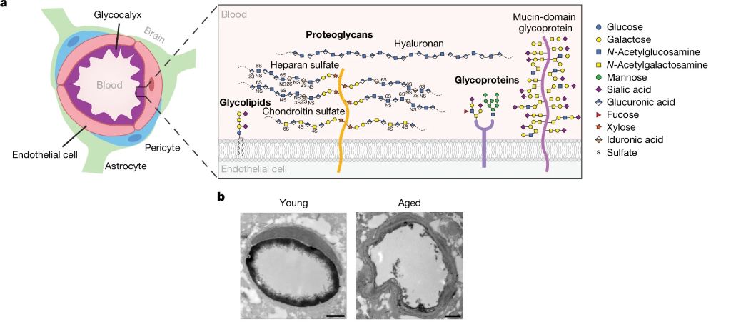
The BBB is a highly specialised safeguard keeping the brain separate from harmful factors, such as toxins and also albumin, IgG and fibrinogen (and, unfortunately, many medications which could otherwise treat brain disease). The leakage of such blood-derived molecules into the brain has been shown to trigger neuroinflammatory changes and create a neurotoxic brain environment. The part of the BBB directly in contact with the blood is the endothelial glycocalyx layer, a carbohydrate-rich meshwork mostly composed of proteoglycans, glycoproteins and glycolipids that coats the BBB lumen. Yet the endothelial glycocalyx’s composition and role is poorly understood despite it being the first layer of interface between the blood and brain.

The researchers found that the brain endothelial glycocalyx is highly dysregulated during ageing and neurodegenerative disease. Two mucin-type O-glycan biosynthetic enzymes, C1GALT1 and B3GNT3 were also found to be downregulated mouse models of ageing and in the brains of Alzheimer’s and Huntington’s disease patients. To test these, the researchers used adeno-associated viruses (AAV) in young mice to turn down the expression of C1GALT1 and B3GNT3. These mice showed signs of BBB leakage and in severe cases, brain haemorrhaging occurred in mice.

In samples from the brains of Alzheimer’s patients, the researchers also observed reduced C1GALT1 in microvessels.

To test if it was possible to restore the BBB’s ability to protect the brain against harmful blood-borne molecules, they administered AAVs in aged mice to restore levels of B3GNT3 and C1GALT1.

Assessing cognitive function, they found that aged mice treated with B3GNT3 via an AAV displayed improvements in spatial working memory in a maze test and hippocampal-dependent learning and memory in a fear conditioning test. Aged mice treated with C1GALT1 did not improve in the maze test, and no significant difference was observed in cued freezing in the fear conditioning among the three aged groups.

Although the study shows that increasing C1GALT1 and B3GNT3 reduces BBB permeability and improves brain health, the precise mechanisms that underlie these beneficial effects remain unclear. The researchers believe that by limiting the nonspecific uptake of blood-derived molecules, the brain can be protected. But C1GALT1 and B3GNT3 are also likely to influence a wide range of proteins and glycan structures and in order to further understand brain ageing and rejuvenation it is therefore crucial to understand the molecular pathways affected by them.

The authors concluded: “Cumulatively, our findings provide a detailed compositional and structural mapping of the ageing brain endothelial glycocalyx layer and reveal important consequences of ageing- and disease-associated glycocalyx dysregulation on BBB integrity and brain health.”

Drug More than Doubles Survival Time for Glioblastoma Patients

MRI scan showing brain cancer. Credit: Michelle Monje, MD, PhD, Stanford University

A drug developed at The University of Texas Health Science Center at San Antonio (UT Health San Antonio) has been shown to extend survival for patients with glioblastoma, the most common primary brain tumour in adults.

Results of a trial led by the university and reported in Nature Communications revealed that a unique investigational drug formulation called Rhenium Obisbemeda (186RNL) more than doubled median survival and progression-free time, compared with standard median survival and progression rates, and with no dose-limiting toxic effects.

“As a disease with a pattern of recurrence, resistance to chemotherapies and difficulty to treat, glioblastoma has needed durable treatments that can directly target the tumour while sparing healthy tissue,” said lead author Andrew J. Brenner, MD, PhD, professor and chair of neuro-oncology research with Mays Cancer Center at UT Health San Antonio. “This trial provides hope, with a second trial under way and planned for completion by the end of this year.”

Brenner said that the median overall survival time for patients with glioblastoma after standard treatment fails with surgery, radiation and chemotherapy is only about 8 months. More than 90% of patients have a recurrence of the disease at its original location.

Rhenium Obisbemeda enables very high levels of a specific activity of rhenium-186 (186Re), a beta-emitting radioisotope, to be delivered by tiny liposomes, referring to artificial vesicles or sacs having at least one lipid bilayer. The researchers used a custom molecule known as BMEDA to chelate or attach 186Re and transport it into the interior of a liposome where it is irreversibly trapped.

In this trial, known as the phase 1 ReSPECT-GBM trial, scientists set out to determine the maximum tolerated dose of the drug, as well as safety, overall response rate, disease progression-free survival and overall survival.

After failing one to three therapies, 21 patients who were enrolled in the study between March 5, 2015, and April 22, 2021, were treated with the drug administered directly to the tumours using neuronavigation and convection catheters.

The researchers observed a significant improvement in survival compared with historical controls, especially in patients with the highest absorbed doses, with a median survival and progression-free time of 17 months and 6 months, respectively, for doses greater than 100Gy.

Importantly, they did not observe any dose-limiting toxic effects, with most adverse effects deemed unrelated to the study treatment.

“The combination of a novel nanoliposome radiotherapeutic delivered by convection-enhanced delivery, facilitated by neuronavigational tools, catheter design and imaging solutions, can successfully and safely provide high absorbed radiation doses to tumours with minimal toxicity and potential survival benefit,” Brenner concluded.

Source: University of Texas Health Science Center at San Antonio

Over-the-counter Pain Relievers Linked to Improved Recovery from Concussion

Credit: Pixabay CC0

People who take over-the-counter pain relievers after a concussion may recover faster than those who do not take pain relievers, according to a preliminary study that will be presented at the American Academy of Neurology’s 77th Annual Meeting taking place April 5–9, 2025.

The study does not prove that pain relievers improve recovery after concussion; it only shows an association.

“These results are exciting as there are limited treatment options for concussion, and over-the-counter pain relievers are readily available and inexpensive,” said study author Kyle Arnold, MD, of the University of Washington in Seattle and a member of the American Academy of Neurology.

“If these results can be confirmed by a controlled study, they could guide us to possible treatment options for people after a concussion.”

The cohort study was conducted by the NCAA and US Department of Defense CARE Consortium and looked at NCAA athletes and military cadets who had concussions. A total of 813 people took over-the-counter pain relievers such as acetaminophen or ibuprofen and other non-steroidal anti-inflammatory drugs after their concussion and 848 people did not take any pain relievers.

Researchers looked at the amount of time it took the athletes to be cleared to return to activities with no restrictions at both 50% recovery and 90% recovery, meaning when 50% of the athletes in the study recovered and then later when 90% recovered.

People who took the pain relievers were 20% more likely to have a faster time before they were cleared to return to activities with no restrictions than those who did not take pain relievers. Those who took the medications were cleared at 50% recovery an average of two days faster, and at 90% recovery an average of seven days faster than those who took no medication.

People who took pain relievers were also about 15% more likely to return to having no symptoms more quickly than those who did not take pain relievers. At 50% recovery, those taking the medications had no symptoms one day sooner than those not taking the medications. At 90% recovery, they had no symptoms three days sooner.

Those who took pain relievers also had lower scores on tests of how severe their symptoms were overall and how severe their headaches were. The researchers also found that the earlier people took the pain relievers after the injury, the faster they recovered. For instance, at 50% recovery, those who started using pain relievers on the first day of their injury returned to play and had resolution of symptoms approximately eight days faster than those who started taking them after five or more days.

There was no difference between the type of pain reliever taken and how quickly people recovered.

“Early medication use appeared to be linked to shorter recovery times, but these findings require further validation through controlled trials,” Arnold said. “In the meantime, these preliminary results may help inform potential treatment options for people recovering from concussions, but additional studies are needed to provide more definitive recommendations.”

Source: American Academy of Neurology

Promising Findings in Testing Nasal Spray for TBI Treatment

Source: CC0

A new study led by researchers at Mass General Brigham suggests a nasal spray developed to target neuroinflammation could one day be an effective treatment for traumatic brain injury (TBI). By studying the effects of the nasal anti-CD3 in a mouse model of TBI, researchers found the spray could reduce damage to the central nervous system and behavioural deficits, suggesting a potential therapeutic approach for TBI and other acute forms of brain injury. The results are published in Nature Neuroscience.

“Traumatic brain injury is a leading cause of death and disability – including cognitive decline – and chronic inflammation is one of the key reasons,” said lead author Saef Izzy, MD, FNCS, FAAN, a neurologist and head of the Immunology of Brain Injury Program at Brigham and Women’s Hospital, a founding member of the Mass General Brigham healthcare system. “Currently, there is no treatment to prevent the long-term effects of traumatic brain injury.”

The study examines the monoclonal antibody Foralumab, made by Tiziana, which has been tested in clinical trials for patients with multiple sclerosisAlzheimer’s disease, and other conditions.

“This opens up a whole new area of research and treatment in traumatic brain injury, something that’s almost impossible to treat,” said senior author Howard Weiner, MD, co-director of the Ann Romney Center for Neurologic Diseases at Brigham and Women’s Hospital. “It also means this could work in intracerebral hemorrhage and other stroke patients with brain injury.”

Multiple experiments were done in mouse models with moderate-to-severe traumatic brain injury to explore the communication between regulatory cells induced by the nasal treatment and the microglial immune cells in the brain. Over time, researchers were able to identify how they modulate immune response.

“Modulating the neuroinflammatory response correlated with improved neurological outcomes, including less anxiety, cognitive decline, and improved motor skills,” Izzy said.

In addition to assessing the effects of the treatment, the research team was able to learn about immune response over time and compare the immune responses and effects of TBI in the mice.

The next step in the research is to translate the findings from preclinical models to human patients.

“Our patients with traumatic brain injury still don’t have an effective therapeutic to improve their outcomes, so this is a very promising and exciting time to move forward with something that’s backed up with solid science and get it to patients’ bedsides,” said Izzy.

Once in the clinical setting, Weiner said the hope is this treatment could be used on a variety of traumatic brain injury patients, including football players with repetitive concussions. 

“We envision giving a nasal spray right there on the sidelines,” said Weiner. “It isn’t something we can do yet, but we see the potential.”

Source: Mass General Brigham

Scientists Crack the Puzzle of How Retinoic Acid Works Against Neuroblastoma

Photo by Anna Shvets

Neuroblastoma is a solid tumour that occurs in children. When high-risk, the disease has a poor prognosis. Decades ago, adding the drug retinoic acid to neuroblastoma treatment increased survival by 10–15%. However, this effect was only evident in post-chemotherapy consolidation after bulky primary tumours had largely been eliminated. Why retinoic acid is effective in this setting but not against primary tumours, has been speculated about for nearly 50 years. 

St. Jude Children’s Research Hospital scientists resolved the mystery in a new study, showing retinoic acid uses a novel mechanism to kill metastasised neuroblastoma. The drug “hijacks” a normal developmental pathway to trigger cancer cell death. The findings, which have implications for future combination therapy approaches, appears in Nature Communications

“We’ve come up with an explanation for a decades-long contradiction about why retinoic acid works in post-chemotherapy consolidation but has little impact on primary neuroblastoma tumours,” said senior co-corresponding author Paul Geeleher, PhD. “Retinoic acid’s activity heavily depends on the cellular microenvironment.” 

The cellular microenvironment is the soup of chemicals, proteins and other signals that surround a cell, and which is unique to that part of the body. For example, the bone marrow microenvironment contains signals to grow blood cells and restructure bone. Metastasised neuroblastoma cells often migrate to bone marrow, where the bone morphogenetic protein (BMP) pathway signalling is highly active. The researchers showed that BMP signaling makes neuroblastoma cells much more vulnerable to retinoic acid. 

“Unexpectedly, we found that cells expressing genes from the BMP signaling pathway were very sensitive to retinoic acid,” said co-first and co-corresponding author Min Pan, PhD, St. Jude Department of Computational Biology. “However, since the bone marrow microenvironment causes neuroblastoma cells there to have higher BMP activity, it neatly explained why retinoic acid is very effective at treating those cells during consolidation therapy, but not the primary tumours during up-front treatment.” 

Hijacking development to drive metastatic neuroblastoma cell death 

Using gene editing technology, the scientists uncovered the relationship between BMP signaling and retinoic acid. They assembled a group of neuroblastoma cell lines susceptible to retinoic acid, then cut out genes to find which were responsible for the drug’s activity. Genes in the BMP pathway had the largest effect while providing a plausible explanation for retinoic acid’s varying outcomes in patients.

“We found that, in neuroblastoma, BMP signaling works with retinoic acid signaling in the same way as during development,” said co-first author Yinwen Zhang, PhD, who characterised how transcription factors, the proteins that bind DNA to regulate gene expression, led to different results in highly retinoic acid-sensitive or insensitive neuroblastoma cells. “If there are a lot of BMP-signaling pathway transcription factors already on DNA, then retinoic acid signaling combines with it to promote downstream cell death–related gene expression. This occurs both in normal embryonic development and neuroblastoma cells in certain microenvironments.” 

“We are the first to uncover such an example of ‘hijacking’ a normal embryonic developmental process preserved in cancer that we can exploit therapeutically,” Geeleher said. “Now, we can look for similar processes in other diseases to design less toxic and more effective treatment strategies.” 

Source: St. Jude Children’s Research Hospital

More Evidence Shows that 40Hz Gamma Stimulation is Beneficial for Brain Health

A decade of studies from labs around the world provide a growing evidence base that increasing the power of the brain’s gamma rhythms could help fight Alzheimer’s, and perhaps other, neurological diseases.

Source: Pixabay

A decade after scientists in The Picower Institute for Learning and Memory at MIT first began testing whether sensory stimulation of the brain’s 40Hz “gamma” frequency rhythms could treat Alzheimer’s disease in mice, a growing evidence base supporting the idea that it can improve brain health – in humans as well as animals – has emerged from the work of labs all over the world. A new review article in PLOS Biology describes the state of research so far and presents some of the fundamental and clinical questions at the forefront of the non-invasive gamma stimulation now.

“As we’ve made all our observations, many other people in the field have published results that are very consistent,” said Li-Huei Tsai, Picower Professor at MIT, director of MIT’s Aging Brain Initiative, and senior author of the new review with postdoc Jung Park. “People have used many different ways to induce gamma including sensory stimulation, transcranial alternating current stimulation or transcranial magnetic stimulation, but the key is delivering stimulation at 40 Hz. They all see beneficial effects.”

A decade of discovery at MIT

Starting with a paper in Nature in 2016, a collaboration led by Tsai has produced a series of studies showing that 40Hz stimulation via light, sound, a combination of the two, or tactile vibration reduces hallmarks of Alzheimer’s pathology such as amyloid and tau proteins, prevents neuron death, decreases synapse loss, and sustains memory and cognition in various Alzheimer’s mouse models. The collaboration’s investigations of the underlying mechanisms that produce these benefits has so far identified specific cellular and molecular responses in many brain cell types including neurons, microglia, astrocytes, oligodendrocytes and the brain’s blood vessels. Last year, for instance, the lab reported in Nature that 40Hz audio and visual stimulation induced interneurons in mice to increase release of the peptide VIP, prompting increased clearance of amyloid from brain tissue via the brain’s glymphatic “plumbing” system.

Meanwhile, at MIT and at the MIT spinoff company Cognito Therapeutics, phase II clinical studies have shown that people with Alzheimer’s exposed to 40Hz light and sound experienced a significant slowing of brain atrophy and improvements on some cognitive measures compared to untreated controls. Cognito, which has also measured significant preservation of white matter in volunteers, has been conducting a pivotal, nationwide phase III clinical trial of sensory gamma stimulation for more than a year.

“Neuroscientists often lament that it is a great time to have AD if you are a mouse,” Park and Tsai wrote in the review. “Our ultimate goal, therefore, is to translate GENUS discoveries into a safe, accessible, and non-invasive therapy for AD patients.” The MIT team often refers to 40Hz stimulation as “GENUS” for Gamma Entrainment Using Sensory Stimulation.

A growing field

As Tsai’s collaboration, which includes MIT colleagues Edward Boyden and Emery N. Brown, has published its results, many other labs have produced studies adding to the evidence that various methods of non-invasive gamma sensory stimulation can combat Alzheimer’s pathology. Among many examples cited in the new review, in 2024 a research team in China independently corroborated that 40Hz sensory stimulation increases glymphatic fluid flows in mice. In another example, a Harvard Medical School-based team in 2022 showed that 40Hz gamma stimulation using Transcranial Alternating Current Stimulation significantly reduced the burden of tau in three out of four human volunteers. And in another study involving more than 100 people, researchers in Scotland in 2023 used audio and visual gamma stimulation (at 37.5Hz) to improve memory recall.

Open questions

Amid the growing number of publications describing preclinical studies with mice and clinical trials with people, open questions remain, Tsai and Park acknowledge. The MIT team and others are still exploring the cellular and molecular mechanisms that underlie GENUS’s effects. Tsai said her lab is looking at other neuropeptide and neuromodulatory systems to better understand the cascade of events linking sensory stimulation to the observed cellular responses. Meanwhile the nature of how some cells, such as microglia, respond to gamma stimulation and how that affects pathology remains unclear, Tsai added.

Even with a national Phase III clinical trial underway, it is still important to investigate these fundamental mechanisms, Tsai said, because new insights into how non-invasive gamma stimulation affects the brain could improve and expand its therapeutic potential.

“The more we understand the mechanisms, the more we will have good ideas about how to further optimize the treatment,” Tsai said. “And the more we understand its action and the circuits it affects, the more we will know beyond Alzheimer’s disease what other neurological disorders will benefit from this.”

Indeed the review points to studies at MIT and other institutions providing at least some evidence that GENUS might be able to help with Parkinson’s disease, stroke, anxiety, epilepsy, and the cognitive side effects of chemotherapy and conditions that reduce myelin such as multiple sclerosis. Tsai’s lab has been studying whether it can help with Down syndrome as well.

The open questions may help define the next decade of GENUS research.

Source: Picower Institute at MIT

VZV Reactivation Is Driving CNS Infections

Varicella zoster vires (VZV). Credit: NIH/NIAID

The varicella zoster virus (VZV), an infectious virus from the herpes virus family, is primarily known to cause varicella in children and shingles in adults. But lately, this virus has also been reported to trigger severe complications like central nervous system (CNS) infections. Researchers from Fujita Health University, Japan, conducted a comprehensive study spanning 10 years (2013–2022), to identify the VZV-related infections affecting the CNS. Their study reveals a marked increase in adult VZV-related CNS infections, particularly since 2019. The findings were published in the journal Emerging Infectious Diseases.

The study was led by Professor Tetsushi Yoshikawa, along with Hiroki Miura and Ayami Yoshikane from the Department of Pediatrics, Fujita Health University School of Medicine. The researchers analysed cerebrospinal fluid samples of 615 adult patients with suspected CNS infections. VZV DNA was most frequently detected in these patients, with its presence in 10.2% of the cases, and aseptic meningitis being the most common infection.

The data from 2019 to 2022 revealed that there was a noticeable rise in VZV DNA-positive cases, forming a distinct temporal cluster during this period. Professor Yoshikawa highlighted the results of the patient demographic analysis, reporting that “the proportion of aseptic meningitis increased from 50% between 2013 and 2018 to 86.8% between 2019 and 2022.” He further adds, “Similar to the rise in herpes zoster cases through VZV reactivation in the elderly, we believe this increase is also linked to VZV reactivation.”

The universal varicella vaccination, introduced in Japan in 2014, has reduced the natural booster effects from re-exposure to the virus. This potentially accelerates the immunity decline, leading to VZV reactivation, especially in cases like shingles. The researchers highlight the connection between the vaccination and the current scenario, saying, “The increase in VZV-induced CNS infections coincides with changes in varicella vaccination programs and emphasises the need for better preventive strategies.”

Furthermore, the researchers examined trends in VZV-induced CNS infection throughout the observation period using Kulldorff’s circular spatial scan statistics. As a result, it was confirmed that there was an accumulation of VZV-related CNS infections from 2019 to 2022. Although no direct causation was established, six patients did develop CNS infections after receiving COVID-19 vaccines.

“Further studies are needed to understand these interactions,” Yoshikawa notes. None of the eligible patients in this study had received the zoster vaccine, which was introduced in Japan in 2016. Increasing the number of VZV-related CNS infections underscores the importance of zoster vaccination in adults.

The research team stresses the broader implications of their findings, stating that the reactivation of VZV in the CNS is linked to an increased risk of dementia, including Alzheimer’s disease. They hypothesize, “If the prevention of VZV-related aseptic meningitis through herpes zoster vaccination is possible, these vaccinations could play a pivotal role in mitigating these risks of dementia.”

To address the growing concern, the research team advocates expanding public health initiatives to promote zoster vaccination among at-risk populations. “Our research underscores the necessity of proactive measures to prevent not just shingles, but also severe neurological complications associated with VZV,” explains Yoshikawa.

With the rise of the aging population and CNS infections, the study calls for urgent action to evaluate and implement comprehensive vaccination strategies to prevent CNS infections in the future.

Source: Fujita Health University

Scientists Discover that GLP-1 is Involved in Cocaine Addiction

Photo by Colin Davis on Unsplash

Cocaine use disorder casts a long shadow, trapping individuals in a cycle of dependence and leaving limited options for effective treatment. A new study in Science Advances delves deep into the brain, offering crucial insights into the underlying mechanisms of this complex disorder. By understanding how this intricate circuitry functions, scientists can pave the way for the development of more effective therapies, offering new hope to those struggling with this debilitating disorder.

At the heart of this discovery lies the role of glucagon-like peptide-1 (GLP-1), a hormone known for its involvement in regulating food intake and blood sugar. The study reveals that chronic cocaine use is associated with reduced GLP-1 levels, effects that suggest that increasing central GLP-1 signalling could reduce cocaine seeking.

Further investigation pinpointed a specific brain circuit: GLP-1-producing neurons in the nucleus tractus solitarius (NTS) that project to the ventral tegmental area (VTA), a key brain region involved in reward and motivation. By manipulating this circuit, researchers were able to significantly reduce cocaine-seeking behavior in animal models.

The study also sheds light on the specific cells involved. GLP-1 receptors were found to be primarily located on GABA neurons within the VTA. GABA, an inhibitory neurotransmitter, plays a crucial role in regulating brain activity. Importantly, activating these GLP-1 receptors increases the activity of GABA neurons, which in turn reduces the activity of dopamine neurons, a key neurotransmitter involved in reward and addiction.

“This research provides exciting new insights into the brain mechanisms underlying cocaine seeking,” said Schmidt, the Killebrew-Censits Chair of Undergraduate Education and a Professor of Neuroscience and Pharmacology in the Department of Biobehavioral Health Sciences. “By understanding how GLP-1 signaling influences brain activity in this context, we can potentially develop new GLP-1-based treatments to treat cocaine use disorder.”

This research opens a new chapter in the fight against cocaine use disorder. The findings offer a promising avenue for developing innovative therapies that target this critical brain circuit, potentially offering a lifeline to individuals struggling to break free from the grip of this devastating disorder.

Source: University of Pennsylvania School of Nursing

These Newly Discovered Brain Cells Enable us to Remember Objects

Discovery of ‘ovoid cells’ reshapes our understanding of how memory works, and could open the door to new treatments for Alzheimer’s disease, epilepsy and more.

Ovoid cells. Photo credit: Dr. Mark Cembrowski

Take a look around your home and you’ll find yourself surrounded by familiar comforts – photos of family and friends on the wall, well-worn tekkies by the door, a shelf adorned with travel mementos.

Objects like these are etched into our memory, shaping who we are and helping us navigate environments and daily life with ease. But how do these memories form? And what if we could stop them from slipping away under a devastating condition like Alzheimer’s disease?

Scientists at the University of British Columbia have just uncovered a crucial piece of the puzzle. In a study published in Nature Communications, the researchers have discovered a new type of brain cell that plays a central role in our ability to remember and recognise objects. 

Called ‘ovoid cells,’ these highly-specialised neurons activate each time we encounter something new, triggering a process that stores those objects in memory and allowing us to recognise them months, even years, later.

“Object recognition memory is central to our identity and how we interact with the world,” said Dr Mark Cembrowski, the study’s senior author, and an associate professor of cellular and physiological sciences at UBC and investigator at the Djavad Mowafaghian Centre for Brain Health. “Knowing if an object is familiar or new can determine everything from survival to day-to-day functioning, and has huge implications for memory-related diseases and disorders.”

Hiding in plain sight

Named for the distinct egg-like shape of their cell body, ovoid cells are present in relatively small numbers within the hippocampus of humans, mice and other animals.

Adrienne Kinman, a PhD student in Dr Cembrowski’s lab and the study’s lead author, discovered the cells’ unique properties while analysing a mouse brain sample, when she noticed a small cluster of neurons with highly distinctive gene expression.

“They were hiding right there in plain sight,” said Kinman. “And with further analysis, we saw that they are quite distinct from other neurons at a cellular and functional level, and in terms of their neural circuitry.”

To understand the role ovoid cells play, Kinman manipulated the cells in mice so they would glow when active inside the brain. The team then used a miniature single-photon microscope to observe the cells as the mice interacted with their environment.

The ovoid cells lit up when the mice encountered an unfamiliar object, but as they grew used to it, the cells stopped responding. In other words, the cells had done their jobs: the mice now remembered the objects.

“What’s remarkable is how vividly these cells react when exposed to something new. It’s rare to witness such a clear link between cell activity and behaviour,” said Kinman. “And in mice, the cells can remember a single encounter with an object for months, which is an extraordinary level of sustained memory for these animals.”

New insights for Alzheimer’s disease, epilepsy

The researchers are now investigating the role that ovoid cells play in a range of brain disorders. The team’s hypothesis is that when the cells become dysregulated, either too active or not active enough, they could be driving the symptoms of conditions like Alzheimer’s disease and epilepsy.

“Recognition memory is one of the hallmarks of Alzheimer’s disease – you forget what keys are, or that photo of a person you love. What if we could manipulate these cells to prevent or reverse that?” said Kinman. “And with epilepsy, we’re seeing that ovoid cells are hyperexcitable and could be playing a role in seizure initiation and propagation, making them a promising target for novel treatments.”

For Dr Cembrowski, discovering the highly specialised neuron upends decades of conventional thinking that the hippocampus contained only a single type of cell that controlled multiple aspects of memory.

“From a fundamental neuroscience perspective, it really transforms our understanding of how memory works,” he said. “It opens the door to the idea that there may be other undiscovered neuron types within the brain, each with specialised roles in learning, memory and cognition. That creates a world of possibilities that would completely reshape how we approach and treat brain health and disease.”

Source: University of British Columbia

‘Healthy’ Vitamin B12 Levels not Enough to Ward off Neuro Decline

Created with Gencraft. CC4.0

Meeting the minimum requirement for vitamin B12, needed to make DNA, red blood cells and nerve tissue, may not actually be enough – particularly if for older adults. It may even put them at risk for cognitive impairment, according to a study published in Annals of Neurology.

The research found that older, healthy volunteers, with lower concentrations of B12, but still in the normal range, showed signs of neurological and cognitive deficiency. These levels were associated with more damage to the brain’s white matter – the nerve fibres that enable communication between areas of the brain – and test scores associated with slower cognitive and visual processing speeds, compared to those with higher B12.

The UC San Francisco researchers, led by senior author Ari J. Green, MD, of the Departments of Neurology and Ophthalmology and the Weill Institute for Neurosciences, said that the results raise questions about current B12 requirements and suggest the recommendations need updating.

“Previous studies that defined healthy amounts of B12 may have missed subtle functional manifestations of high or low levels that can affect people without causing overt symptoms,” said Green, noting that clear deficiencies of the vitamin are commonly associated with a type of anaemia. “Revisiting the definition of B12 deficiency to incorporate functional biomarkers could lead to earlier intervention and prevention of cognitive decline.”

Lower B12 correlates with slower processing speeds, brain lesions

In the study, researchers enrolled 231 healthy participants without dementia or mild cognitive impairment, whose average age was 71. They were recruited through the Brain Aging Network for Cognitive Health (BrANCH) study at UCSF.

Their blood B12 amounts averaged 414.8pmol/L, well above the U.S. minimum of 148pmol/L. Adjusted for factors like age, sex, education and cardiovascular risks, researchers looked at the biologically active component of B12, which provides a more accurate measure of the amount of the vitamin that the body can utilize. In cognitive testing, participants with lower active B12 were found to have slower processing speed, relating to subtle cognitive decline. Its impact was amplified by older age. They also showed significant delays responding to visual stimuli, indicating slower visual processing speeds and general slower brain conductivity.

MRIs revealed a higher volume of lesions in the participants’ white matter, which may be associated with cognitive decline, dementia or stroke.

While the study volunteers were older adults, who may have a specific vulnerability to lower levels of B12, co-first author Alexandra Beaudry-Richard, MSc, said that these lower levels could “impact cognition to a greater extent than what we previously thought, and may affect a much larger proportion of the population than we realize.” Beaudry-Richard is currently completing her doctorate in research and medicine at the UCSF Department of Neurology and the Department of Microbiology and Immunology at the University of Ottawa.

“In addition to redefining B12 deficiency, clinicians should consider supplementation in older patients with neurological symptoms even if their levels are within normal limits,” she said. “Ultimately, we need to invest in more research about the underlying biology of B12 insufficiency, since it may be a preventable cause of cognitive decline.”

Source: University of California – San Francisco