Category: Neurodegenerative Diseases

Taming Parkinson’s Disease with Adaptive Deep Brain Stimulation
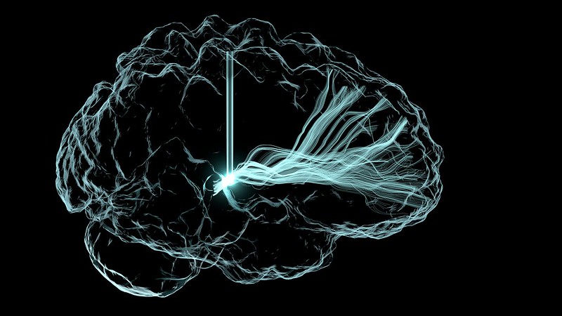
Deep brain stimulation illustration. Credit: NIH

Two new studies from UC San Francisco are pointing the way toward round-the-clock personalised care for people with Parkinson’s disease through an implanted device that can treat movement problems during the day and insomnia at night. 

The approach, called adaptive deep brain stimulation, or aDBS, uses methods derived from AI to monitor a patient’s brain activity for changes in symptoms. 

When it spots them, it intervenes with precisely calibrated pulses of electricity. The therapy complements the medications that Parkinson’s patients take to manage their symptoms, giving less stimulation when the drug is active, to ward off excess movements, and more stimulation as the drug wears off, to prevent stiffness.

It is the first time a so-called “closed loop” brain implant technology has been shown to work in Parkinson’s patients as they go about their daily lives. The device picks up brain signals to create a continuous feedback mechanism that can curtail symptoms as they arise. Users can switch out of the adaptive mode or turn the treatment off entirely with a hand-held device.

For the first study, researchers conducted a clinical trial with four people to test how well the approach worked during the day, comparing it to an earlier brain implant DBS technology known as constant or cDBS. 

To ensure the treatment provided the maximum relief to each participant, the researchers asked them to identify their most bothersome symptom. The new technology reduced them by 50%. Results appear August 19 in Nature Medicine.

“This is the future of deep brain stimulation for Parkinson’s disease,” said senior author Philip Starr, MD, PhD, the Dolores Cakebread Professor of Neurological Surgery, co-director of the UCSF Movement Disorders and Neuromodulation Clinic

Starr has been laying the groundwork for this technology for more than a decade. In 2013, he developed a way to detect and then record the abnormal brain rhythms associated with Parkinson’s. In 2021, his team identified specific patterns in those brain rhythms that correspond to motor symptoms.

“There’s been a great deal of interest in improving DBS therapy by making it adaptive and self-regulating, but it’s only been recently that the right tools and methods have been available to allow people to use this long-term in their homes,” said Starr, who was recruited by UCSF in 1998 to start its DBS program.

Earlier this year, UCSF researchers led by Simon Little, MBBS, PhD, demonstrated in Nature Communications that adaptive DBS has the potential to alleviate the insomnia that plagues many patients with Parkinson’s. 

“The big shift we’ve made with adaptive DBS is that we’re able to detect, in real time, where a patient is on the symptom spectrum and match it with the exact amount of stimulation they need,” said Little, associate professor of neurology and a senior author of both studies. Both Little and Starr are members of the UCSF Weill Institute for Neurosciences.

Restoring movement

Parkinson’s disease affects about 10 million people around the world. It arises from the loss of dopamine-producing neurons in deep regions of the brain that are responsible for controlling movement. The lack of those cells can also cause non-motor symptoms, affecting mood, motivation and sleep.

Treatment usually begins with levodopa, a drug that replaces the dopamine these cells are no longer able to make. However, excess dopamine in the brain as the drug takes effect can cause uncontrolled movements, called dyskinesia. As the medication wears off, tremor and stiffness set in again.  

Some patients then opt to have a standard cDBS device implanted, which provides a constant level of electrical stimulation. Constant DBS may reduce the amount of medication needed and partially reduce swings in symptoms. But the device also can over- or undercompensate, causing symptoms to veer from one extreme to the other during the day.

Closing the loop

To develop a DBS system that could adapt to a person’s changing dopamine levels, Starr and Little needed to make the DBS capable of recognising the brain signals that accompany different symptoms. 

Previous research had identified patterns of brain activity related to those symptoms in the subthalamic nucleus, or STN, the deep brain region that coordinates movement. This is the same area that cDBS stimulates, and Starr suspected that stimulation would mute the signals they needed to pick up.

So, he found alternative signals elsewhere in the brain – the motor cortex – that wouldn’t be weakened by the DBS stimulation. 

The next challenge was to work out how to develop a system that could use these dynamic signals to control DBS in an environment outside the lab. 

Building on findings from adaptive DBS studies that he had run at Oxford University a decade earlier, Little worked with Starr and the team to develop an approach for detecting these highly variable signals across different medication and stimulation levels.  

Over the course of many months, postdoctoral scholars Carina Oerhn, PhD, Lauren Hammer, PhD, and Stephanie Cernera, PhD, created a data analysis pipeline that could turn all of this into personalised algorithms to record, analyse and respond to the unique brain activity associated with each patient’s symptom state.

John Ngai, PhD, who directs the Brain Research Through Advancing Innovative Neurotechnologies® initiative (The BRAIN Initiative®) at the National Institutes of Health, said the study promises a marked improvement over current Parkinson’s treatment. 

“This personalised, adaptive DBS embodies The BRAIN Initiative’s core mission to revolutionise our understanding of the human brain,” he said. 

A better night’s sleep

Continuous DBS is aimed at mitigating daytime movement symptoms and doesn’t usually alleviate insomnia.

But in the last decade, there has been a growing recognition of the impact that insomnia, mood disorders and memory problems have on Parkinson’s patients. 

To help fill that gap, Little conducted a separate trial that included four patients with Parkinson’s and one patient with dystonia, a related movement disorder. In their paper published in Nature Communications, first author Fahim Anjum, PhD, a postdoctoral scholar in the Department of Neurology at UCSF, demonstrated that the device could recognise brain activity associated with various states of sleep. He also showed it could recognise other patterns that indicate a person is likely to wake up in the middle of the night. 

Little and Starr’s research teams, including their graduate student Clay Smyth, have started testing new algorithms to help people sleep. Their first sleep aDBS study was published last year in Brain Stimulation.  

Scientists are now developing similar closed-loop DBS treatments for a range of neurological disorders. 

“We see that it has a profound impact on patients, with potential not just in Parkinson’s but probably for psychiatric conditions like depression and obsessive-compulsive disorder as well,” Starr said. “We’re at the beginning of a new era of neurostimulation therapies.”

Source: University of California San Francisco

Shingles Increases Risk of Cognitive Decline in Later Life

The risk was higher for men who were carriers of a gene linked to dementia

Photo by Mari Lezhava on Unsplash

A new study led by investigators from Brigham and Women’s Hospital found that an episode of shingles is associated with about a 20 percent higher long-term risk of subjective cognitive decline. The study’s findings provide additional support for getting the shingles vaccine to decrease risk of developing shingles, according to the researchers. Their results are published in Alzheimer’s Research & Therapy.

“Our findings show long-term implications of shingles and highlight the importance of public health efforts to prevent and promote uptake of the shingles vaccine,” said corresponding author Sharon Curhan, MD, of the Channing Division for Network Medicine at Brigham and Women’s Hospital. “Given the growing number of Americans at risk for this painful and often disabling disease and the availability of a very effective vaccine, shingles vaccination could provide a valuable opportunity to reduce the burden of shingles and possibly reduce the burden of subsequent cognitive decline.”

Shingles, medically known as “herpes zoster,” is a viral infection that often causes a painful rash. Shingles is caused by the varicella zoster virus (VZV), the same virus that causes chickenpox. After a person has chickenpox, the virus stays in their body for the rest of their life. Most of the time, our immune system keeps the virus at bay. Years and even decades later, the virus may reactivate as shingles.

Almost all individuals in the US age 50 years and older have been infected with VZV and are therefore at risk for shingles. There’s a growing body of evidence that herpes viruses, including VZV, can influence cognitive decline. Subjective cognitive decline is an individual’s self-perceived experience of worsening or more frequent confusion or memory loss. It is a form of cognitive impairment and is one of the earliest noticeable symptoms of Alzheimer’s disease and related dementias.

Previous studies of shingles and dementia have been conflicting. Some research indicates that shingles increases the risk of dementia, while others indicate there’s no association or a negative association. In recent studies, the shingles vaccine was associated with a reduced risk of dementia.

To learn more about the link between shingles and cognitive decline, Curhan and her team used data from three large, well-characterized studies of men and women over long periods: The Nurses’ Health Study, the Nurses’ Health Study 2, and the Health Professionals Follow-Up Study. The study included 149,327 participants who completed health status surveys every two years, including questions about shingles episodes and cognitive decline. They compared those who had shingles with those who didn’t.

Curhan designed the study with first author Tian-Shin Yeh, formerly of the Harvard TH Chan School of Public Health. The researchers found that a history of shingles was significantly and independently associated with a higher risk – approximately 20% higher – of subjective cognitive decline in both women and men. That risk was higher among men who were carriers of the gene APOE4, which is linked to cognitive impairment and dementia. That same association wasn’t present in the women.

Researchers don’t know the mechanisms that link the virus to cognitive health, but there are several possible ways it may contribute to cognitive decline. There is growing evidence linking VZV to vascular disease, called VZV vasculopathy, in which the virus causes damage to blood vessels in the brain or body. Curhan’s group previously found that shingles was associated with higher long-term risk of stroke or heart disease.

Other mechanisms that may explain how the virus may lead to cognitive decline include causing inflammation in the brain, directly damaging the nerve and brain cells, and the activation of other herpesviruses.

The limitations of this research include that it was an observational study, information was based on self-report, and included a mostly white, highly educated population. In future studies, the researchers hope to learn more about preventing shingles and its complications.

“We’re evaluating to see if we can identify risk factors that could be modified to help reduce people’s risk of developing shingles,” Curhan said. “We also want to study whether the shingles vaccine can help reduce the risk of adverse health outcomes from shingles, such as cardiovascular disease and cognitive decline.” 

Source: Brigham and Women’s Hospital

Antioxidants in Seaweed Could help Prevent Parkinson’s Disease

Photo by Kampus Production on Pexels

Parkinson’s disease is induced by neuronal damage due to excessive production of reactive oxygen species. Suppression of reactive oxygen species generation is essential because it is fatal to dopaminergic neurons that manage dopamine neurotransmitters. Currently, only symptomatic treatment is available, so the development of treatment regimens and prevention methods is necessary.

Fortunately, Associate Professor Akiko Kojima-Yuasa of Osaka Metropolitan University’s Graduate School of Human Life and Ecology led a research group that has verified the physiological effect of Ecklonia cava polyphenols, seaweed antioxidants, on the prevention of Parkinson’s disease.

In this study, published in the journal Nutrients, two types of motor function tests were conducted using Parkinson’s disease model mice that were orally fed the antioxidants daily for one week and then administered rotenone. Results showed that motor function, which was decreased by rotenone, was restored. There was also improvement in intestinal motor function and the colon mucosa structure, a special tissue that covers the colon.

Further, cellular experiments using Parkinson’s disease model cells verified the biochemical interaction of the preventive effect of Ecklonia cava. Validation results showed that the antioxidants activate the AMPK enzyme (adenosine monophosphate-activated protein kinase), an intracellular energy sensor, and inhibit the production of reactive oxygen species that cause neuronal cell death.

“This study suggests that Ecklonia cava antioxidants may reduce neuronal damage by AMPK activation and inhibiting intracellular reactive oxygen species production,” stated Professor Kojima-Yuasa. “It is hoped that Ecklonia cava will be an effective ingredient in the prevention of Parkinson’s disease.”

Source: Osaka Metropolitan University

Brain’s Support Cells Contribute to Alzheimer’s Disease by Producing Toxic Peptide

Targeting oligodendrocytes could help reduce amyloid beta production

Neurons in the brain of an Alzheimer’s patient, with plaques caused by tau proteins. Credit: NIH

Oligodendrocytes are an important source of amyloid beta (Aβ) and play a key role in promoting neuronal dysfunction in Alzheimer’s disease (AD), according to a study published July 23, 2024 in the open-access journal PLOS Biology by Rikesh Rajani and Marc Aurel Busche from the UK Dementia Research Institute at University College London, and colleagues.

AD is a devastating neurodegenerative disorder affecting millions of people worldwide. Accumulation of Aβ – peptides consisting of 36 to 43 amino acids – is an early critical hallmark of the disease. Recent clinical trials demonstrating a slowing of cognitive and functional decline in individuals with AD who are treated with anti-Aβ antibodies reinforce the important role of Aβ in the disease process. Despite the key cellular effects of Aβ and its essential role in AD, the traditional assumption that neurons are the primary source of toxic Aβ in the brain has remained untested.

In the study, Rajani and Busche showed that non-neuronal brain cells called oligodendrocytes produce Aβ. They further demonstrated that selectively suppressing Aβ production in oligodendrocytes in an AD mouse model is sufficient to rescue abnormal neuronal hyperactivity. The results provide evidence for a critical role of oligodendrocyte-derived Aβ for early neuronal dysfunction in AD. Collectively, the findings suggest that targeting oligodendrocyte Aβ production could be a promising therapeutic strategy for treating AD.

According to the authors, the functional rescue is remarkable given the relatively modest reduction in plaque load that results from blocking oligodendrocyte Aβ production, while blocking neuronal Aβ production leads to a near elimination of plaques – another hallmark of the disease. This small contribution of oligodendrocytes to plaque load could suggest that a main effect of oligodendrocyte-derived Aβ is to promote neuronal dysfunction.

Together with the data showing an increased number of Aβ-producing oligodendrocytes in deeper cortical layers of the brains of individuals with AD, these results indicate that oligodendrocyte-derived Aβ plays a pivotal role in the early impairment of neuronal circuits in AD, which has important implications for how the disease progresses and is treated. The increased number of oligodendrocytes in human AD brains also raises the intriguing possibility that these cells could potentially offset reduced Aβ production due to neuronal loss as the disease progresses.

The authors add, “Our study challenges the long-held belief that neurons are the exclusive source of amyloid beta in the brain, one of the key toxic proteins that builds up in Alzheimer’s Disease. In fact, we show that oligodendrocytes, the myelinating cells of the central nervous system, can also produce significant amounts of amyloid beta which impairs neuronal function, and suggests that targeting these cells may be a promising new strategy to treat Alzheimer’s Disease.”

Provided by PLOS

Positive Life Experiences Boost Brain Mitochondria

Photo by Matteo Vistocco on Unsplash

Having more positive experiences in life is associated with lower odds of developing brain disorders like Alzheimer’s disease, slower cognitive decline with age, and even a longer life. But how feelings and experiences are translated into physical changes that protect or harm the brain is still unclear. 

Now, a study from Columbia researchers suggests that the brain’s mitochondria may play a fundamental part. The new study shows that the molecular machinery used by mitochondria to transform energy is boosted in older adults who experienced less psychological stress during their lives compared with individuals who had more negative experiences. 

“We’re showing that older individuals’ state of mind is linked to the biology of their brain mitochondria, which is the first time that subjective psychosocial experiences have been related to brain biology,” says Caroline Trumpff, assistant professor of medical psychology, who led the research with Martin Picard, associate professor of behavioural medicine at Columbia University Vagelos College of Physicians and Surgeons and in the Robert N. Butler Columbia Aging Center. 

“We think that the mitochondria in the brain are like antennae, picking up molecular and hormonal signals and transmitting information to the cell nucleus, changing the life course of each cell,” says Picard. “And if mitochondria can change cell behaviour, they can change the biology of the brain, the mind, and the whole person.” 

Study details 

The new research used data collected by two extensive studies of nearly 450 older adults in the United States. Each study collected detailed psychosocial information from the participants for two decades during their lives. Study participants donated their brains after death for further analysis, which provided data on the state of the participants’ brain cells. 

Trumpff created indices that converted patients’ reports of positive and negative psychosocial factors into a single score of overall psychosocial experience. She also scored each participant on seven domains that represent distinct genetic networks active in mitochondria. 

“The use of multivariate mitotype indices is an important innovation because we could more easily interpret the biological state of the mitochondria with networks of related genes than an analysis of thousands of individual genes,” Picard says. 

Study results 

The results showed that one mitochondrial domain – which assessed the organelle’s energy transformation machinery – was associated with psychosocial scores. 

“Greater well-being was linked to greater abundance of proteins in mitochondria needed to transform energy, whereas negative mood was linked to lower protein content,” Trumpff says. “This may be why chronic psychological stress and negative experiences are bad for the brain, because they damage or impair mitochondrial energy transformation in the dorsolateral prefrontal cortex, the part of the brain responsible for high-level cognitive tasks.” 

The researchers also analysed mitochondria in specific cell types in the brain and found that the associations between mitochondria and psychosocial factors were driven not by the brain’s neurons, but its glia cells, which may be playing more than their traditionally assumed “supportive” roles. 

“This piece of the study, made possible by our collaboration with the Columbia Center for Translational and Computational Neuroimmunology, is what I think makes it particularly significant,” Picard says. “To ask questions at this level of cellular resolution in the brain is unprecedented in the mitochondrial field.

“Neurons have been the focus of neuroscience, but we’re waking up to the fact that other cells in the brain may be driving disease.” 

Do mitochondria change mood, or does mood change mitochondria? 

Though the current study cannot determine if the participant’s psychosocial experiences altered their brain mitochondria or if innate or acquired mitochondrial states contributed to those experiences, other studies suggest that the relationship between mitochondria and mood works both ways. 

In animal studies, the evidence is very strong, Picard says, that chronic stress affects mitochondrial energy transformation. And in people, a recent study conducted by Picard and collaborator Elissa Epel at UCSF found the first evidence that mood may affect mitochondria in humans: In that study, positive mood predicted greater mitochondrial energy production in the participants’ blood cells on subsequent days, but mitochondrial activity did not predict mood on subsequent days. 

A growing body of work in animals and humans also indicates that mitochondria themselves can alter behaviour. 

“It’s possible that these mechanisms reinforce one another,” Trumpff says. “Chronic stress could alter an individual’s mitochondrial biology in ways that subsequently affects their perception of social events, creating more stress. The emerging picture in the literature is that all these pathways are interactive.” 

Next steps 

Though the brain’s energy transformation machinery was greater in participants with higher psychosocial scores, the researchers do not yet know if that leads to greater energy transformation. Trumpff and Picard are currently doing those studies with hundreds of brains from the same cohorts of participants. 

The team is also exploring a way to measure the brain’s mitochondrial health, which could be used in doctors’ offices in the future. 

“Mitochondria are the source of health and life, but we don’t have ways to quantify health, only disease,” Picard says. “We need a science of health. We need tests that show how healthy and resilient someone is.

“This would be valuable clinically to monitor changes in health before the appearance of disease, and it could transform medical research by giving scientists something to target other than decades of accumulated protein deposits or other forms of long-term damage.”

Source: Columbia University Irving Medical Center

How Astrocytes Know to Give the Brain an Energy Boost

Image of an astrocyte, a subtype of glial cells. Glial cells are the most common cell in the brain. Credit: Pasca Lab, Stanford University

A key mechanism by which astrocytes detect when an energy boost is needed for the brain has been elucidated by University College of London researchers using mouse-based and in vitro studies.

The findings, published in Nature, could inform new therapies to maintain brain health and longevity, the researchers say, since other studies have found that brain energy metabolism can become impaired late in life and contribute to cognitive decline and the development of neurodegenerative disease.

Lead author Professor Alexander Gourine (UCL Neuroscience, Physiology & Pharmacology) said: “When our brain is more active, such as when we’re performing a mentally taxing task, our brain needs an immediate boost of energy, but the exact mechanisms that ensure on-demand local supply of metabolic energy to active brain regions are not fully understood.”

First and co-corresponding author Dr Shefeeq Theparambil, who began the study at UCL before moving to Lancaster University, said: “The normal activities of the brain require enormous amounts of energy, comparable to that of a human leg muscle running a marathon. This energy is primarily derived from blood glucose. Neurons in the brain consume more than 75% of this energy.”

Prior research has shown that numerous brain cells called astrocytes appear to play a role in providing the brain neurons with energy they need. Astrocytes, shaped like stars, are a type of glial cell, which are non-neuronal cells found in the central nervous system. When neighbouring neurons need an increase in energy supply, astrocytes jump into action by rapidly activating their own glucose stores and metabolism, leading to the increased production and release of lactate. Lactate supplements the pool of energy that is readily available for use by neurons in the brain.

Professor Gourine explained: “In our study, we have figured out how exactly astrocytes are able to monitor the energy use by their neighbouring nerve cells, and kick-start this process that delivers additional chemical energy to busy brain regions.”

In a series of experiments using mouse models and cell samples, the researchers identified a set of specific receptors in astrocytes that can detect and monitor neuronal activity, and trigger a signalling pathway involving an essential molecule called adenosine. The researchers found that the metabolic signalling pathway activated by adenosine in astrocytes is exactly the same as the pathway that recruits energy stores in the muscle and the liver, for example when we exercise.

Adenosine activates astrocyte glucose metabolism and supply of energy to neurons to ensure that synaptic function (neurotransmitters passing communication signals between cells) continues apace under conditions of high energy demand or reduced energy supply.

The researchers found that when they deactivated the key astrocyte receptors in mice, the animal’s brain activity was less effective, including significant impairments in global brain metabolism, memory and disruption of sleep, thus demonstrating that the signalling pathway they identified is vital for processes such as learning, memory and sleep.

Dr Theparambil said: “Identification of this mechanism may have broader implications as it could be a way of treating brain diseases where brain energetics are downregulated, such as neurodegeneration and dementia.”

Professor Gourine added: “We know that brain energy homeostasis is progressively impaired in ageing and this process is accelerated during the development of neurodegenerative diseases such as Alzheimer’s disease. Our study identifies an attractive readily druggable target and therapeutic opportunity for brain energy rescue for the purpose of protecting brain function, maintaining cognitive health, and promoting brain longevity.”

New Pulsatility Metric in Brain Blood Vessels for Studying Dementia

Photo by Anna Shvets on Pexels

Researchers from the Mātai Institute and the Auckland Bioengineering Institute have developed a new metric from measured blood circulation in the brain. The new metric opens up new research avenues for brain conditions, including Alzheimer’s disease and other forms of dementia. The research has been published in the leading research journal Scientific Reports Nature.

Each time the heart beats, it pumps blood through the brain vessels, causing them to expand slightly and then relax. This pulsation in the brain helps distribute blood evenly across different areas of the brain, ensuring that all parts receive the oxygen and nutrients they need to function properly.

In healthy vessels, the pulse wave is dampened before it reaches the smallest vessels, where high pulsatility could be harmful. The new metric provides a comprehensive measure of the small vessel pulsatility risk.

The new metric is based on 4D flow MRI technology, and is particularly crucial because increased vascular pulsatility is linked to several brain conditions, including Alzheimer’s disease and other forms of dementia.

By accurately measuring how pulsatility is transmitted in the brain, researchers can better understand the underlying mechanisms of these diseases and potentially guide the development of new treatments.

Current MRI methods face limitations due to anatomical variations and measurement constraints. The new technique removes this issue by integrating thousands of measurements across all brain vessels, rather than the traditional method of looking at one spot. This provides a richer metric representative of the entire brain.

“The ability to measure how pulsatility is transmitted through the brain’s arteries could revolutionise our approach to neurological diseases, and support research in vascular damage hypotheses,” says first author Sergio Dempsey.

“Our method allows for a detailed assessment of the brain’s vascular health, which is often compromised in neurodegenerative disorders.”

The study also highlighted the potential to enhance clinical assessments and research on brain health. By integrating this new metric into routine diagnostic procedures, healthcare providers can offer more precise and personalised care plans for individuals at risk of or suffering from cognitive impairments.

To make the most of the new metric’s implications for patient care, the researchers have made their tools publicly available, integrating them into pre-existing open-source software. This enables scientists and clinicians worldwide to adopt the advanced methodology, fostering further research and collaboration in the field of neurology.

Results from the initial study of the metric also identified important sex differences in vascular dynamics which has initiated a new study focusing on sex-related dynamics.

The research team is planning further studies to explore the applications of this technique in larger and more diverse populations.

Source: University of Auckland

Could Drugs for Enlarged Prostate also Protect against Lewy Body Dementia?

Credit: Darryl Leja National Human Genome Research Institute National Institutes Of Health

A new study published in Neurology suggests that certain drugs commonly used to treat enlarged prostate may also decrease the risk for dementia with Lewy bodies (DLB). This observational finding may seem surprising, but it mirrors previous work by the University of Iowa Health Care team that links the drugs to a protective effect in another neurodegenerative condition: Parkinson’s disease. 

The UI researchers think that a specific side effect of the drugs targets a biological flaw shared by DLB and Parkinson’s disease, as well as other neurodegenerative diseases, raising the possibility that they may have broad potential for treating a wide range of neurodegenerative conditions. 

“Diseases like dementia with Lewy bodies, or Parkinson’s disease, or Alzheimer’s disease are debilitating, and we don’t really have any good treatments that can modify the disease progression. We can treat symptoms, but we can’t actually slow the disease,” explains lead study author Jacob Simmering, PhD, UI assistant professor of internal medicine. “One of the most exciting things about this study is that we find that same neuroprotective effect that we saw in Parkinson’s disease. If there is a broadly protective mechanism, these medications could potentially be used to manage or prevent other neurodegenerative diseases.” 

Large observational study links prostate drugs to lower risk of dementia with Lewy bodies

DLB is a neurodegenerative disease that causes substantial and rapid cognitive decline and dementia. It affects about one in 1000 people per year, accounting for 3 to 7% of all dementia cases. 

For the new study, the UI researchers used a large database of patient information to identify more than 643 000 men with no history of DLB who were newly starting one of six drugs used to treat benign prostatic hyperplasia (enlarged prostate). 

Three of the drugs, terazosin, doxazosin, and alfuzosin (Tz/Dz/Az), have an unexpected side effect; they can boost energy production in brain cells. Preclinical studies suggest that this ability may help slow or prevent neurodegenerative diseases like PD and DLB.  

The other drugs, tamsulosin and two 5-alpha-reductase inhibitors (5ARIs) called finasteride and dutasteride, do not enhance energy production in the brain and therefore provide a good comparison to test the effect of the Tz/Dz/Az drugs. 

The team then followed the data on these men from when they started taking the medication until they left the database or developed dementia with Lewy bodies, whichever happened first. On average, the men were followed for about three years. 

Because all the participants were selected to start a drug that treats the same condition, the researchers reasoned that the men were likely similar to each other at the outset of the treatment. The men were all propensity score-matched for characteristics like age, year of medication start, and other illnesses they had before starting the treatment, to further reduce the differences between the groups. 

“We found that men who took Tz/Az/Dz drugs were less likely to develop a diagnosis of dementia with Lewy bodies,” Simmering says. “Overall, men taking terazosin-type medications had about a 40% lower risk of developing a DLB diagnosis compared to men taking tamsulosin, and about a 37% reduction in risk compared to men taking five alpha reductase inhibitors.” 

Meanwhile, there was no statistically significant difference in risk between men taking tamsulosin and alpha reductase inhibitors. 

Approved drugs show potential

Since this was an observational study, causation cannot be established, only an association. In addition, the study only included men because the drugs are prescribed for prostate problems, which means that the researchers don’t know if the findings would apply to women. However, Simmering and his colleagues are excited by the potential of these drugs, which are already FDA approved, inexpensive, and have been used safely for decades. 

“If terazosin and these similar medications can help slow this progression – if not outright preventing the disease – this would be important to preserving cognitive function and quality of life in people with DLB,” Simmering says. 

Source: University of Iowa Carver College of Medicine

New Ultrasound and Genetics Combination Precisely Targets Neurons in Diseased Regions

McKelvey School of Engineering researchers have developed a noninvasive technology combining a holographic acoustic device with genetic engineering that allows them to precisely target affected neurons in the brain, creating the potential to precisely modulate selected cell types in multiple diseased brain regions. (Credit: Yaoheng Yang)

Brain diseases such as Parkinson’s disease involve damage in more than one region of the brain, requiring technology that could precisely and flexibly address all affected regions simultaneously. Researchers have developed a noninvasive technology combining a holographic acoustic device with genetic engineering that allows them to precisely target affected neurons in the brain. This has the potential to precisely modulate selected cell types in multiple diseased brain regions. 

Hong Chen, associate professor of biomedical engineering and neurosurgery at Washington University in St. Louis and her team created AhSonogenetics, or Airy-beam holographic sonogenetics, a technique that uses a noninvasive wearable ultrasound device to alter genetically selected neurons in the brains of mice. Results of the proof-of-concept study were published in Proceedings of the National Academy of Sciences

AhSonogenetics brings together several of Chen’s group’s recent advances into one technology. In 2021, she and her team launched Sonogenetics, a method that uses focused ultrasound to deliver a viral construct containing ultrasound-sensitive ion channels to genetically selected neurons in the brain. They use low-intensity focused ultrasound to deliver a small burst of warmth, which opens the ion channels and activates the neurons. Chen’s team was the first to show that sonogenetics could modulate the behaviour of freely moving mice.

In 2022, she and members of her lab designed and 3D-printed a flexible and versatile tool known as an Airy beam-enabled binary acoustic metasurface that allowed them to manipulate ultrasound beams. She also is developing Sonogenetics 2.0, which combines the advantage of ultrasound and genetic engineering to modulate defined neurons noninvasively and precisely in the brains of humans and animals. AhSonogenetics brings them together as a potential method to intervene in neurodegenerative diseases. 

“By enabling precise and flexible cell-type-specific neuromodulation without invasive procedures, AhSonogenetics provides a powerful tool for investigating intact neural circuits and offers promising interventions for neurological disorders,” Chen said. 

Sonogenetics gives researchers a way to precisely control the brains, while airy-beam technology allows researchers to bend or steer the sound waves to generate arbitrary beam patterns inside the brain with a high spatial resolution. Yaoheng (Mack) Yang, a postdoctoral research associate who earned a doctorate in biomedical engineering from McKelvey Engineering in 2022, said the technology gives the researchers three unique advantages.

“Airy beam is the technology that can give us precise targeting of a smaller region than conventional technology, the flexibility to steer to the targeted brain regions, and to target multiple brain regions simultaneously,” Yang said.

Chen and her team, including first authors Zhongtao Hu, a former postdoctoral research associate, and Yang, designed each Airy-beam metasurface individually as the foundation for wearable ultrasound devices that were tailored for different applications and for precise locations in the brain.

Chen’s team tested the technique on a mouse model of Parkinson’s disease. With AhSonogenetics, they were able to stimulate two brain regions simultaneously in a single mouse, eliminating the need for multiple implants or interventions. This stimulation alleviated Parkinson’s-related motor deficits in the mouse model, including slow movements, difficulty walking and freezing behaviours.

The team’s Airy-beam device overcomes some of the limits of sonogenetics, including tailoring the design of the device to target specific brain locations, as well as incorporating the flexibility to adjust target locations in a single brain.

Hu said the device, which costs roughly $50 to make, can be tailored in size to fit various brain sizes, expanding its potential applications. 

“This technology can be used as a research platform to speed neuroscience research because of the capability to flexibly target different brain regions,” Hu said. “The affordability and ease of fabrication lower the barriers to the widespread adoption of our proposed devices by the research community for neuromodulation applications.”

Source: Washington University in St. Louis

Gut Bacteria in Parkinson’s Disease Produce Fewer B Vitamins

In Parkinson’s disease, a reduction in the gut bacteria of genes responsible for synthesising the essential B vitamins B2 and B7 was found. Credit: Reiko Matsushita

A study led by Nagoya University in Japan has revealed a link between gut microbiota and Parkinson’s disease (PD). The researchers found that the gut bacteria genes responsible for synthesising vitamins B2 and B7 were reduced. This gene reduction was also linked to low levels of agents that help maintain the integrity of the intestinal barrier, which when weakened causes the inflammation seen in PD. Their findings, published in npj Parkinson’s Disease, suggest that treatment with B vitamins to address these deficiencies can be used to treat PD. 

PD is characterized by a variety of physical symptoms that hinder daily activities and mobility, such as shaking, slow movement, stiffness, and balance problems. While the frequency of PD may vary between different populations, it is estimated to affect approximately 1-2% of individuals aged 55 years or older. 

Various physiological processes are heavily influenced by the microorganisms found in the gut, which are collectively known as gut microbiota. In ideal conditions, gut microbiota produce SCFAs and polyamines, which maintain the intestinal barrier that prevents toxins entering the bloodstream. Toxins in the blood can be carried to the brain where they cause inflammation and affect neurotransmission processes that are critical for maintaining mental health.

To better understand the relationship between the microbial characteristics of the gut in PD, Hiroshi Nishiwaki and Jun Ueyama from the Nagoya University Graduate School of Medicine conducted a metanalysis of stool samples from patients with PD from Japan, the United States, Germany, China, and Taiwan. They used shotgun sequencing, a technique that sequences all genetic material in a sample. This is an invaluable tool because it offers researchers a better understanding of the microbial community and genetic makeup of the sample.

They observed a decrease in the bacterial genes responsible for the synthesising of riboflavin (vitamin B2) and biotin (vitamin B7) in patients diagnosed with PD. Riboflavin and biotin, derived from both food and gut microbiota, have anti-inflammatory properties, which may counteract the neuroinflammation seen in diseases like PD. 

B vitamins play crucial roles in the metabolic processes that influence the production and functions of short-chain fatty acids (SCFAs) and polyamines, two agents that help maintain the integrity of the intestinal barrier, preventing toxins entering the bloodstream. An examination of fecal metabolites revealed decreases of both in patients with PD. 

The findings indicate a potential explanation for the progression of PD. “Deficiencies in polyamines and SCFAs could lead to thinning of the intestinal mucus layer, increasing intestinal permeability, both of which have been observed in PD,” Nishiwaki explained. “This higher permeability exposes nerves to toxins, contributing to abnormal aggregation of alpha-synuclein, activating the immune cells in the brain, and leading to long-term inflammation.” 

He added, “Supplementation therapy targeting riboflavin and biotin holds promise as a potential therapeutic avenue for alleviating PD symptoms and slowing disease progression.”

The results of the study highlight the importance of understanding the complex relationship among gut microbiota, metabolic pathways, and neurodegeneration. In the coming years, customised therapy could potentially be based on patients’ unique microbiome profiles. By altering bacterial levels in the microbiome, doctors can potentially delay the onset of symptoms associated with diseases like PD.

“We could perform gut microbiota analysis on patients or conduct faecal metabolite analysis,” Nishiwaki said. “Using these findings, we could identify individuals with specific deficiencies and administer oral riboflavin and biotin supplements to those with decreased levels, potentially creating an effective treatment.”

Source: Nagoya University

The study, “Meta-analysis of shotgun sequencing of gut microbiota in Parkinson’s disease,” was published in npj Parkinson’s Disease on May 21, 2024, at DOI:10.1038/s41531-024-00724-z.