Past research has suggested that inflammation may contribute to the development and progression of dementia and that non-steroidal anti-inflammatory (NSAID) medications may help protect against dementia due to their anti-inflammatory effects. A new large prospective study published in the Journal of the American Geriatrics Society provides additional evidence, showing that long-term NSAID use is linked to a decreased risk of developing dementia.
In the population-based study of 11 745 adults with an average follow-up of 14.5 years, 9520 participants had used NSAIDs at any given time, and 2091 participants developed dementia. Long-term NSAID use was associated with a 12% reduced risk of developing dementia. Short- and intermediate-term use did not provide benefits. Also, the cumulative dose of NSAIDs was not associated with decreased dementia risk.
The findings suggest that prolonged, rather than intensive, use of anti-inflammatory medications may help protect against dementia.
“Our study provides evidence on possible preventive effects of anti-inflammatory medication against the dementia process. There is a need for more studies to further consolidate this evidence and possibly develop preventive strategies,” said corresponding author M. Arfan Ikram, MSc, MD, PhD, of Erasmus MC University Medical Center Rotterdam, in the Netherlands.
A decade of studies from labs around the world provide a growing evidence base that increasing the power of the brain’s gamma rhythms could help fight Alzheimer’s, and perhaps other, neurological diseases.
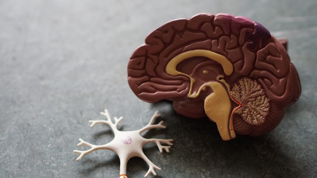
Source: Pixabay
A decade after scientists in The Picower Institute for Learning and Memory at MIT first began testing whether sensory stimulation of the brain’s 40Hz “gamma” frequency rhythms could treat Alzheimer’s disease in mice, a growing evidence base supporting the idea that it can improve brain health – in humans as well as animals – has emerged from the work of labs all over the world. A new review article in PLOS Biology describes the state of research so far and presents some of the fundamental and clinical questions at the forefront of the non-invasive gamma stimulation now.
“As we’ve made all our observations, many other people in the field have published results that are very consistent,” said Li-Huei Tsai, Picower Professor at MIT, director of MIT’s Aging Brain Initiative, and senior author of the new review with postdoc Jung Park. “People have used many different ways to induce gamma including sensory stimulation, transcranial alternating current stimulation or transcranial magnetic stimulation, but the key is delivering stimulation at 40 Hz. They all see beneficial effects.”
A decade of discovery at MIT
Starting with a paper in Nature in 2016, a collaboration led by Tsai has produced a series of studies showing that 40Hz stimulation via light, sound, a combination of the two, or tactile vibration reduces hallmarks of Alzheimer’s pathology such as amyloid and tau proteins, prevents neuron death, decreases synapse loss, and sustains memory and cognition in various Alzheimer’s mouse models. The collaboration’s investigations of the underlying mechanisms that produce these benefits has so far identified specific cellular and molecular responses in many brain cell types including neurons, microglia, astrocytes, oligodendrocytes and the brain’s blood vessels. Last year, for instance, the lab reported in Nature that 40Hz audio and visual stimulation induced interneurons in mice to increase release of the peptide VIP, prompting increased clearance of amyloid from brain tissue via the brain’s glymphatic “plumbing” system.
Meanwhile, at MIT and at the MIT spinoff company Cognito Therapeutics, phase II clinical studies have shown that people with Alzheimer’s exposed to 40Hz light and sound experienced a significant slowing of brain atrophy and improvements on some cognitive measures compared to untreated controls. Cognito, which has also measured significant preservation of white matter in volunteers, has been conducting a pivotal, nationwide phase III clinical trial of sensory gamma stimulation for more than a year.
“Neuroscientists often lament that it is a great time to have AD if you are a mouse,” Park and Tsai wrote in the review. “Our ultimate goal, therefore, is to translate GENUS discoveries into a safe, accessible, and non-invasive therapy for AD patients.” The MIT team often refers to 40Hz stimulation as “GENUS” for Gamma Entrainment Using Sensory Stimulation.
A growing field
As Tsai’s collaboration, which includes MIT colleagues Edward Boyden and Emery N. Brown, has published its results, many other labs have produced studies adding to the evidence that various methods of non-invasive gamma sensory stimulation can combat Alzheimer’s pathology. Among many examples cited in the new review, in 2024 a research team in China independently corroborated that 40Hz sensory stimulation increases glymphatic fluid flows in mice. In another example, a Harvard Medical School-based team in 2022 showed that 40Hz gamma stimulation using Transcranial Alternating Current Stimulation significantly reduced the burden of tau in three out of four human volunteers. And in another study involving more than 100 people, researchers in Scotland in 2023 used audio and visual gamma stimulation (at 37.5Hz) to improve memory recall.
Open questions
Amid the growing number of publications describing preclinical studies with mice and clinical trials with people, open questions remain, Tsai and Park acknowledge. The MIT team and others are still exploring the cellular and molecular mechanisms that underlie GENUS’s effects. Tsai said her lab is looking at other neuropeptide and neuromodulatory systems to better understand the cascade of events linking sensory stimulation to the observed cellular responses. Meanwhile the nature of how some cells, such as microglia, respond to gamma stimulation and how that affects pathology remains unclear, Tsai added.
Even with a national Phase III clinical trial underway, it is still important to investigate these fundamental mechanisms, Tsai said, because new insights into how non-invasive gamma stimulation affects the brain could improve and expand its therapeutic potential.
“The more we understand the mechanisms, the more we will have good ideas about how to further optimize the treatment,” Tsai said. “And the more we understand its action and the circuits it affects, the more we will know beyond Alzheimer’s disease what other neurological disorders will benefit from this.”
Indeed the review points to studies at MIT and other institutions providing at least some evidence that GENUS might be able to help with Parkinson’s disease, stroke, anxiety, epilepsy, and the cognitive side effects of chemotherapy and conditions that reduce myelin such as multiple sclerosis. Tsai’s lab has been studying whether it can help with Down syndrome as well.
The open questions may help define the next decade of GENUS research.
Getting at least 30 minutes of daily summer sun in the first year of life may mean a lower relapse risk for children who are diagnosed with multiple sclerosis (MS) later, according to a study published in Neurology® Neuroimmunology & Neuroinflammation, an official journal of the American Academy of Neurology. The study also found if a child’s biological mother had at least 30 minutes of daily sun during the second trimester of pregnancy, the child had a lower risk of MS relapses.
The study does not prove that sun lowers relapse risk for children with MS, it only shows an association. “It is important not to spend too much time in the sun without sun protection, however greater exposure to sun has been tied in previous research to a lower risk of developing MS in childhood,” said Gina Chang, MD, MPH, of The Children’s Hospital of Philadelphia and member of the American Academy of Neurology. “It’s encouraging that our study found that greater sun exposure during early development may also be beneficial in helping to reduce disease activity in children who are later diagnosed with MS.”
For the study, researchers looked at health records from 18 MS clinics across the United States to identify 334 children and young people with childhood-onset MS age four to 21. Participants were within four years of experiencing their first symptoms. The median follow-up time was 3.3 years. To determine sun exposure, participants’ parents or guardians completed questionnaires that asked how much time the participant and their biological mother had spent in the sun at various periods of life, what kind of clothing they typically wore and how often they used sunscreen.
Of the total group, 206, or 62%, experienced at least one relapse during the study. Relapses were defined as new or returning symptoms lasting for at least 24 hours and separated by at least 30 days from the last MS attack, without a fever or infection. They found that of 75 participants who had 30 minutes to an hour of daily summer sun during their first year of life, 34 children, or 45%, had a relapse.
Of the 182 participants who had less than 30 minutes of daily summer sun during their first year of life, 118 children, or 65%, had a relapse. After adjusting for factors such as tobacco exposure in the first year of life, season of birth, the type of MS medication taken and use of sun protection such as sunscreen, hats and clothing, researchers found that 30 or more minutes of daily summer sun during the first year of life was associated with a 33% lower risk of relapse compared to less than 30 minutes of daily summer sun.
Researchers also looked at sun exposure for the biological mothers of the children. They found that 30 minutes or more of daily sun during the second trimester of pregnancy was associated with a 32% reduced risk of relapse for their child with MS.
“Our findings suggest that sun exposure in early childhood may have long-lasting benefits on the progression of childhood-onset MS,” said Chang. “Future studies should look at how time in the sun at other time periods before and after MS diagnosis affects disease course, to better guide sun exposure recommendations for children with MS and to help design potential clinical trials.” A limitation of the study was that it relied on participants’ parents or guardians reporting their sun exposure and use of sun protection, which they may not have remembered accurately.
Electrical stimulation of the sensory spinal nerves targets the root cause of progressive loss of neural function in spinal muscle atrophy (SMA), an inherited neuromuscular disease. The intervention can gradually reawaken functionally silent motor neurons in the spinal cord and improve leg muscle strength and walking in adults with SMA. The findings were reported by University of Pittsburgh School of Medicine researchers in Nature Medicine.
Early results from a pilot clinical trial in three human volunteers with SMA show that one month of regular neurostimulation sessions improved motoneuron function, reduced fatigue and improved strength and walking in all participants, regardless of the severity of their symptoms.
“To counteract neurodegeneration, we need two things – stop neuron death and restore function of surviving neurons,” said co-corresponding author Marco Capogrosso, assistant professor of neurological surgery at Pitt School of Medicine. “In this study we proposed an approach to treat the root cause of neural dysfunction, complementing existing neuroprotective treatments with a new approach that reverses nerve cell dysfunction.”
Doug McCullough, one of three participants in the study, says his SMA had progressed to the point that even walking on smooth surfaces was difficult when he started the trial in 2023. The research team kept him blind to most of the quantitative data but showed him video to reveal how effective the treatment was proving to be. The team captured footage of McCullough at various points during the trial to monitor his progress.
“Because my hip flexors are so weak, I basically have this waddling gait where my hips sway back and forth and I swing my legs out to the side because I can’t pick them straight up,” he says. “You could clearly see from the video that my walk was improved and that I was walking faster. I had a little more natural gait. It still wasn’t completely normal, but it was better than what it was before the study.”
SMA is a genetic neurodegenerative disease that manifests in progressive death and functional decline of motor neurons – nerve cells that control movement by transmitting signals from the brain and the spinal cord to the muscles. Over time, the loss of motor neurons causes gradual muscle weakness and leads to a variety of motor deficits, including for the participants in this trial, difficulty in walking, climbing stairs and standing up from chairs.
While there is no cure for SMA, several promising neuroprotective treatments have become available in the last decade. These include gene replacement therapies and medications, both of which stimulate the production of motoneuron-supporting proteins that prevent neuronal death and that slow down, though not reverse, disease progression.
Studies show that movement deficits in SMA emerge before widespread motoneuron death, suggesting that underlying dysfunction in spinal nerve circuitry may contribute to disease onset and symptom development. Earlier research on animal models of SMA by study coauthor George Mentis of Columbia University, showed that surviving motor neurons receive fewer stimulation inputs from sensory nerves. Compensating for this deficit in neural feedback could, therefore, improve communication between the nervous system and the muscles, aid muscle movement and combat muscle wasting.
Pitt researchers hypothesised that a targeted epidural electrical stimulation therapy could be used to rescue lost nerve cell function by amplifying sensory inputs to the motor neurons and engaging the degenerated neural circuits. These cellular changes could, in turn, translate into functional improvements in movement capacity.
The Pitt study was conducted as part of a pilot clinical trial that enrolled three adults with milder forms of SMA (Type 3 or 4 SMA). During a study period of 29 days, participants were implanted with two spinal cord stimulation (SCS) electrodes that were placed in the lower-back region on each side of the spinal cord, directing the stimulation exclusively to sensory nerve roots. Testing sessions lasted four hours each and were conducted five times a week for a total of 19 sessions, until the stimulation device was explanted.
After confirming that the stimulation worked as intended and engaged spinal motor neurons, researchers performed a battery of tests to measure muscle strength and fatigue, changes in gait, range of motion and walking distance, as well as motoneuron function.
“Because SMA is a progressive disease, patients do not expect to get better as time goes on. But that is not what we saw in our study. Over the four weeks of treatment, our study participants improved in several clinical outcomes with improvements in activities of daily living. For instance, toward the end of the study, one patient reported being able to walk from their home to the lab without becoming tired,” said co-corresponding author Elvira Pirondini, assistant professor of physical medicine and rehabilitation at Pitt School of Medicine.
All participants increased their 6-Minute Walk Test score (a measure of muscle endurance and fatigue) by at least 20m, compared to a mean improvement of 1.4m over three months of comparable exercise regimen unaided by SCS and a median increase of 20m after 15 months of SMA-specific neuroprotective pharmacologic therapy.
These functional gains were mirrored by improved neural function, including a boost in motoneurons’ capacity to generate electrical impulses and transmit them to the muscles.
Antibiotics, antivirals, vaccinations and anti-inflammatory medication are associated with reduced risk of dementia, according to new research that looked at health data from over 130 million individuals.
The study, led by researchers from the universities of Cambridge and Exeter, identified several drugs already licensed and in use that have the potential to be repurposed to treat dementia.
Dementia is a leading cause of death in the UK and can lead to profound distress in the individual and among those caring for them. It has been estimated to have a worldwide economic cost in excess of US$1 trillion dollars.
Despite intensive efforts, progress in identifying drugs that can slow or even prevent dementia has been disappointing. Until recently, dementia drugs were effective only for symptoms and have a modest effect. Recently, lecanemab and donanemab have been shown to reduce the build-up in the brain of amyloid plaques – a key characteristic of Alzheimer’s disease – and to slow down progression of the disease, but the National Institute for Health and Care Excellence (NICE) concluded that the benefits were insufficient to justify approval for use within the NHS.
Scientists are increasingly turning to existing drugs to see if they may be repurposed to treat dementia. As the safety profile of these drugs is already known, the move to clinical trials can be accelerated significantly.
Dr Ben Underwood, from the Department of Psychiatry at the University of Cambridge and Cambridgeshire and Peterborough NHS Foundation Trust, said: “We urgently need new treatments to slow the progress of dementia, if not to prevent it. If we can find drugs that are already licensed for other conditions, then we can get them into trials and – crucially – may be able to make them available to patients much, much faster than we could do for an entirely new drug. The fact they are already available is likely to reduce cost and therefore make them more likely to be approved for use in the NHS.”
In a study published today in Alzheimer’s and Dementia: Translational Research & Clinical Interventions, Dr Underwood, together with Dr Ilianna Lourida from the University of Exeter, led a systematic review of existing scientific literature to look for evidence of prescription drugs that altered the risk of dementia. Systematic reviews allow researchers to pool several studies where evidence may be weak, or even contradictory, to arrive at more robust conclusions.
In total, the team examined 14 studies that used large clinical datasets and medical records, capturing data from more than 130 million individuals and 1 million dementia cases. Although they found a lack of consistency between studies in identifying individual drugs that affect the risk of dementia, they identified several drug classes associated with altered risk.
One unexpected finding was an association between antibiotics, antivirals and vaccines, and a reduced risk of dementia. This finding supports the hypothesis that common dementias may be triggered by viral or bacterial infections, and supports recent interest in vaccines, such as the BCG vaccine for tuberculosis, and decreased risk of dementia.
Anti-inflammatory drugs such as ibuprofen were also found to be associated with reduced risk. Inflammation is increasingly being seen to be a significant contributor to a wide range of diseases, and its role in dementia is supported by the fact that some genes that increase the risk of dementia are part of inflammatory pathways.
The team found conflicting evidence for several classes of drugs, with some blood pressure medications and anti-depressants and, to a lesser extent, diabetes medication associated with a decreased risk of dementia and others associated with increased risk.
Dr Ilianna Lourida from the National Institute for Health and Care Research Applied Research Collaboration South West Peninsula (PenARC), University of Exeter, said: “Because a particular drug is associated with an altered risk of dementia, it doesn’t necessarily mean that it causes or indeed helps in dementia. We know that diabetes increases your risk of dementia, for example, so anyone on medication to manage their glucose levels would naturally also be at a higher risk of dementia – but that doesn’t mean the drug increases your risk.
“It’s important to remember that all drugs have benefits and risks. You should never change your medicine without discussing this first with your doctor, and you should speak to them if you have any concerns.”
The conflicting evidence may also reflect differences in how particular studies were conducted and how data was collected, as well as the fact that different medications even within the same class often target different biological mechanisms.
The UK government is supporting the development of an Alzheimer’s trial platform to evaluate drugs rapidly and efficiently, including repurposed drugs currently used for other conditions.
“Pooling these massive health data sets provides one source of evidence which we can use to help us focus on which drugs we should try first,” said Dr Underwood. “We’re hopeful this will mean we can find some much-needed new treatments for dementia and speed up the process of getting them to patients.”
Subtle changes in the brain, detectable through advanced imaging, blood and spinal fluid analysis, happen approximately twenty years before a clinical motor diagnosis in people with Huntington’s disease, finds a new study led by UCL researchers which appears in Nature Medicine.
The team found that although functions such as movement, thinking or behaviour remained normal for a long time before the onset of symptoms in Huntington’s disease, subtle changes to the brain were taking place up to two decades earlier. These findings pave the way for future preventative clinical trials, offer hope for earlier interventions that could preserve brain function and improve outcomes for individuals at risk of Huntington’s disease.
Huntington’s disease is a devastating neurodegenerative condition affecting movement, thinking and behaviour. It is a genetic disease and people with an affected parent have a 50% chance of inheriting the Huntington’s disease mutation, meaning they will develop disease symptoms – typically in mid-adulthood.
The disease is caused by repetitive expansions of three DNA blocks (C, A and G) in the huntingtin gene. This sequence tends to continually expand in certain cells over a person’s life, in a process known as somatic CAG expansion. This ongoing expansion accelerates neurodegeneration, making brain cells more vulnerable over time.
For the new study, the researchers studied 57 people with the Huntington’s disease gene expansion, who were calculated as being on average 23.2 years from a predicted clinical motor diagnosis.
They were examined at two time points over approximately five years to see how their bodies and brains changed over time. Their results were compared to 46 control participants, matched closely for age, sex and educational level.
As part of the study, all participants volunteered to undergo comprehensive assessments of their thinking, movement and behaviour, alongside brain scans and blood and spinal fluid sampling.
Importantly, the group with Huntington’s disease gene expansion showed no decline in any clinical function (thinking, movement or behaviour) during the study period, compared to the closely matched control group.
However, compared to the control group, subtle changes were detected in brain scans and spinal fluid biomarkers of those with Huntington’s disease gene expansion. This indicates that the neurodegenerative process begins long before symptoms are evident and before a clinical motor diagnosis.
Specifically, the researchers identified elevated levels of neurofilament light chain (NfL), a protein released into the spinal fluid when neurons are injured, and reduced levels of proenkephalin (PENK), a neuropeptide marker of healthy neuron state that could reflect changes in the brain’s response to neurodegeneration.
Lead author, Professor Sarah Tabrizi (UCL Huntington’s Disease Research Centre, UCL Queen Square Institute of Neurology, and UK Dementia Research Institute at UCL), said: “Our study underpins the importance of somatic CAG repeat expansion driving the earliest neuropathological changes of the disease in living humans with the Huntington’s disease gene expansion. I want to thank the participants in our young adult study as their dedication and commitment over the last five years mean we hope that clinical trials aimed at preventing Huntington’s disease will become a reality in the next few years.”
The findings suggest that there is a treatment window, potentially decades before symptoms are present, where those at risk of developing Huntington’s disease are functioning normally despite having detectable measures of subtle, early neurodegeneration. Identifying these early markers of disease is essential for future clinical trials in order to determine whether a treatment is having any effect.
Co-first author of the study, Dr Rachael Scahill (UCL Huntington’s Disease Research Centre and UCL Queen Square Institute of Neurology) said: “This unique cohort of individuals with the Huntington’s disease gene expansion and control participants provides us with unprecedented insights into the very earliest disease processes prior to the appearance of clinical symptoms, which has implications not only for Huntington’s disease but for other neurodegenerative conditions such as Alzheimer’s disease.”
This study is the first to establish a direct link between somatic CAG repeat expansion, measured in blood, and early brain changes in humans, decades before clinical motor diagnosis in Huntington’s disease.
While somatic CAG expansion was already known to accelerate neurodegeneration, this research demonstrates how it actively drives the earliest detectable changes in the brain: specifically in the caudate and putamen, regions critical to movement and thinking.
By showing that somatic CAG repeat expansion changes measured in blood predicts brain volume changes and other markers of neurodegeneration, the findings provide crucial evidence to support the hypothesis that somatic CAG expansion is a key driver of neurodegeneration.
With treatments aimed at suppressing somatic CAG repeat expansion currently in development, this work validates this mechanistic process as a promising therapeutic target and represents a critical advance towards future prevention trials in Huntington’s disease.
Co-first author of the study, Dr Mena Farag (UCL Huntington’s Disease Research Centre and UCL Queen Square Institute of Neurology) added: “These findings are particularly timely as the Huntington’s disease therapeutic landscape expands and progresses toward preventive clinical trials.”
The research was done in collaboration with experts at the Universities of Glasgow, Gothenburg, Iowa, and Cambridge.
Most treatments being pursued today to protect against Alzheimer’s disease focus on amyloid plaques and tau tangles that accumulate in the brain, but new research from Mass General Brigham and Washington University School of Medicine in St. Louis points to a novel – and noble – approach: using xenon gas. The study found that xenon gas inhalation suppressed neuroinflammation, reduced brain atrophy, and increased protective neuronal states in mouse models of Alzheimer’s disease. Results are published in Science Translational Medicine, and a phase 1 clinical trial of the treatment in healthy volunteers will begin in early 2025.
“It is a very novel discovery showing that simply inhaling an inert gas can have such a profound neuroprotective effect,” said senior and co-corresponding author Oleg Butovsky, PhD, at Brigham and Women’s Hospital (BWH). “One of the main limitations in the field of Alzheimer’s disease research and treatment is that it is extremely difficult to design medications that can pass the blood-brain barrier – but senon gas does. We look forward to seeing this novel approach tested in humans.”
“It is exciting that in both animal models that model different aspects of Alzheimer’s disease, amyloid pathology in one model and tau pathology in another model, that Xenon had protective effects in both situations,” said senior and co-corresponding author David M. Holtzman, MD, from Washington University School of Medicine in St. Louis.
The causes of Alzheimer’s disease are not fully understood; there is currently no cure, and more effective treatments are desperately needed. Characterised by protein buildups in the brain, including tau and amyloid, Alzheimer’s disease disrupts nerve cell communication and causes progressive brain abnormalities that lead to neuronal damage and ultimately to death. Microglia, the brain’s primary and most prominent immune cells, act as ‘first responders’ when something goes awry in the brain, and they play a key role in regulating brain function in all phases of development. Microglial dysregulation is a key component of Alzheimer’s disease. Butovsky’s lab previously designed a way to study how microglia respond to neurodegeneration and confirmed that a specific phenotype of microglia can be modulated in a way that is protective in Alzheimer’s disease.
In this study, mouse models of Alzheimer’s disease were treated with xenon gas that has been used in human medicine as an anesthetic and as a neuroprotectant for treating brain injuries. Xenon gas penetrates the blood-brain barrier, passing from the bloodstream directly into the fluid surrounding the brain. The team found that xenon gas inhalation reduced brain atrophy and neuroinflammation and improved nest-building behaviours in the Alzheimer’s disease mouse models. It also induced and increased a protective microglial response that is associated with clearing amyloid and improving cognition. Together, these findings identify the promising potential of xenon inhalation as a therapeutic approach that could modify microglial activity and reduce neurodegeneration in Alzheimer’s disease.
The clinical trial at Brigham and Women’s Hospital, which will initially only enrol healthy volunteers, is set to begin in the next few months.
As early phases of the clinical trial get underway to establish safety and dosage, the research team plans to continue to study the mechanisms by which xenon gas achieves its effects in addition to its potential for treating other diseases such as multiple sclerosis, amyotrophic lateral sclerosis, and eye diseases that involve the loss of neurons. The team is also devising technologies to help use xenon gas more efficiently as well as potentially recycle it.
“If the clinical trial goes well, the opportunities for the use of Xenon gas are great,” said co-author Howard Weiner, MD, co-director of the Ann Romney Center for Neurologic Diseases at BWH and principal investigator of the upcoming clinical trial. “It could open the door to new treatments for helping patients with neurologic diseases.”
There may be a link between hearing impairment and an increased risk of developing Parkinson’s according to research led by Lancaster University. This is one of the first studies to examine whether sensory impairments, such as hearing loss, might increase the risk for Parkinson’s or serve as an early warning sign.
Researchers analysed data from the UK Biobank, a biomedical database containing data from half a million participants across the UK. They looked at data from 159,395 individuals who had previously undergone a hearing test measuring their ability to detect speech in noisy environments and had no history of Parkinson’s at the time of the assessment.
Over an average follow-up period of 14.24 years, 810 participants were subsequently diagnosed with Parkinson’s disease. The analysis revealed a 57% increased risk of Parkinson’s for every 10-decibel increase in baseline hearing impairment.
Dr Megan Readman, ESRC Post Doctoral Research Fellow from Lancaster University’s Department of Psychology, led the study.
She said: “These findings are incredibly important; first, this is one of the first studies to look at how hearing impairments may increase risk for Parkinson’s or be an early warning sign of Parkinson’s.
“Secondly, as our findings suggest, hearing loss is intricately related to Parkinson’s so it may be beneficial for auditory functioning and the management of auditory impairment to be considered at the time of diagnosis and follow-up care.”
However, Dr Readman stressed that it is not clear if the link between Parkinson’s and hearing loss is causal or if there is simply a correlation.
“We do not know whether hearing loss can cause Parkinson’s, or if there is a common underlying cause for both conditions.”
The other authors included Yang Wang and Fang Wan, Sally Linkenauger, Trevor Crawford and Christopher Plack plus Ian Fairman who has Parkinson’s and hearing impairment.
Professor Plack said: “It is increasingly clear that hearing loss is not an isolated condition but is associated with several other disorders. Understanding these links is vital if we are to provide effective patient care, improving independence and quality of life for the individuals concerned.”
By identifying factors that might contribute to its onset, such as hearing impairment, researchers hope to pave the way for new strategies in prevention and care.
Dr Readman said: “Our findings suggest hearing impairment is intricately related to Parkinson’s and underscore the potential benefits of addressing auditory function in Parkinson’s diagnosis and follow-up care.”
Professor Trevor Crawford said: “This important study is the latest discovery in a decade-long series of research on neurodegenerative disorders, conducted by our team at Lancaster University in collaboration with colleagues across the UK.”
People who eat more red meat, especially processed red meat like bacon, sausage and bologna, are more likely to have a higher risk of cognitive decline and dementia when compared to those who eat very little red meat, according to a study published in the January 15, 2025, online issue of Neurology®, the medical journal of the American Academy of Neurology.
“Red meat is high in saturated fat and has been shown in previous studies to increase the risk of type 2 diabetes and heart disease, which are both linked to reduced brain health,” said study author Dong Wang, MD, ScD, of Brigham and Women’s Hospital in Boston. “Our study found processed red meat may increase the risk of cognitive decline and dementia, but the good news is that it also found that replacing it with healthier alternatives, like nuts, fish and poultry, may reduce a person’s risk.”
To examine the risk of dementia, researchers included a group of 133 771 people (65.4% female) with an average age of 49 who did not have dementia at the start of the study. They were followed up to 43 years. Of this group, 11 173 people developed dementia.
Participants completed a food diary every two to four years, listing what they ate and how often.
Researchers defined processed red meat as bacon, hot dogs, sausages, salami, bologna and other processed meat products. They defined unprocessed red meat as beef, pork, lamb and hamburger. A serving of red meat is three ounces (85gm), about the size of a deck of cards.
For processed red meat, they divided participants into three groups. The low group ate an average of fewer than 0.10 servings per day; the medium group ate between 0.10 and 0.24 servings per day; and the high group, 0.25 or more servings per day.
After adjusting for factors such as age, sex and other risk factors for cognitive decline, researchers found that participants in the high group had a 13% higher risk of developing dementia compared to those in the low group.
For unprocessed red meat, researchers compared people who ate an average of less than one half serving per day to people who ate one or more servings per day and did not find a difference in dementia risk.
To measure subjective cognitive decline, researchers looked at a different group of 43,966 participants with an average age of 78. Subjective cognitive decline is when a person reports memory and thinking problems before any decline is large enough to show up on standard tests.
The subjective cognitive decline group took surveys rating their own memory and thinking skills twice during the study.
After adjusting for factors such as age, sex and other risk factors for cognitive decline, researchers found that participants who ate an average of 0.25 servings or more per day of processed red meat had a 14% higher risk of subjective cognitive decline compared to those who ate an average of fewer than 0.10 servings per day.
They also found people who ate one or more servings of unprocessed red meat per day had a 16% higher risk of subjective cognitive decline compared to people who ate less than a half serving per day.
To measure objective cognitive function, researchers looked at a different group of 17 458 female participants with an average age of 74. Objective cognitive function is how well your brain works to remember, think and solve problems.
This group took memory and thinking tests four times during the study.
After adjusting for factors such as age, sex and other risk factors for cognitive decline, researchers found that eating higher processed red meat was associated with faster brain aging in global cognition with 1.61 years with each additional serving per day and in verbal memory with 1.69 years with each additional serving per day.
Finally, researchers found that replacing one serving per day of processed red meat with one serving per day of nuts and legumes was associated with a 19% lower risk of dementia and 1.37 fewer years of cognitive aging. Making the same substitution for fish was associated with a 28% lower risk of dementia and replacing with chicken was associated with a 16% lower risk of dementia.
“Reducing how much red meat a person eats and replacing it with other protein sources and plant-based options could be included in dietary guidelines to promote cognitive health,” said Wang. “More research is needed to assess our findings in more diverse groups.”
A limitation of the study was that it primarily looked at white health care professionals, so the results might not be the same for other race, ethnic and non-binary sex and gender populations.
Neurons in the brain of an Alzheimer’s patient, with plaques caused by tau proteins. Credit: NIH
University of Pittsburgh researchers uncovered a surprising link between Alzheimer’s disease and herpes simplex virus-1 (HSV-1), suggesting that viral infections may play a role in the disease. The study results were published in Cell Reports.
The study also revealed how tau protein, often viewed as harmful in Alzheimer’s, might initially protect the brain from the virus but contribute to brain damage later. These findings could lead to new treatments targeting infections and the brain’s immune response.
“Our study challenges the conventional view of tau as solely harmful, showing that it may initially act as part of the brain’s immune defence,” said senior author Or Shemesh, assistant professor in the Department of Ophthalmology at Pitt. “These findings emphasise the complex interplay between infections, immune responses and neurodegeneration, offering a fresh perspective and potential new targets for therapeutic development.”
The scientists identified forms of HSV-1-related proteins in Alzheimer’s brain samples, with greater amounts of viral proteins co-localised with tangles of phosphorylated tau—one of the hallmarks of Alzheimer’s pathology—in brain regions especially vulnerable to Alzheimer’s across disease stages.
Further studies on miniature models of human brains in a Petri dish suggested that HSV-1 infection could modulate levels of brain tau protein and regulate its function, a protective mechanism that seemed to decrease post-infection death of human neurons.
While the precise mechanisms by which HSV-1 influences tau protein and contributes to Alzheimer’s disease are still unknown, Shemesh and his colleagues plan to explore those questions in future research. They aim to test potential therapeutic strategies that target viral proteins or fine-tune the brain’s immune response and investigate whether similar mechanisms are involved in other neurodegenerative diseases, such as Parkinson’s disease and amyotrophic lateral sclerosis.