Adhering to medications may no longer be a “bitter pill to swallow”
Photo by Danilo Alvesd on Unsplash
The bitter taste of certain drugs is a barrier to taking some medications as prescribed, especially for people who are particularly sensitive to bitter taste. Published in Clinical Therapeutics, a team from the Monell Chemical Senses Center found that the diabetes drug rosiglitazone could partially block the bitter taste of some especially bad-tasting medications.
“To our knowledge, there are no previous reports on the bitter-blocking effect of this diabetes drug,” said first author Ha Nguyen, PhD, Monell Postdoctoral Fellow. Rosiglitizone was identified as a potential bitter blocker using tests of human cells from taste tissue.
The team conducted taste-testing experiments on research participants in the United States and Poland, and they found that adding rosiglitazone to the medicines reduced bitterness for many, but not all, research participants.
“People differ, and we need to test many types of people from different parts of the world to make sure that efforts to reduce bitterness and make medicines easier to take work well for all people,” said senior author Danielle Reed, PhD, Monell Chief Science Officer.
These results suggest having more blockers to choose from will help entirely suppress the bitterness of many types of medicines for a wide range of populations and ancestries. Mixtures of several blockers may help attain a low-to-zero-bitterness standard for even the most bitter-tasting medicines.
“Although rosiglitazone was only partially effective as a bitter blocker in this study, modifying these drugs to improve potency, palatability, and efficacy may allow us to find a better version of this drug,” said Nguyen.
“Rosiglitazone is valuable as a bitter blocker because it is potentially effective in most people and is part of a class of drugs already approved worldwide for treating diabetes.”
Next steps in this line of research include a similar study that measures bitter blocking in several hundred African and Asian immigrants to add to the diversity of participants’ ancestries with regard to bitter taste.
ES cells can now be used to induce structures with regionalised maxillary and mandibular primordia through the neural crest cell state, allowing for the recapitulation of jaw development in vitro. (POU3F3+ for maxillary and HAND2+ for mandibular)
CREDIT: KyotoU/Mototsugu Eiraku and Yusuke Seto
The highly complex shapes of animal faces originate from their respective transient neural crest cells. These embryonic pluripotent cells within the facial primordium – the early development form – may be necessary for forming proper facial structures. They migrate from their dorsal origin to the ventral craniofacial primordium and contribute to the cartilage, bones, and connective tissues. Analysing the molecular mechanisms in such early stages of development however poses many technical challenges.
Now, a group of Kyoto University researchers have produced neural crest cell-rich aggregates from human pluripotent stem cells and also developed a method to differentiate them in cell populations with a branchial arch-like gene expression pattern. Their research is published in Nature Communications.
“After the cell populations differentiate into precursors of maxillary and mandibular cells in response to external signalling factors, these populations spontaneously form patterns of the facial primordium,” explains Yusuke Seto of KyotoU’s Institute for Life and Medical Sciences.
This cartilage-like structure, reminiscent of Meckel’s cartilage, is formed locally within the aggregates.
“We aim to establish a model for studying early facial development by using the properties of human pluripotent stem cells to generate in vitro tissue resembling the bronchial arch of the primordial face,” adds Ryoma Ogihara, also of the Institute.
Researchers are examining the various developmental processes that cause interspecific and individual differences in facial structure to explain conditions such as craniofacial disorders.
“Using our in vitro model could help us better understand and control signal integration during the fate determination of the branchial arch and cartilage formation in the face and elsewhere. We hope our technology can contribute to the development of cellular materials for new regenerative medicine,” adds Mototsugu Eiraku, also of the Institute.
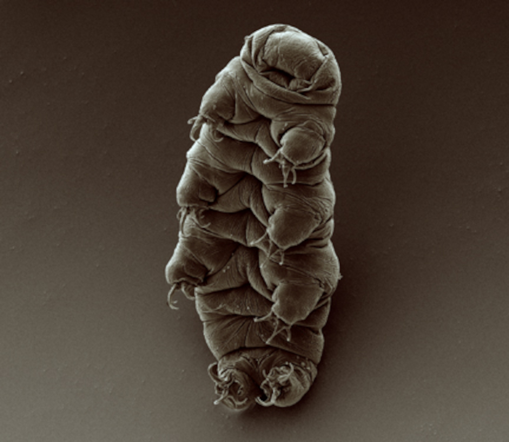
Scanning electron micrograph of an adult tardigrade. Source: Wikimedia Commons
University of Wyoming researchers have gained further insight into how tardigrades survive extreme conditions and shown that proteins from the microscopic creatures expressed in human cells can slow down molecular processes.
This makes the tardigrade proteins potential candidates in technologies centred on slowing the aging process and in long-term storage of human cells.
The new study, published in the journal Protein Science, examines the mechanisms used by tardigrades to enter and exit from suspended animation when faced by environmental stress.
Led by Senior Research Scientist Silvia Sanchez-Martinez in the lab of UW Department of Molecular Biology Assistant Professor Thomas Boothby, the research provides additional evidence that tardigrade proteins eventually could be used to make life-saving treatments available to people where refrigeration is not possible — and enhance storage of cell-based therapies, such as stem cells.
Measuring less than half a millimetre long, tardigrades can survive being completely dried out; being frozen to just above absolute zero; heated to more than 150°C; survive radiation of several thousand times a human’s lethal dose; and even survive the vacuum of outer space.
They survive by entering a state of suspended animation called biostasis, using proteins that form gels inside of cells and slow down life processes, according to the new UW-led research.
Co-authors of the study are from institutions including the University of Bristol in the United Kingdom, Washington University in St. Louis, the University of California-Merced, the University of Bologna in Italy and the University of Amsterdam in the Netherlands.
Sanchez-Martinez, who came from the Howard Hughes Medical Institute to join Boothby’s UW lab, was the lead author of the paper.
“Amazingly, when we introduce these proteins into human cells, they gel and slow down metabolism, just like in tardigrades,” Sanchez-Martinez says.
“Furthermore, just like tardigrades, when you put human cells that have these proteins into biostasis, they become more resistant to stresses, conferring some of the tardigrades’ abilities to the human cells.”
Importantly, the research shows that the whole process is reversible: “When the stress is relieved, the tardigrade gels dissolve, and the human cells return to their normal metabolism,” Boothby says.
“Our findings provide an avenue for pursuing technologies centred on the induction of biostasis in cells and even whole organisms to slow aging and enhance storage and stability,” the researchers concluded.
Previous research by Boothby’s team showed that natural and engineered versions of tardigrade proteins can be used to stabilize an important pharmaceutical used to treat people with hemophilia and other conditions without the need for refrigeration.
Tardigrades’ ability to survive being dried out has puzzled scientists, as the creatures do so in a manner that appears to differ from a number of other organisms with the ability to enter suspended animation.
Forming social bonds facilitates effective communication and neural synchronisation across individuals of different social status within a group
When small hierarchical groups bond, neural activity between leaders and followers aligns, promoting quicker and more frequent communication, according to a study published on March 19th in the open-access journal PLOS Biology by Jun Ni from Beijing Normal University, China, and colleagues.
Social groups are often organised hierarchically, where status differences and bonds between members shape the group’s dynamic. To better understand how bonding influences communication within hierarchical groups and which brain regions are involved in these processes, the researchers recorded 176 three-person groups of human participants (who had never met before) while they communicated with each other, sitting face-to-face in a triangle. Participants wore caps with fNIRS (functional near-infrared spectroscopy) electrodes to non-invasively measure brain activity while they communicated with their group members. Each group democratically selected a leader, so each group of three ultimately included one leader and two followers. After strategising together, groups played two economic games designed to test their willingness to make sacrifices to benefit their group (or harm other groups).
Experimenters assigned some triads to go through a bonding session, where they were grouped according to colour preferences, given uniforms, and led through an introductory chat session to build familiarity. Bonded groups spoke more freely and bounced between speakers more frequently and rapidly, relative to groups that didn’t experience this bonding session. This bonding effect was stronger between leaders and followers than between two followers. Neural activity in two brain regions linked to social interaction, the right dorsolateral prefrontal cortex (rDLPFC) and the right temporoparietal junction (rTPJ), aligned between leaders and followers if they had bonded. The authors state that this neural synchronisation suggests that leaders may be anticipating followers’ mental states during group decision-making, though they acknowledge that their findings are restricted to East Asian Chinese individuals communicating via text (without non-verbal cues), whose culture emphasises group cohesion and commitment towards group leaders.
The authors add, “Social bonding increases information exchange and prefrontal neural synchronisation selectively among individuals with different social statuses, providing a potential neurocognitive explanation for how social bonding facilitates the hierarchical structure of human groups.”
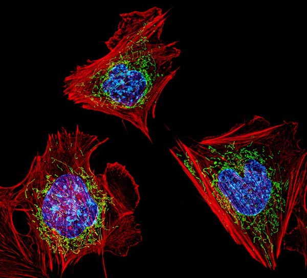
Cells with nuclei in blue, energy factories in green and the actin cytoskeleton in red. Credit: NIH
A new discovery, which was published in Nature Cell Biology, reveals how genetic material can escape mitochondria, prompting the body to launch a damaging immune response, setting off diseases such as lupus and rheumatoid arthritis. By developing therapies to target this process, doctors may one day be able to stop the harmful inflammation and prevent the toll it takes on our bodies.
“When mitochondria don’t correctly replicate their genetic material, they try to eliminate it. However, if this is happening too often and the cell can’t dispose of all of it, it can cause inflammation, and too much inflammation can lead to disease, including autoimmune and chronic diseases,” said researcher Laura E. Newman, PhD, of the University of Virginia School of Medicine. “Now that we are beginning to understand how this inflammation starts, we might be able to prevent this process, with the ultimate goal of limiting inflammation and treating disease.”
Powering inflammation
Mitochondria have their own set of genetic material, separate from the DNA that serves as the operating instructions for our cells. Scientists have known that this mitochondrial DNA, known as mtDNA, can escape into our cells and cause inflammation. But exactly what caused this has been a mystery until now.
“We knew that mtDNA was escaping mitochondria, but how was still unclear,” said Gerald Shadel, PhD, director of the San Diego-Nathan Shock Center of Excellence in the Basic Biology of Aging at the Salk Institute. “Using imaging and cell biology approaches, we’re able to trace the steps of the pathway for moving mtDNA out of the mitochondria, which we can now try to target with therapeutic interventions to hopefully prevent the resulting inflammation.”
Shadel and Newman, then a postdoctoral researcher in Shadel’s lab, and their collaborators used sophisticated imaging techniques to determine what was happening inside the leaky mitochondria. They found that the leak was triggered by a malfunction in mtDNA replication. This caused the accumulation of protein masses caused nucleoids.
To try to fix this problem, the cell containing the faulty mitochondrion begins to export the excess nucleoids to its cellular trash bins. But the trash bins, called endosomes, can become overwhelmed by the volume of debris, the scientists found. These overburdened endosomes respond by releasing mtDNA into the cell — in short, the trash can overflows.
“We had a huge breakthrough when we saw that mtDNA was inside of a mysterious membrane structure once it left mitochondria. After assembling all of the puzzle pieces, we realised that structure was an endosome,” Newman said. “That discovery eventually led us to the realisation that the mtDNA was being disposed of and, in the process, some of it was leaking out.”
The cell responds to this hazardous waste spill by flagging the nucleoids as foreign DNA, like a virus, and launches an immune response that results in harmful inflammation, the scientists determined.
“Using our cutting-edge imaging tools for probing mitochondria dynamics and mtDNA release, we have discovered an entirely novel release mechanism for mtDNA,” said researcher Uri Manor, PhD, former director of the Waitt Advanced Biophotonics Core at Salk and current assistant professor at UC San Diego. “There are so many follow-up questions we cannot wait to ask, like how other interactions between organelles control innate immune pathways, how different cell types release mtDNA, and how we can target this new pathway to reduce inflammation during disease and aging.”
Newman will continue to seek these answers in her new role at the UVA School of Medicine’s Department of Cell Biology. “We want to understand the physiological and disease contexts where this process can become activated,” she said. “For example, many viruses attack mitochondria during infection, so we will be testing whether mitochondria purposely use this pathway to sound the alarm against invading viruses, and whether over-reliance on this pathway to fight off infection can later trigger chronic diseases.”
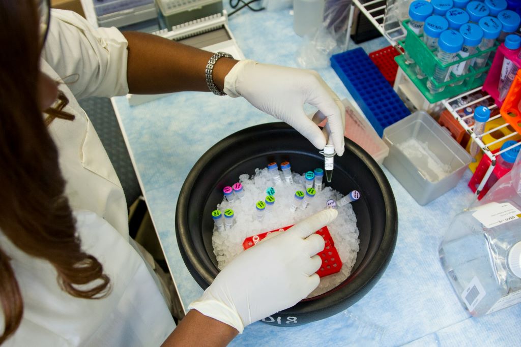
During the 2023 Christmas holidays, a freezer failure occurred at the Karolinska Institutet’s Neo building, where the automatic refilling of cryotanks with liquid nitrogen was interrupted for some reason. As a result, the temperature in 16 of 19 cryogenic tanks rose and large amounts of biological research material have been destroyed, including medical research samples which stretch back for decades.
An investigation with internal and external experts is now underway to find out how this failure could have happened, although there are no signs of sabotage. On the evening of 22 December, the level of liquid nitrogen in the Neo building’s cryo tanks, which contain biomaterial and cell lines from multiple departments, was due to be routinely topped up from an external tank.
The automatic refilling of nitrogen ensures that the correct temperature of -190°C can be maintained in the isothermal (cryo) tanks.
However, for reasons unknown, the flow of nitrogen from the external storage tank malfunctioned that evening, and the temperature in 16 of the tanks rose.
Automatic alarm
The cryotanks are able to maintain a sufficiently low temperature for up to 96 hours without refilling. During the Christmas break, they remained un-refilled for around 120 hours, and their internal temperature increased.
“When the flow of nitrogen ceased on 22 December, an automatic alert was supposed to be sent out, both by email and SMS, to registered owners of material in the freezers. However, a malfunction in the alarm unit meant that the alerts did not work properly. The email reached the recipients, but the texts got stuck in the server and never arrived,” explains Elisabeth Raschperger, researcher and senior lab manager at Neo.
According to Dr Raschperger, there has been a history of false alarms from freezers and cryotanks at Neo, partly caused by overly sensitive settings for when alarms should go off.
The alarm supplier inspected the system in 2023 and gave approval for its continued use in November.
Troubleshooting by the suppliers
Five days after the incident when the Neo service team found out what had happened, they called in the suppliers to make an initial check of the valves, pipes and pressure regulation tanks. The alarm was also tested.
“The companies went through every part of the system and found no faults or indications that any piece of equipment was faulty or broken, with the exception of the SMS alarm,” says Dr Raschperger. “We also looked through the operational logbook for the external nitrogen tank for October, November and December, and the refilling system had been working perfectly.”
The affected departments
From a research perspective, the Department of Medicine, Huddinge (MedH), was most affected, but so too were researchers at the Department of Biosciences and Nutrition (BioNut).
“At MedH, the research areas of haematology, endocrinology and cardiology have been particularly affected by the crash. It involves a very large amount of irreplaceable research material with samples, cell lines and biomaterials collected over decades,” says Professor Petter Höglund, head of MedH. He continues:
“The affected research teams are now working to take stock of the full extent of the losses. The analyses made so far speak for themselves: the malfunction will have far-reaching consequences for the department’s research in the affected areas.”
The staff at Neo, BioNut and MedH receive regular updates on the incident and the steps being taken to investigate the cause. They are also receiving crisis support from HR.
Expert inquiry
An inquiry to ensure that the incident never happens again is now underway. The inquiry will take a technical and procedural – rather than a personal – approach to chart and analyse the incident and look into how KI can build sustainable, robust systems going forward. Pending the inquiry’s report, it is important not to make a bad situation worse.
“Rumours are circulating that the malfunction was an act of sabotage,” says Dr Raschperger. “I would like to emphasise that there are at the present no such indications, and urge everyone to wait for the experts’ conclusions.”
Amongst the intricacies of South Africa’s healthcare landscape, a silent but significant challenge lurks – the prevalence of rare diseases. Behind the curtain of mainstream medical discourse, millions grapple with the complexities of these often overlooked conditions, a stark reality often overshadowed by the glare of more prevalent health concerns.
With more than 7000 identified rare diseases to date, they affect as many as 4.2 million South Africans, of which 50 – 70% are children1. These conditions are more prevalent than predicted, each posing unique and often debilitating challenges for patients and families alike.
With 29 February commemorated as Rare Diseases Day, Rare Diseases South Africa (RDSA), is hosting its third biennial rare diseases conference, Rare-X 2024, at the Indaba Hotel in Fourways, from 14 to 17 February.
More than just a conference, Rare-X 2024 will focus on patient advocacy, education, policy reform, and improving equitable access to ensure better outcomes and support for individuals living with rare diseases.
As the first in-person conference since the COVID-19 pandemic, the event brings together patients, policymakers, academics, government and pharmaceutical companies to discuss the plight of rare diseases and find collaborative ways to improve patients’ lives and treatment efforts.
The conference will comprise several activities, including keynote speeches by renowned experts in rare diseases; interactive panel discussions; workshops and training sessions; scientific presentations; networking opportunities and policy roundtables.
Some of the renowned speakers to share their insights and global developments on rare diseases include Prof Alex van den Heever, Chair of Social Security Systems Administration and Management Studies at the Wits School of Governance; Professor Fatima Suleman, Professor in the School of Health Sciences at the University of KwaZulu-Natal; and Professor Chris Hendriksz, Global Clinical Development Lead for Rare Diseases at Nestle Health Science, amongst others. Bringing a wealth of practical experience following his work with health professionals, will be traditional health practitioners (THPs), Mr Elliot Makhathini and Dr Conradie from North-West University’s Centre for Human Metabolomics, to name a few.
A rare disease relates to a condition that is considered rare when it affects one person in 20002. Currently, South Africa does not have its own definition of a rare disease, which is one of the major issues that need to be addressed by the government3.
As a patient-focused non-profit organisation, RDSA was launched in 2013 by CEO and Rare-X Director, Kelly du Plessis. The mother of a child with a rare condition, du Plessis realised the dire need for support for a highly under-acknowledged community, with the organisation advocating that people living with rare diseases and congenital disorders experience greater recognition, support, improved health services, and overall, a better quality of life.
“Despite the need for increased representation, the rare diseases community remains vulnerable from a medical and policy perspective,” says du Plessis. “As part of our mandate, RDSA brings together international best practice and local medical innovation, driving a collective voice and playing a fundamental role in bridging the gap between vulnerable communities and medical advancement.”
To date, RDSA has successfully launched initiatives that have positively impacted the lives of over 6500 patients including engaging with various governmental departments, organs of state, industry players and strategic stakeholders to raise awareness and move rare disease policy forward.
For more information on the Rare-X conference, kindly visit www.rare-x.co.za
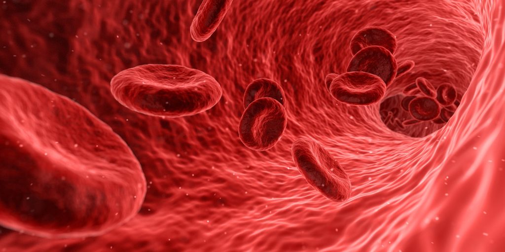
Research conducted by a team of scientists from Kaunas universities, Lithuania, revealed that low-frequency ultrasound influences blood parameters. The findings suggest that ultrasound’s effect on haemoglobin can improve oxygen’s transfer from the lungs to bodily tissues.
The research was undertaken on 300 blood samples collected from 42 pulmonary patients.
The samples were exposed to six different low-frequency ultrasound modes at the Institute of Mechatronics of Kaunas University of Technology (KTU). The calculations were made at the KTU Artificial Intelligence Centre.
Improved oxygen circulation and reduced blood pressure
KTU professors Vytautas Ostasevicius and Vytautas Jurenas say that the ongoing research papers are related to blood platelet aggregation.
The research of the KTU team revealed that the ultrasound affects not only platelet count but also red blood cells (RBC), which can result in better oxygen circulation and lowered blood pressure.
“During exposure to low-frequency ultrasound, aggregated RBCs are dissociated into single RBCs, whose haemoglobin molecules interact with oxygen over the entire surface area of RBCs, which is larger than that of aggregated RBCs and improves oxygen saturation in blood. The number of dissociated single RBCs per unit volume of blood decreases due to the spaces between them, compared to aggregates, which reduces blood viscosity and affects blood pressure,” explains Prof Ostasevicius, the Head of KTU Institute of Mechatronics.
The scientists claim that the effect of ultrasound on the haemoglobin in RBCs was higher than its impact on platelet aggregation, which is responsible for blood clotting.
Their findings have been supported by an additional analysis made at the LSMU Laboratory of Molecular Cardiology.
“This means that low-frequency ultrasound can be potentially used for improving oxygen saturation in lungs for pulmonary hypertension patients. Keeping in mind the recent COVID-19 pandemic, we see a huge potential in exploring the possibilities of our technology further,” says Prof Ostasevicius.
Partnership between medical and engineering scientists
In medicine, high-frequency ultrasound from 2 to 12MHz is used for both diagnostic and therapeutic purposes.
“Acoustic waves emitted by high-frequency ultrasound have a limited penetration depth into the body, so external tissues are more affected by high-frequency ultrasound than internal organs. Low-frequency ultrasound acoustic waves, penetrate deeper into the internal organs with a more uniform sound pressure distribution,” explains Prof Jurenas.
There are numerous applications for ultrasound in medical settings.
“For example, focused ultrasonic waves are used to break kidney stones, and to kill cancer cells. Maybe ultrasound can be used to activate certain medications. Or, to alleviate the delivery of antibiotics to the inflamed areas?” says Prof Jurenas.
The technology used in the above-described study is only one illustration of many successful working partnerships between engineers and physicians.
For example, just recently, the researchers of KTU Institute of Mechatronics have created the frame for immobilising the Gamma Knife radiosurgery patients at the Clinics of the Lithuanian University of Health Sciences.
“We believe, that using the know-how of different areas one can achieve greater results,” say KTU researchers about interinstitutional and interdisciplinary cooperation.
Freezing is one of the most common and debilitating symptoms of Parkinson’s disease, when they suddenly lose the ability to move their feet, often mid-stride, resulting in a series of staccato stutter steps that get shorter until the person stops altogether. These episodes are one of the biggest contributors to falls among people living with Parkinson’s disease.
Today, freezing is treated with a range of pharmacological, surgical or behavioural therapies, none of which are particularly effective. What if there was a way to stop freezing altogether?
In a Nature Medicine report, researchers used a soft, wearable robot to help a person living with Parkinson’s walk without freezing. The robotic garment, worn around the hips and thighs, gives a gentle push to the hips as the leg swings, helping the patient achieve a longer stride. The device completely eliminated the participant’s freezing while walking indoors, allowing them to walk faster and further.
“We found that just a small amount of mechanical assistance from our soft robotic apparel delivered instantaneous effects and consistently improved walking across a range of conditions for the individual in our study,” said Conor Walsh, professor at SEAS and co-corresponding author of the study.
For over a decade, Walsh’s Biodesign Lab at SEAS has been developing assistive and rehabilitative robotic technologies to improve mobility for individuals’ post-stroke and those living with ALS or other diseases that impact mobility. Some of that technology, specifically an exosuit for post-stroke gait retraining, received support to develop and commercialise the technology.
“Leveraging soft wearable robots to prevent freezing of gait in patients with Parkinson’s required a collaboration between engineers, rehabilitation scientists, physical therapists, biomechanists and apparel designers,” said Walsh, whose team collaborated closely with that of Terry Ellis, Professor and Physical Therapy Department Chair and Director of the Center for Neurorehabilitation at Boston University.
The team spent six months working with a 73-year-old man with Parkinson’s disease, who, despite using both surgical and pharmacologic treatments, endured substantial and incapacitating freezing episodes more than 10 times a day, causing him to fall frequently. These episodes prevented him from walking around his community and forced him to rely on a scooter to get around outside.
In previous research, Walsh and his team leveraged human-in-the-loop optimization to demonstrate that a soft, wearable device could be used to augment hip flexion and assist in swinging the leg forward to provide an efficient approach to reduce energy expenditure during walking in healthy individuals.
Here, the researchers used the same approach but to address freezing. The wearable device uses cable-driven actuators and sensors worn around the waist and thighs. Using motion data collected by the sensors, algorithms estimate the phase of the gait and generate assistive forces in tandem with muscle movement.
The effect was instantaneous. Without any special training, the patient was able to walk without any freezing indoors and with only occasional episodes outdoors. He was also able to walk and talk without freezing, a rarity without the device.
“Our team was really excited to see the impact of the technology on the participant’s walking,” said Jinsoo Kim, former PhD student at SEAS and co-lead author on the study.
During the study visits, the participant told researchers: “The suit helps me take longer steps and when it is not active, I notice I drag my feet much more. It has really helped me, and I feel it is a positive step forward. It could help me to walk longer and maintain the quality of my life.”
“Our study participants who volunteer their time are real partners,” said Walsh. “Because mobility is difficult, it was a real challenge for this individual to even come into the lab, but we benefited so much from his perspective and feedback.”
The device could also be used to better understand the mechanisms of gait freezing, which is poorly understood.
“Because we don’t really understand freezing, we don’t really know why this approach works so well,” said Ellis. “But this work suggests the potential benefits of a ‘bottom-up’ rather than ‘top-down’ solution to treating gait freezing. We see that restoring almost-normal biomechanics alters the peripheral dynamics of gait and may influence the central processing of gait control.”
Of more than four hundred phase 2 and 3 randomised trials of cancer drugs registered in China between 2016 and 2017, about sixty had suboptimal control arms
More than one-eighth of the randomised trials of cancer drugs seeking regulatory approval in China in recent years used inappropriate controls to test the effectiveness and safety of the drugs, according to a new study published December 12th in the open access journal PLOS Medicineby Professor Xiaodong Guan of Peking University, China, and colleagues.
In randomised trials, patients are assigned to either a control arm, in which they receive the current optimal treatment, or an experimental arm, in which they receive the new drug being tested. However, studies have previously found that control arms in cancer clinical trials (including in the United States) are not supported by relevant guidelines, instead using treatments other than the standard-of-care. Adopting a suboptimal control group may bias a study’s results in favour of the experimental arm, potentially exposing patients to substandard therapy and producing unreliable results of clinical efficacy.
In the new study, researchers analysed the control arms of 453 Phase II/III and Phase III randomised oncology trials authorised by Chinese institutional review boards between 2016 and 2021, supporting investigational new drug applications of these drugs in China.
Overall, 60 trials (13.2%) used suboptimal control arms. Of those suboptimal trials, 35 (58.3%) used comparators that were not recommended by a prior guideline. In total, 18 610 people enrolled in clinical trials (15.1% of the total number in all samples trials) were exposed to suboptimal treatments due to the control arms. Trials using suboptimal controls were more likely to report a positive result for the experimental arm. In addition, the researchers found an overall upward trend in the number of trials using inappropriate control arms.
“Trial sponsors, ethical review boards, and oncologists should make collaborative efforts to protect patients from unnecessary harm and drugs with uncertain clinical benefits over the existing standard of care,” the authors say. “Regulatory agencies should be cautious when reviewing investigational new drug applications whose supporting trial used a suboptimal control.”
The authors add, “This research highlights the necessity to refine the design of randomised trials to generate optimal clinical evidence for new cancer therapies. In November 2021, China issued the Guidance on Clinical Value-Oriented Oncology Drug Research and Development, aiming to promote a better generation of clinically relevant novel oncology drugs in China. We hope our research findings can provide empirical evidence to the stakeholders and draw regulators’ attention to this matter so that the guideline can be delivered in the manner that it set out to be.”