View of the spinal cord. Credit: Scientific Animations CC4.0
In a recent study published in Nature, researchers prevented T cells from causing the normal autoimmune damage that comes with spinal cord injury, sparing neurons and successfully aiding recovery in mouse models.
In spinal cord injury, the wound site attracts a whole host of peripheral immune cells, including T cells, which result in both beneficial and deleterious effects. Notably, antigen-presenting cells activate CD4+ T cells to release cytokines, ultimately leading to neuroinflammation and tissue destruction. This neuroinflammation is notably most pronounced during the acute phase of spinal cord injury. The problem is that these same T cells have a neuroprotective effect initially, only later developing autoimmunity and attacking the injury site.
Using single cell RNA sequencing, the researchers found that CD4+ T cell clones in mice showed antigen specificity towards self-peptides of myelin and neuronal proteins. Self-peptides have been implicated in a wide range of autoimmune conditions.
Using mRNA techniques, the researchers edited the T cell receptor, so that they shut off after a few days. In mouse models of spinal cord injury, they showed notable neuroprotective efficacy, partly as a result of modulating myeloid cells via interferon-γ.
Their findings provided insights into the mechanisms behind the neuroprotective function of injury-responsive T cells. This will help pave the way for the future development of T cell therapies for central nervous system injuries, and perhaps treatments for neurodegenerative diseases such as Alzheimer’s.
Spinal cord injury survivor is a capable and helpful big brother
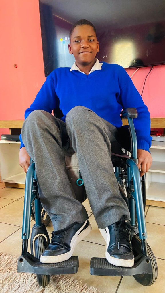
Kamogelo Sodi, who was injured in a car crash when he was just six years old, says he learned valuable skills on how to regain his independence at the Netcare Rehabilitation Hospital. The teenager enjoys cooking for himself, taking care of his three younger brothers, and playing basketball when he’s not studying hard to achieve his dream of being a medical practitioner one day.
5 September 2024: At 14 years old, Kamogelo Sodi of Alberton enjoys listening to music, chatting with his friends on social media and working hard at school towards his dream of becoming a neurosurgeon one day. He cooks for himself when he’s hungry and loves looking after his three little brothers. He also likes playing basketball. The difference between him and most other teenagers is that he does all this from his wheelchair.
“Since I’ve been in a wheelchair, I’ve become more confident,” says the vivacious teenager. “I was extremely shy, and I didn’t have a lot of friends, but now I have loads of friends.”
In 2016, when he was just six years old, Kamogelo’s life changed forever. He was in a devastating car crash, which left him with fractures in the lumbar region of his spine, resulting in complete paraplegia.
Once discharged from the hospital, where he had emergency surgery, Kamogelo was sent to the Netcare Rehabilitation Hospital to learn how to cope with, as his mother Reshoketswe Sodi calls it, his new normal. He was to stay there for almost six months.
Mrs Sodi, a radiation therapist, says the enduring care of the doctors, occupational therapists and physiotherapists there helped support Kamogelo and their family on their journey towards accepting and learning to cope with this difficult transition in his life. “It was important for me that he continued his schoolwork while there. When the social worker asked me what I wanted to happen, the first thing I said was that I didn’t want to break the routine of what he had been doing and that I wanted him to continue with school.
“It’s been a struggle, but with the help of the occupational therapists and physiotherapists, it has been an easier journey. We saw real progress when they taught Kamogelo something, and he grasped it, putting all his energy into it by thinking positively about it. It’s been hard, but with the support of the team from Netcare Rehabilitation Hospital, we managed it,” she says.
“After he was discharged, initially, we lived in a flat on the seventh floor. When the lifts weren’t working, like during load shedding, I’d have to carry him upstairs on my back – there was no other way to take him up. I’m so fortunate that I had a lot of support from my family and friends who’ve been pillars of strength for us.”
Kamogelo remembers his first visit to the Netcare Rehabilitation Hospital in Auckland Park. “When I first got to the hospital, I was lost. I didn’t know how to use a wheelchair. I was still so young. But they were so kind and taught me everything I needed to know.
“At first, I struggled to move around. I battled to transfer myself from place to place, but they showed me what to do, and over time, I started getting used to it. I managed to start moving myself around, and I began to enjoy it. From that day forward, I didn’t like people pushing me around. The staff also taught me how to transfer myself from my wheelchair to the car. It was a bit difficult at first, but I learned to push myself up properly so my bottom wouldn’t scrape on the wheelchair.
“It does help you become more independent, but you must be consistent. You don’t need to complain about things, you just need to listen to the people who want to help you learn to be independent.”
Later, in 2022, when he was 12 years old, Kamogelo returned to the Netcare Rehabilitation Hospital after he developed a severe pressure sore.
Dr Anrie Carstens, a doctor at the Netcare Rehabilitation Hospital, said Kamogelo was operated on at Netcare Milpark Hospital under the care of a plastic surgeon who did a flap to close the wound. “When the doctor was happy with his progress, Kamogelo came to us to help him because you get weak after surgery. The wound had healed, but the skin was delicate, so we had a graded seating approach for him to build up his strength and so that the areas of the skin didn’t break down. Another area of focus for Kamogelo was spasticity at the ankles. We worked on relaxing the ankles to get to a ninety-degree angle so he could sit better in his chair with his feet positioned well in the footrest.”
When homesickness inevitably struck, the staff comforted Kamogelo. “I began to miss home, and I cried and said I wanted to go home. They spoke nicely to me and said they first had to help me so I could go back home with no problems so my parents wouldn’t have to worry about me because of the pressure sore.”
Kamogelo said the staff also taught him valuable techniques to help him empty his bladder and bowels and assisted him in his journey to independence. “I was worried it would be painful and was a bit hesitant to try them out. But, doing it daily helped my routine and helped me become independent.”
Charne Cox, a physiotherapist at Netcare Rehabilitation Hospital, describes Kamogelo as bubbly, intelligent and with lovely manners. “He’s so motivated and tried so hard in therapy. He manages to go to school each day, not because of us, but because of his character.”
She says as children grow, their needs change. “The pressure sore developed because his seating in his wheelchair was not adequate because he had grown so much. We collaborated with the wheelchair manufacturer to re-evaluate and reassess the wheelchair seating, and they made him a new wheelchair. He was getting heavier, and his feet weren’t in alignment, so it was trickier for him to safely transfer from the wheelchair to the bed, for instance. It was good to re-educate him on pressure relief and pressure sores. It’s vital that adolescents are taught to take responsibility for themselves.”
Cox also helped Kamogelo work towards getting his feet in a better position.
“Children are so good about learning to use a wheelchair. Kamogelo was so motivated to move and be independent. He absorbed the information we gave him to enable him to go up ramps, turn and even do wheelies because he liked to explore.
“Children want to learn and have fun. They want to be independent. It’s amazing to help give them the tools to be the best new person they can be. Unfortunately, sometimes we can’t fix the injury, but we can give them the best opportunity to be as independent as possible. It’s so satisfying to know that Kamogelo is going to school and playing basketball.”
Kamogelo is determined to pursue a career as a neurosurgeon. “As long as I follow the path that I want to do and enjoy it, I will continue pursuing that path. Academically, I was the top achiever from grade four to grade six at my school.”
When he’s not at school, he loves going around the estate he lives in, getting fresh air, and being a good big brother to his three younger brothers. “They’re a handful, but what can I say – they’re my brothers, and I love them,” he says with a laugh.
Asked who his hero is, Kamogelo is quick to say his mother and father are both his heroes. His mom clearly thinks he’s a hero too. She’s smiling as she speaks about her son. “He’s playful and has a great sense of humour. He’s helpful in the house. Instead of wanting us to help him, thanks to the skills he learned at Netcare Rehabilitation Hospital, Kamogelo always says, ‘Let me give you a hand. Let me help you.’”
5 September 2024, International Spinal Cord Injury Day is commemorated on Thursday 5 September, drawing attention to the many ways people can be affected by spinal cord injury, creating awareness of prevention, and highlighting the possibilities for a fulfilling life after injury.
According to the World Health Organization, globally, over 15 million people are living with spinal cord injuries. Most of these cases are due to trauma, including falls, road traffic injuries or violence.
Jessica Morris, an occupational therapist at the Netcare Rehabilitation Hospital in Auckland Park, says one of the most critical aspects of care for those who’ve been impacted by spinal cord injuries is the importance of successful rehabilitation through a holistic, integrated approach from a multidisciplinary team.
“Many people just think it’s just about mobility. It’s so much more than that. Rehabilitation is complex because many different areas of our patients’ lives are affected.” Morris says they are fortunate that their team has so many different practitioners who can contribute to treating spinal cord injury patients, helping them regain a level of independence, which is vital to their self-confidence and sense of empowerment.
Dr Anrie Carstens, a general practitioner with a particular interest in physical medicine and rehabilitation who practises at the Netcare Rehabilitation Hospital, says the message of Spinal Cord Injury Awareness Day has relevance all year round, as people with spinal cord injuries need to be incorporated into society.
“It’s an opportunity to tell people not to be nervous to talk to someone in a wheelchair. They’re just like you or me, and they just have special ways of moving around and managing their pain and different aspects of their bodies. With the help of proper rehabilitation, the person can be better integrated as a functional, contributing member of society.”
Dr Carstens says people should also be aware that if they or their loved ones are ever impacted by a spinal cord injury, professional support is available. “Don’t just go straight home after your hospital stay and try to do everything on your own. Instead, come to a specialised spinal cord injury unit like ours, with therapists, doctors and nursing staff who are well versed in spinal cord injury and know the finer nuances necessary to optimally treat the person and show them how best to cope with their injury.
“In the multidisciplinary approach, every practitioner has a role in getting the person back into the real world, whether it means going back home, back to school, back to work or wherever they were before their injury occurred.”
From doctors and nurses with specialised skills to physiotherapists, occupational therapists, social workers and psychologists, speech therapists, a prosthetist and dieticians, the team provides a broad person focussed rehabilitation service to both adults and children. Their aim is to optimise their patients’ independence level using specialised equipment and teaching specific techniques to help overcome the obstacles a person may face.
Dr Carstens says it’s rewarding work for the staff at the hospital, who build up enduring relationships with those they care for. “One of the highlights is to compare and see what the patient was like when you admitted them and then see on discharge how much they’ve grown, how they’ve gained confidence and become more independent. What’s even better is to see them after they’ve been discharged and observe how well they’ve coped and how they’ve integrated and adjusted to their environment. We build a relationship with our patients because they stay with us for quite a while, and we usually have checkups every year after the person is discharged, often for life. We get to see them grow and thrive outside the healthcare setting, and we need more awareness about how much it is possible for people with spinal cord injuries to achieve.”
Coup and contrecoup brain injury. Credit: Scientific Animations CC4.0
Researchers at Johns Hopkins explored a potential alternative and less-invasive approach to evaluate intracranial pressure (ICP) in patients with serious neurological conditions. This research, using artificial intelligence (AI) to analyse routinely captured ICU data, was published in Computers in Biology and Medicine.
ICP is a physiological variable that can increase abnormally if one has severe traumatic brain injury, stroke or obstruction to the flow of cerebrospinal fluid. Symptoms of elevated ICP may include headaches, blurred vision, vomiting, changes in behaviour and decreased level of consciousness. It can be life-threatening, hence the need for ICP monitoring in selected patients who are at increased risk. But the current standard for ICP monitoring is highly invasive: it requires the placement of an external ventricular drain (EVD) or an intraparenchymal brain monitor (IPM) in the functional tissue in the brain consisting of neurons and glial cells by drilling through the skull.
“ICP is universally accepted as a critical vital sign – there is an imperative need to measure and treat ICP in patients with serious neurological disorders, yet the current standard for ICP measurement is invasive, risky, and resource-intensive. Here we explored a novel approach leveraging Artificial Intelligence which we believed could represent a viable noninvasive alternative ICP assessment method,” says senior author Robert Stevens, MD, MBA, associate professor of anaesthesiology and critical care medicine.
EVD procedures carry a number of risks including catheter misplacement, infection, and haemorrhaging at 15.3 %, 5.8 %, and 12.1 %, respectively, according to recent research. EVD and IPM procedures also require surgical expertise and specialised equipment that is not consistently available in many settings thus underscoring the need for an alternative method in examining and monitoring ICP in patients.
The Johns Hopkins team, a group that included faculty and students from the School of Medicine and Whiting School of Engineering, hypothesised that severe forms of brain injury, and elevations in ICP in particular, are associated with pathological changes in systemic cardiocirculatory function due, for example, to dysregulation of the central autonomic nervous system. This hypothesis suggests that extracranial physiological waveforms can be studied to better understand brain activity and ICP severity.
In this study, the Johns Hopkins team set out to explore the relationship between the ICP waveform and the three physiological waveforms that are routinely captured in the ICU: invasive arterial blood pressure (ABP), photoplethysmography (PPG) and electrocardiography (ECG). ABP, PPG and ECG data were used to train deep learning algorithms, resulting in a level of accuracy in determining ICP that rivals or exceeds other methodologies.
Overall study findings suggest a completely new, noninvasive alternative to monitor ICP in patients.
Stevens says, “with validation, physiology-based AI solutions, such as the one used here, could significantly expand the proportion of patients and health care settings in which ICP monitoring and management can be delivered.”
Fibrotic scar 14d after spinal cord injury, red – Col1a1+ perivascular fibroblast derived cells Photo: Daniel Holl
New research has found that scar formation after spinal cord injuries is more complex than previously thought. Scientists at Karolinska Institutet have identified two types of perivascular cells as key contributors to scar tissue, which hinders nerve regeneration and functional recovery. These findings, published in Natural Neuroscience, are also relevant for other brain and spinal cord injuries and could lead to targeted therapies for reducing scarring and improving outcomes.
The central nervous system (CNS) has very limited healing abilities. Injuries or autoimmune diseases like multiple sclerosis often lead to permanent functional deficits.
Regardless of the injury’s cause, the body responds by forming a boundary around the damaged tissue, which eventually becomes permanent scar tissue.
Two contributing cell types
While scar tissue seals the damaged area, it also prevents functional repair. After spinal cord injuries, scar tissue blocks the regeneration of nerve fibers that connect the brain with the body, resulting in paralysis after severe injuries.
The research team led by Christian Göritz at Karolinska Institutet has made significant progress in understanding how scar tissue forms in the CNS. The group now identified two distinct types of perivascular cells, which line different parts of blood vessels, as the major contributors to fibrotic scar tissue after spinal cord injury. Depending on the lesion’s location, the two identified cell types contribute differently.
“We found that damage to the spinal cord activates perivascular cells close to the damaged area and induces the generation of myofibroblasts, which consequently form persistent scar tissue,” explains first author Daniel Holl, researcher at the Department of Cell and Molecular Biology.
By examining the process of scar formation in detail, the researchers hope to identify specific therapeutic targets to control fibrotic scarring.
Zebrafish have a remarkable ability to heal their spinal cord after injury. Now, researchers at Karolinska Institutet have uncovered an important mechanism behind this phenomenon – a finding that could have implications for the treatment of spinal cord injury in humans.
In a new study published in Nature Communications,researchers show that the neurons of adult zebrafish immediately start to cooperate after a spinal cord injury, keeping the cells alive and stimulating the healing process.
“We have shown that the neurons form small channels called gap junctions, which create a direct connection between the neurons and enable the exchange of important biochemical molecules, allowing the cells to communicate and protect each other,” explains Konstantinos Ampatzis, a researcher in the Department of Neuroscience at Karolinska Institutet, who led the study.
The researchers will further investigate the exact mechanisms behind this protective strategy in zebrafish and hope this knowledge will lead to new ways of treating spinal cord injury in humans.
“Spinal cord injuries are a major burden for sufferers and their families,” says Konstantinos Ampatzis. “What if we could get human neurons to adopt the same survival strategy and behave like zebrafish neurons after an injury? This could be the key to developing new effective treatments.”
People who suffer from headaches after experiencing concussions may also be more likely to have higher levels of iron in areas of the brain – a sign of injury to brain cells, according to a preliminary study presented at the American Academy of Neurology’s 76th Annual Meeting.
“These results suggest that iron accumulation in the brain can be used as a biomarker for concussion and post-traumatic headache, which could potentially help us understand the underlying processes that occur with these conditions,” said study author Simona Nikolova, PhD, of the Mayo Clinic in Phoenix, Arizona, and a member of the American Academy of Neurology.
The study involved 120 participants, 60 of whom who had post-traumatic headache (PTH) due to mild traumatic brain injury (mTBI), and 60 healthy controls. The injuries were due to a fall for 45% of the people, 30% were due to a motor vehicle accident and 12% were due to a fight. Other causes were the head hitting against or by an object and sports injuries. A total of 46% of the people had one mild traumatic brain injury in their lifetime, 17% had two, 16% had three, 5% had four and 16% had five or more mild traumatic brain injuries.
Participants underwent 3T brain magnetic resonance imaging (T2* maps). T2* differences were determined using age-matched paired t-tests. For the PTH group, scans were done an average of 25 days after injury. T2* correlations with headache frequency, number of lifetime mTBIs, time since most recent mTBI, and Sport Concussion Assessment Tool (SCAT) severity scale scores,
The researchers observed lower T2* values in PTH participants relative to HC in the right supramarginal area, left occipital, bilateral precuneus, right cuneus, right cerebellum, right temporal, bilateral caudate, genu of the corpus callosum, right anterior cingulate cortex and right rolandic operculum (p < 0.001).
Within PTH subjects, there were positive correlations with iron accumulation between lifetime mTBIs, the time since most recent mTBI and headache frequency in certain areas of the brain. For example, T2* levels in headache frequency with T2* in the posterior corona radiata, bilateral temporal, right frontal, bilateral supplemental motor area, left fusiform, right hippocampus, sagittal striatum, and left cerebellum were associated with headache frequency.
“Previous studies have shown that iron accumulation can affect how areas of the brain interact with each other,” Nikolova said. “This research may help us better understand how the brain responds and recovers from concussion.”
Nikolova said that using the indirect measure of iron burden also means that the change in that measure could be due to other factors such as haemorrhage or changes in tissue water rather than iron accumulation.
The Achilles tendon, although considered the toughest in the body, can rupture, with many such injuries involving sports enthusiasts in their 30s or 40s. Surgery might be required, and a prolonged period of rest, immobilisation, and treatment can be difficult to endure. Researchers in Japan have developed an approach using irradiation with plasma to accelerate healing.
A research team led by Osaka Metropolitan University Graduate School of Medicine’s Katsumasa Nakazawa, a graduate student in the Department of Orthopedic Surgery, Associate Professor Hiromitsu Toyoda, and Professor Hiroaki Nakamura, and Graduate School of Engineering Professor Jun-Seok Oh has focused on non-thermal atmospheric-pressure plasma (the electrically-charged gas such as found in a neon lamp – not blood plasma!) as a treatment method for tendon repair.
Their study, published in PLOS ONE, is the first to show that such plasma irradiation can accelerate tendon repair.
The team ruptured then sutured the Achilles tendon of lab rats. For one group of rats, the sutured area was irradiated with a helium plasma jet.
The plasma-irradiated group exhibited faster tendon regeneration and increased strength at two, four, and six weeks after surgery compared to the untreated group.
“We have previously discovered that irradiation of non-thermal atmospheric-pressure plasma has the effect of promoting bone regeneration. In this study, we discovered that the technology also promotes tendon regeneration and healing, showing that it has applications for a wide range of fields,” Professor Toyoda declared. “Combined with current tendon treatments, it is expected to contribute to more reliable tendon regeneration and shorter treatment time.”
New research indicates that various features assessed through imaging tests can reveal an individual’s risk of developing meniscus tears, which is one of the most common knee injuries.
The study, which is published in the Journal of Orthopaedic Research, was based on the use of radiomics, which unveils imperceptible patterns in medical images. Investigators used magnetic resonance images from 215 people with intact menisci at the start of the study who had 4-year meniscal status data.
Over 4 years, 34 participants developed meniscus tears. Use of radiomics at the start of the study correctly classified 24 of these 34 cases and 172 of 181 controls with a sensitivity of 70.6% and a specificity of 95.0%. Therefore, the technique provides sensitive and quantitative measures of meniscus alterations that could help clinicians know when to intervene to safeguard against meniscus tears.
“Understanding meniscus tear risk through radiomics opens new possibilities for proactive knee health management, offering clinicians a valuable tool to anticipate and prevent such injuries,” said corresponding author Matthew Harkey, PhD, ATC, of Michigan State University.
Coup and contrecoup brain injury. Credit: Scientific Animations CC4.0
Important brain structures that are key for signalling in the brain are narrower and less dense in females, and more likely to be damaged by brain injuries, such as concussion. Long-term cognitive deficits occur when the signals between brain structures weaken due to the injury. These structural differences in male and female brains might explain why females are more prone to concussions and experience longer recovery from the injury than their male counterparts, according to a University of Pennsylvania-led preclinical study published in Acta Neuropathologica.
Each year, approximately 50 million individuals worldwide suffer a concussion, also referred to as mild traumatic brain injury (TBI). For more than 15% of individuals who suffer persisting cognitive dysfunction, which includes difficulty concentrating, learning and remembering new information, and making decisions.
Although males make up the majority of emergency department visits for concussion, this has been primarily attributed to their greater exposure to activities with a risk of head impacts compared to females. In contrast, it has recently been observed that female athletes have a higher rate of concussion and appear to have worse outcomes than their male counterparts participating in the same sport.
“Clinicians have observed for a long time that females suffer from concussion at higher rates than males in the same sports, and that they take longer to recover cognitive function, but couldn’t explain the underlying mechanisms of this phenomenon,” said senior author Douglas Smith, MD, a professor of Neurosurgery and director of Penn’s Center for Brain Injury and Repair. “The variances in brain structures of females and males not only illuminate why this disparity exists, but also exposes biomarkers, such as axon protein fragments, that can be measured in the blood to determine injury severity, monitor recovery, and eventually help identify and develop treatments that help patients repair these damaged structures and restore cognitive function.”
Axons connect neurons, allowing communication across the brain. These axons form bundles that make up white matter in the brain and play a large role in learning and communication between different brain regions. Axons are delicate structures and are vulnerable to damage from concussion.
Communication between axons in the brain is powered by sodium channels that serve as the brain’s electric grid. When axons are damaged, these sodium channels are also impaired, which causes loss of signaling in the brain. The loss of signaling causes the cognitive impairment experienced by individuals after concussion.
In this study, researchers used large animal models of concussion to identify differences in brains of males and females after a concussion. They found that females had a higher population of smaller axons, which researchers demonstrated are more vulnerable to injury. They also reported that in these models, females had greater loss of sodium channels after concussion.
“The differences in brain structure not only tell us a lot about how brain injury affects males and females differently but could offer insights in other brain conditions that impact axons, like Alzheimer’s and Parkinson’s disease,” said Smith. “If female brains are more vulnerable to damage from concussion, they might also be more vulnerable to neurodegeneration, and it’s worth further research to understand how sex influences the structure and functions of the brain.”