Category: Injury & Trauma

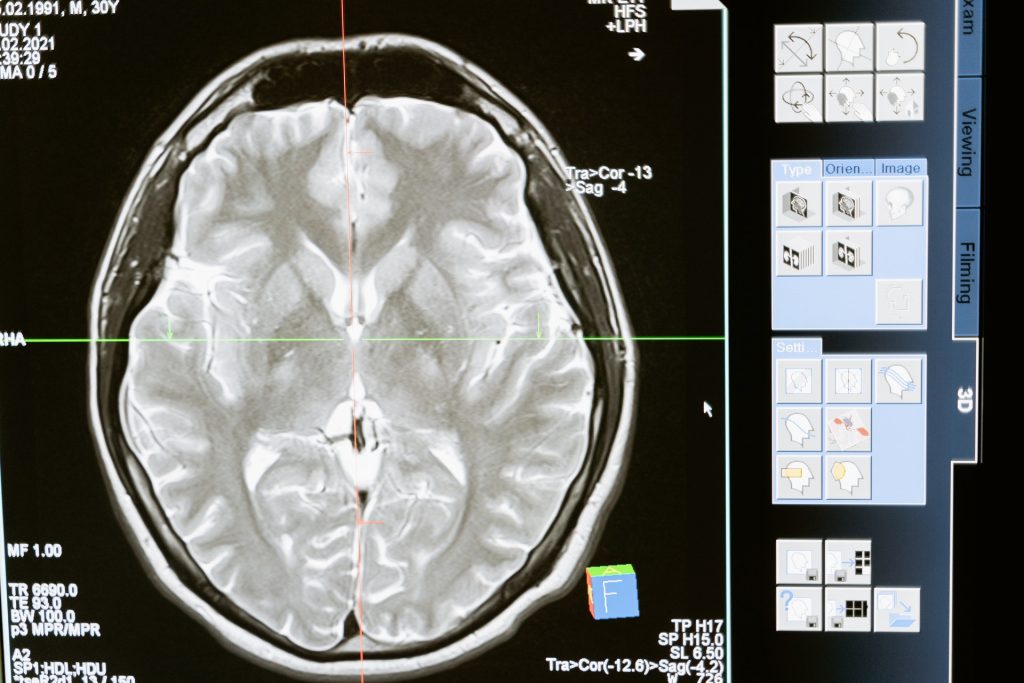
CT Scans Improve Outcomes for Concussion Patients

Photo by MART PRODUCTION from Pexels

A study found that CT scans for concussion patients provide crucial information on their risk for long-term impairment and their potential to make a complete recovery, and points to the need for more follow-up.

In the UC San Francisco-led study, researchers examined CT scans of 1935 patients, aged 17 and over, whose neurological exams met criteria for concussion, or mild traumatic brain injury (TBI). Outcomes for moderate and severe TBI have been linked to CT imaging features, but this may be the first time this link has been identified in patients with concussion. This contradicts previous research which had found no prognostic significance of specific types of CT abnormalities.

“Radiologists who routinely read trauma scans know intuitively that patterns of intracranial injury on CT are not random,” said first author Esther Yuh, MD, PhD, of the UCSF Department of Radiology and Biomedical Imaging. “We showed there are patterns of injury, that some of these are associated with worse outcome than others, and that they provide a window into mechanisms of injury that is reproducible across large studies.”

The study was published online in JAMA Neurology.

“Although concussions are referred to as mild traumatic brain injuries, there is nothing mild about some concussions,” explained senior author Geoffrey Manley, MD, PhD, professor and vice chair of neurological surgery at UCSF and chief of neurosurgery at Zuckerberg San Francisco General Hospital. “Patients with concussion may suffer from prolonged headache, poor sleep and impaired concentration, and they are at higher risk of self-medicating with drugs and alcohol. Concussion can also contribute to depression and anxiety, and increase the risk for suicide. We need to view concussion not as an event but as a disease requiring physician follow-up after a patient is discharged from the hospital.”

The participants were enrolled by the brain injury research initiative TRACK-TBI, of which Manley is the principal investigator. To enrich the number of so-called complicated concussions, the researchers drew exclusively from patients who had been seen at hospitals with level 1 trauma centres. This meant 37 percent of study participants had a positive CT, significantly more than the 9 percent of positive CTs from patients in US emergency departments.

The most common patterns of injury, affecting more than half of CT-positive patients, were combinations of subarachnoid haemorrhage (SAH), subdural haematoma (SDH), and/or contusion, which may be caused by injuries such as falls from standing. About 7 percent had intraventricular haemorrhage (IVH) or petechial haemorrhage, caused by head rotation as in some sporting, scooter and automobile accidents; and 5 percent had epidural haematoma (EDH), often seen in sports injuries such as being hit with a baseball.

Average age of the patients was 41 and 66 percent were male. They were followed-up at two weeks, and at three-, six- and 12 months following injury. Patients in the SAH/SDH/contusion group failed to make a complete recovery at 12 months post-injury and had a range of outcome impairments, from mild to more severe.

Patients in the IVH/petechial haemorrhage group tended toward more severe impairments, in the lower-moderate disability range, a level potentially affecting multiple areas of function, such as employment, social and leisure activities, up to 12 months post-injury. Patients with EDH fared significantly better and demonstrated complete recovery by their six-month assessment.

Results from CENTER-TBI, a parallel brain injury research group that had enrolled 2594 participants at European trauma centres. validated the findings. “The confirmation of the findings in an independent cohort confirms the fidelity of our results,” said Manley, adding that patients with EDH were one exception, with incomplete recovery lingering for months longer than those patients followed by TRACK-TBI. However, more severe outcomes were not seen at any point in either study.

The researchers noted that even among concussion patients with positive CT scans, only 39 percent get follow-up care, which should be routine. They also cautioned that their findings are not a call for increased CT use, which has radiation dose concerns and is restricted to known or suspected concussions.

Indeed, a recently approved rapid hand-held blood test may reduce the amount of CT scans. Manley found this test was more sensitive than CT in detecting concussion. The blood test measures biomarkers associated with TBI, which were nearly 52 times higher in MRI-identified concussion patients than in healthy participants.

In addition to challenging the belief that CT features in concussion are not relevant, the researchers are also challenging the idea that concussion is “what the patient brings to the injury,” said Manley, who is also affiliated with the UCSF Weill Institute for Neurosciences. “In moderate and severe TBI, it is anecdotally taught that outcome is determined by ‘what the injury brings to the patient,’ while concussion is determined by baseline characteristics like age, sex and years of education. While the study confirms the importance of these characteristics, we show that in some concussion cases, poor outcomes are also attributed to ‘what the injury brings to the patient.'”

Source: University of California, San Francisco

Journal information: Yuh EL et al., Pathological computed tomography features associated with adverse outcomes after mild traumatic brain injury, JAMA Neurology, July 19, 2021.

New Biomarker for Soft Tissue Infections

This illustration depicted a three-dimensional (3D), computer-generated image, of a group of Gram-positive, Streptococcus pneumoniae bacteria. The artistic recreation was based upon scanning electron microscopic (SEM) imagery. Photo by CDC on Unsplash
This illustration depicted a three-dimensional (3D), computer-generated image, of a group of Gram-positive, Streptococcus pneumoniae bacteria. The artistic recreation was based upon scanning electron microscopic (SEM) imagery. Image by CDC on Unsplash

Researchers have identified a new and very promising biomarker for bacterial soft tissue infections, which previously lacked one. 

In bacterial soft tissue infections, rapid diagnosis is crucial in reducing the risk of severe injury or amputation. Vague symptoms and a varied patient presentations increase the risk of misdiagnosis.The study, by  Researchers at Karolinska Institutet in Sweden  and other research institutions, and published in the Journal of Clinical Investigation, may have implications for both diagnosis and treatment.

Last author Anna Norrby-Teglund, Professor, Department of Medicine, Karolinska Institutet, said: “There are currently no tools for safe, rapid diagnosis in life-threatening soft tissue infections. Our findings are consequently very interesting as the biomarkers identified are possible candidates for improved diagnostics. The results are also relevant for individualised treatment in the future.”

Necrotising soft tissue infections (NSTI) are bacterial infections which are characterised by rapid tissue degradation. Such infections, often caused by streptococci, while relatively uncommon, are extremely serious. In most cases they necessitate intensive care and can quickly become life-threatening.

Extensive surgery, intravenous antibiotics are often required to prevent the infection from spreading, and amputation may be required in extreme situations. Many patients also develop sepsis, which further complicates the course of the condition.

Early, correct diagnosis is crucial to save lives and avoid amputation, but this is complicated by factors such as vague symptoms including vomiting, fever and severe pain, as well as the heterogeneous group of patients. Despite recommendations for surgical evaluation in suspected NSTI, there is a considerable risk of misdiagnosis.

Currently, various laboratory tests, including white blood cell counts, are used as diagnostic tools, but suffer from low sensitivity. NSTI-specific biomarkers are therefore needed. The condition is classified into four types depending on the infecting organism.

Researchers at Karolinska Institutet, Haukeland University Hospital, Norway, and Copenhagen University Hospital, Denmark, have now been able to identify biomarkers specific to different patient groups with soft tissue infections.

Using machine learning, the researchers analysed 36 soluble factors in blood plasma from the 311 NSTI patients included in the international INFECT study. Control groups included patients with suspected NSTI and sepsis, respectively.

The analyses showed a new biomarker that accurately identifies patients with tissue necrosis.

“The new biomarker, thrombomodulin, proved to be superior to the laboratory parameters used clinically today. The analyses also identified biomarkers for patients with soft tissue infection caused by different types of bacteria, as well as patients who developed septic shock,” said first author Laura Palma Medina, researcher at the Department of Medicine, Karolinska Institutet (Huddinge).

Source: Karolinska Institutet

Journal information: Palma Medina, L.M., et al. (2021) Discriminatory plasma biomarkers predict specific clinical phenotypes of necrotizing soft-tissue infections. Journal of Clinical Investigation. doi.org/10.1172/JCI149523.

Self-inflicted Firearm Injuries Among Rural Youth Three Times Urban Rates

Photo by Annie Spratt on Unsplash

Emergency Department visits by youth for self-inflicted firearm injuries were three times more common in rural areas compared to urban ones, a national study has found.
The study, published in the Journal of Pediatrics found that Emergency Department (ED) visits by youth for self-harm were nearly 40 percent higher in rural areas compared to urban settings. Youth from rural areas presenting to the ED for suicidal ideation or self-harm also were more likely to need to be transferred to another hospital for care, which underscores the insufficient mental health resources in rural hospitals.

“Our study used pre-pandemic data, and we know that increased attention to youth mental health is even more pressing now everywhere, but especially in rural settings to prevent self-harm in youth,” said lead author Jennifer Hoffmann, MD, pediatric emergency medicine physician at Ann & Robert H. Lurie Children’s Hospital of Chicago and Assistant Professor of Pediatrics at Northwestern University Feinberg School of Medicine. “We need universal screening for suicidal ideation for all children and adolescents age 10 and up who present in the ED to identify youth at risk and intervene before tragedy occurs.”

The study drew on national data on suicidal ideation or self-harm in youth (ages 5-19 years) from a sample of EDs across the country, including those in general hospitals and children’s hospitals. The researchers extrapolated the results to arrive at national estimates.

Dr Hoffmann explained that a number of factors contribute to higher suicide rates and self-harm in rural youth. One of these is access to mental healthcare, which she said is a huge challenge. A lack of paediatric mental health professionals in rural areas is another factor, forcing patients to travel long distances for help. In addition, poor insurance coverage resulting from lower family income and unemployment. Small towns also have anonymity concerns, possibly delaying seeking care until a crisis brings the child to the ED. Firearm ownership is higher in rural firearms, so increased access to firearms may account for the high degree of disparity in self-inflicted firearm injuries.

“We need to improve mental health training for ED providers, allocate more resources and implement policies in rural hospitals on managing young patients who present with suicidal ideation or self-harm,” said Dr Hoffmann. “More widespread use of tele-psychiatry also might help prevent unnecessary transfers to other hospitals. But even more importantly, we need to train primary care providers to help diagnose and treat mental health issues earlier, so we can prevent self-inflicted injuries and death.”

Source: Ann & Robert H. Lurie Children’s Hospital of Chicago

A New Snake Venom ‘Super Glue’ For Wound Closures

Photo by David Clode on Unsplash

A novel snake venom ‘super glue’ has been developed, that can stop life-threatening bleeding in under a minute.

Over the past 20 years, bioengineer Kibret Mequanint, a professor at the University, has developed an array of biomaterials-based medical devices and therapeutic technologies – some of which are either now licensed to medical companies or are in the advanced stage of preclinical testing.

This latest work focuses on a blood clotting enzyme called reptilase or batroxobin, which is found in the venom of lancehead snakes (Bothrops atrox), which are amongst the most venomous snakes in South America.

Prof Mequanint and the international research team designed a body tissue adhesive that takes advantage of the clotting property of this enzyme, incorporating it into a modified gelatin that can be packaged into a small, handy tube for easy application.

“During trauma, injury and emergency bleeding, this ‘super glue’ can be applied by simply squeezing the tube and shining a visible light, such as a laser pointer, over it for a few seconds. Even a smartphone flashlight will do the job,” said Prof Mequanint.

Compared to the industry gold standard for clinical and field surgeons, clinical fibrin glue, the new tissue sealant has 10 times the adhesive strength to resist detachment or washout from bleeding. The blood clotting time is also much shorter, halving the 90 seconds for fibrin glue to 45 seconds for this new adhesive.

This novel biotechnology could reduce blood loss and save more lives. Tests were performed in models of major bleeding, such as deep skin cuts, ruptured aortae, and severely injured livers.

“We envision that this tissue ‘super glue’ will be used in saving lives on the battlefield, or other accidental traumas like car crashes,” said Prof Mequanint. “The applicator easily fits in first aid kits too.”

Besides its trauma application, the new snake venom ‘super glue’ can be used in surgical wound closures.

The study was published in the journal Science Advances..

“The next phase of study which is underway is to translate the tissue ‘super glue’ discovery to the clinic,” said Prof Mequanint.

Source: University of Western Ontario

Journal information: Guo, Y., et al. (2021) Snake extract–laden hemostatic bioadhesive gel cross-linked by visible light. Science Advances.doi.org/10.1126/sciadv.abf9635.

Trauma Patients with COVID at Great Risk

Photo by Nate Isaac on Unsplash

The COVID pandemic has placed a great strain on healthcare resources, with a number of indirect impacts ranging from increased incidence of heart attacks to decreased cancer screenings, but also increased the risk of complications and death among trauma patients with COVID. 

The study revealed that the risk of death for COVID-positive patients in trauma centres across the US state of Pennsylvania was six times higher than non-COVID-negative patients with similar injuries. Complication risk in COVID-positive patients was doubled for venous thromboembolism, renal failure, need for intubation, and unplanned ICU admission, and was five times greater for pulmonary complications. In patients over age 65, the risks were even higher. The findings were recently published in The Journal of Trauma and Acute Surgery.  

“COVID had the largest impact on patients whose injuries were relatively minor, and who we would have otherwise expected to do well,” said lead author Elinore Kaufman, MD, MSHP, an assistant professor in the Division of Trauma, Surgical Critical Care and Emergency Surgery at Penn Medicine. “Our findings underscore how important it is for hospitals to consistently test admitted patients, so that providers can be aware of this additional risk and treat patients with extra care and vigilance.”

Researchers conducted a retrospective study of 15 550 patients admitted to Pennsylvania trauma centers from March 21, 2020, (when non-essential businesses statewide were ordered close) to July 31, 2020. Of the 15 550 patients, 8170 were tested for the virus, and 219 tested positive. During this period, the researchers evaluated length of stay, complications, and overall outcomes for patients who tested positive for COVID, compared to patients who did not have the virus. They found that rates of testing increased over time, from 34% in April 2020 to 56% in July. Centres had a great variability in testing, a median of 56.2% of the time with a range of 0 to 96.4%.

“First, we need to investigate how to best care for these high-risk patients, and establish standard protocols to minimise risks,” said senior author Niels D Martin, MD, chief of Surgical Critical Care and an associate professor in the division of Trauma, Surgical Critical Care and Emergency Surgery. “Second, we need more data on the risks associated with patients who present symptoms of COVID, versus those who are asymptomatic, so we can administer proven treatments appropriately and increase the likelihood of survival with minimal complications.”

Source: University of Pennsylvania

Treating Brain Injuries with Sex-specific Interventions

New research has identified a sex-specific window of opportunity to treat traumatic brain injuries (TBIs), which scientists are exploiting in a project to create a sex-targeted drug delivery for TBI.

The study, a collaboration of The University of Texas Health Science Center at Houston (UTHealth) and Arizona State University will be used to help design nanoparticle delivery systems targeting both sexes for treatment of TBI.

“Under normal circumstances, most drugs, even when encapsulated within nanoparticles, do not reach the brain at an effective concentration due to the presence of the blood-brain barrier. However, after a TBI this barrier is compromised, allowing us a window of opportunity to deliver those drugs to the brain where they can have a better chance of exerting a therapeutic effect,” said Rachael Sirianni, PhD, associate professor of neurosurgery at McGovern Medical School at UTHealth. Dr Sirianni’s collaborator and co-lead investigator on this grant, Sarah Stabenfeldt, PhD, was the first to demonstrate that the window of opportunity created in the blood-brain barrier differed between men and women, and it was this key finding that led them to apply for funding.

TBI results from blows to the head, and in the most severe form of TBI, the entirety of the brain is affected by a diffuse type of injury and swelling. The body responds with an acute response to the injury, followed by a chronic phase as it tries to heal.

“In this second phase, a variety of abnormal processes create additional injury that go well beyond the original physical damage to the brain,” Dr Sirianni said.

Normally, the blood vessels maintain a very carefully controlled blood-brain barrier to prevent the entry of harmful substances. However, during this second phase of healing following a TBI, those blood vessels are compromised, possibly allowing substances to seep in.

One of the numerous differences between female and male patients is varying levels and cycles of sex hormones such as oestrogen, progesterone, and testosterone. While these levels already differ in healthy people, additional hormone disruption for both sexes can result from a brain injury.

Dr Sirianni explained that this work is extremely important as presently TBIs have no effective treatment options. Current treatments for TBI vary widely based on injury severity and range from daily cognitive therapy sessions to radical surgery such as bilateral decompressive craniectomies. 

“The goal of this research is to develop different nanoparticle delivery systems that can target the unique physiological state of males versus females following a TBI. Through this research, we hope to develop an optimum distribution system for these drugs to be delivered to the brain and can hopefully find an effective treatment plan for TBIs,” Sirianni said.

Drugs that previously perceived as unsafe or ineffective when given systemically can instead be targeted directly to the injury microenvironment through nanoparticle delivery systems.

“With these nanoparticle systems, we’re looking at how we can revisit a drug that showed promise in preclinical studies or clinical trials but then failed,” Stabenfeldt said.

Source: The University of Texas Health Science Center at Houston

White Matter Changes Uncovered in Repeated Brain Injury

Photo by MART PRODUCTION from Pexels

A new study has uncovered insights into white matter changes that occur during chronic traumatic encephalopathy (CTE), a progressive brain disease associated with repetitive head impacts. This discovery may help in identifying new targets for therapies.

CTE been diagnosed after death in the brains of American football players and other contact sport athletes as well as members of the armed services. The disease has been identified as causing impulsivity, explosivity, depression, memory impairment and executive dysfunction.

Though much prior research focused on repetitive head trauma leading to the development of abnormal tau, this study focused on white matter changes, particularly the oligodendrocytes which myelinate nerve sheaths. The results have been published online [PDF] in the journal Acta Neuropathologica.

“Research to date has focused on the deposition of abnormal tau in the gray matter in CTE. This study shows that the white matter undergoes important alterations as well.  There is loss of oligodendrocytes and alteration of oligodendrocyte subtypes in CTE that might provide new targets for prevention and therapies,” explained corresponding author Ann McKee, MD, chief of neuropathology at VA Boston Healthcare, director of the BU CTE Center.

Dr McKee and her team isolated cellular nuclei from the postmortem dorsolateral frontal white matter in eight cases of CTE and eight matched controls. They conducted single-nucleus RNA-seq (snRNA-seq) with these nuclei, revealing transcriptomic, cell-type-specific differences between the CTE and control cases. In doing so, they discovered that the white matter in CTE had fewer oligodendrocytes and the oligodendroglial subtypes were altered compared to control tissue.

Since previous studies have largely focused on the CTE-specific tau lesion located in the cortex in the brain, these findings are particularly informative as they explain a number of features of the disease. “In comparison, the cellular death process occurring in white matter oligodendrocytes in CTE appears to be separate from the accumulation of hyperphosphorylated tau,” she said. “We know that the behavioural and mood changes that occur in CTE are not explained by tau deposition. This study suggests that white matter alterations are also important features of the disease, and future studies will determine whether these white matter changes play a role in the production of behavioral or mood symptoms in CTE, such as explosivity, violence, impulsivity, and depression.”

Source: Boston University School of Medicine

Journal information: Chancellor, K. B., et al. (2021) Altered oligodendroglia and astroglia in chronic traumatic Encephalopathy. Acta Neuropathologica. doi.org/10.1007/s00401-021-02322-2.

Why Nerves Fail to Regenerate

Image source: Pixabay

Though there are many reasons why nerves fail to regenerate, researchers from Ruhr-Universität Bochum (RUB) have made a breakthrough in their discovery a new mechanism which could lead to effective treatments.

Damage to nerve fibers in the central nervous system – brain, spinal cord, or optic nerve– often results in lifelong and severe disabilities, such as paraplegia or blindness. Though there are various known reasons why nerves fail to regenerate, treating them has not thus far not resulted in success.

Now, the RUB researchers have discovered that nerves release a protein at the injury site that attracts growing nerve fibres — and keeps them entrapped there. This prevents them from growing in the right direction to bridge the injury. Their findings are published in the journal Proceedings of the National Academy of Science (PNAS).

There are three known main causes for the inability of injured nerves of the central nervous system (CNS) to regenerate: insufficient activation of a regeneration program in injured nerve cells that stimulates the growth of fibres, so-called axons; scar formation at the injury site that is difficult for nerve fibres to penetrate; and an inhibitory effect of molecules in the nerve on regrowing axons. “Although experimental approaches have been found in recent decades to address these individual aspects by therapeutic means, even combinatorial approaches have shown only little success,” said Fischer. “So there must be other yet unknown causes for why nerve fibres in the CNS don’t regenerate.”

Using the optic nerve as a model, the research time has now shown another — quite surprising — cause for the regenerative failure in the CNS. The underlying mechanism is not based on inhibition of axon growth, as in the previously identified causes, but instead on a positive effect of a protein at the injury site on the nerve. This molecule is a so-called chemokine known as CXCL12. “The protein actually promotes the growth of axons and attracts regenerating fibers. It is, therefore, chemoattractive,” explained lead investigator Professor Dietmar Fischer. However, this chemoattraction turned out to be more hindrance than help after nerve injury in living animals.

Nerve fibres are trapped

The scientists showed that this protein is released at the nerve’s lesion site and, as a result, keeps the axons at the injured area through the chemoattractive effect. As a result, even some fibres that had already regenerated across the injury site reversed direction, growing backwards to the injury site. The regrowing fibers thus remained trapped due to CXCL12’s attractive effect.

The researchers figured out this effect when they knocked out the receptor for CXCL12 in the retinal nerve cells, rendering them blind to this protein. “Surprisingly, this led to greatly increased fibre growth in the injured optic nerves, and axons showed significantly less regrowth back to the injury site,” Dietmar Fischer points out.

New drug possibilities

The researchers then investigated where at the injury site the CXCL12 originated. They found out that about eight percent of the nerve cells in the retina produce this protein themselves, transport it along their fibers to the injury site in the optic nerve, and release it there from the severed axons. “It is still unknown why some of these nerve cells make CXCL12 and others make the receptor,” said Prof Fischer. “We don’t yet understand the physiological role of the protein, but we can see that it is a major inhibitor of neural repair.”

In further experiments, the researchers showed that knocking out CXCL12 in retinal nerve cells to prevent its release at the injury site equally improved axonal regeneration into the optic nerve. “These new findings open the opportunity to develop pharmacological approaches aimed at disrupting the interaction of CXCL12 and its receptor on the nerve fibres, to free them from their captivity at the site of injury,” concluded Prof Fischer.

His team is now investigating whether similar approaches can also promote the regeneration of axons in other areas of the injured brain or spinal cord.

Source: Ruhr-Universität Bochum

Journal information: Alexander M. Hilla, et al. CXCR4/CXCL12-mediated entrapment of axons at the injury site compromises optic nerve regeneration, in: PNAS, 2021, DOI: 10.1073/pnas.2016409118

Head Injuries Widespread Among Female Prisoners

Photo by RODNAE Productions from Pexels

New research has found that 78% of women prisoners in Scotland have a history of significant head injury – most of which occurred in the context of years of domestic abuse.

The University of Glasgow-led study also found 66% of women prisoners had suffered repeat head injuries for many years. The majority of the study participants were from the most deprived 20% of the population. One US study of male prisoners found 63.7% of at least one traumatic brain injury, and 32.5% had experienced multiple such injuries.

Of those with a history of head injury, the most common cause (89%) of repeat head injury was domestic violence. Only five women had experienced a single incident of moderate-severe head injury. Of those with a history of significant head injury, a first head injury before the age of 15 was reported by 69% of women.

For the study, researchers interviewed around a quarter of women in Scottish prisons, 109 women in total, between 2018 and 2019. They were assessed for a history of head injury, including its causes, a history of abuse, as well as for disability and mental and physical health conditions.

Of the 78% with a history of significant head injury, 40% also had an associated disability. Previous research has reported that many women in prison have a history of head injury, but none looked at disability.

Those with a history of significant head injury were three times more likely to have violent criminal behaviour, and also spent three times longer in prison.

Nearly all participants (95%) reported a history of abuse, with over half reporting sexual abuse in childhood and 46% reporting sexual abuse in adulthood. Physical abuse in childhood was reported by 39%, while 81% of participants reported physical abuse in adulthood.

Alcohol or drug misuse history was common, with substantially higher rates in the group who reported significant head injuries. Almost all, 92%, complained of mental health difficulties, with anxiety and depression the most commonly reported. Although the participants had 12 years of education on average, schooling was often disrupted by exclusion or truanting and many required special schooling or support.

Professor Tom McMillan, Professor of Clinical Neuropsychology at the University of Glasgow and lead author of the study, said: “It is already recognised that women in prison are vulnerable because of histories of abuse and substance misuse. However, this research shows that a history of significant head injury is also a vulnerability and needs to be included when considering mental health needs and in developing criminal justice policy given the relationships with associated disabilities, abuse and violent crime’’.

“Our findings suggest that interventions to reduce mental health morbidity, and assessment and management of risk of violent offending should include history of significant head injury. There is a need to recognise these vulnerabilities at an early stage, including at the first contact with the criminal justice system, to assess these women and provide long term support.”

Common persistent effects of significant head injury include impairments in information processing and emotional changes associated with impulsivity, irritability and egocentricity. These effects can impair judgement and self-control, increase the risk of offending. Significant head injury can also impair the maturation of the developing brain if occurring before adulthood.

The characteristics of significant head injury in women in prison differ from women with significant head injury in the general population. Domestic violence was the most common cause of these injuries in women in prison, whereas  in the general population falls are most common. In addition head injury occurred repeatedly in around two-thirds of women in prison with significant head injury, whereas single incident head injury from an accident is more common in the general population.

Source: University of Glasgow

Journal information: Tom M McMillan et al. Associations between significant head injury and persisting disability and violent crime in women in prison in Scotland, UK: a cross-sectional study, The Lancet Psychiatry (2021). DOI: 10.1016/S2215-0366(21)00082-1

Girls at Almost Double The Risk of Concussion in American Football

In high school American football, girls are at nearly double the risk of concussion compared to boys, according to a new study. Girls are also less likely to be removed from play and take longer to recover from the injury than their male counterparts.

Researchers at the University of Pennsylvania and Michigan State University along with Prof Willie Stewart, Honorary Professor at the University of Glasgow reviewed three years of injury data for a population of around 40 000 female high school American footballers in the Michigan High School Athletic Association, comparing them to a similar number of their male counterparts.

They found that, as seen in previous studies, the risk of sports related concussion among female footballers was 1.88 times higher than among males. The researchers also identified several sex-associated differences in sports concussion mechanism and management. This provides new insight into this kind of injury in this demographic. 

Male footballers tended to be injured in collisions with another player and were 1.5 times more likely than females to be removed from play on the day of injury. Female footballers were most often injured from impacts involving equipment, such as the ball or a goalpost, and also took on two days longer on average to recover from injury and return to play. A recent study however showed no differences for concussion recovery time among male and female college athletes, but there could be differences depending on the type of sport.

These sex-associated differences among teenage athletes in mechanism of injury and in management and outcomes of concussion raise the question of whether sports should consider the adoption of sex specific approaches to both participation and concussion management.

Lead author Dr Abigail Bretzin, postdoctoral fellow and certified athletic trainer at the University of Pennsylvania, said: “This is the first study to look in this detail at sex-associated differences in concussion management and outcomes in teenage footballers. Our findings add to research showing that female athletes are at increased concussion risk compared to male athletes, and highlight the importance of sex-specific research in this field.”

Senior author Prof Stewart, said: “Given we know the importance of immediate removal from play for any athlete with suspected concussion, it is notable that “if in doubt, sit them out” appears more likely to happen for boys than girls. This, together with the finding that mechanism of injury appears different between boys and girls, suggests that there might be value in sex-specific approaches to concussion education and management in this age group.”

Source: Medical Xpress

Journal information: Abigail C. Bretzin et al. Association of Sex With Adolescent Soccer Concussion Incidence and Characteristics, JAMA Network Open (2021). DOI: 10.1001/jamanetworkopen.2021.8191