Category: Injury & Trauma

Controlling Fibrosis with the Right Mechanical Forces

Photo by Kampus Production: https://www.pexels.com/photo/man-in-blue-and-black-crew-neck-shirt-8638036/

The cells in human bodies are subject to both chemical and mechanical forces. But until recently, scientists have not understood much about how to manipulate the mechanical side of that equation. That’s about to change.

“This is a major breakthrough in our ability to be able to control the cells that drive fibrosis,” said Guy Genin, professor of mechanical engineering in the McKelvey School of Engineering at Washington University in St. Louis, whose research was just published in Nature Materials.

Fibrosis is an affliction wherein cells produce excess fibrous tissue. Fibroblast cells do this to close wounds, but the process can cascade in unwanted places. Examples include cardiac fibrosis; kidney or liver fibrosis, which precedes cancer; and pulmonary fibrosis, which can cause major scarring and breathing difficulties. Every soft tissue in the human body, even the brain, has the potential for cells to start going through a wound-healing cascade when they’re not supposed to, according to Genin.

The problem has both chemical and mechanical roots, but mechanical forces seem to play an outsized role. WashU researchers sought to harness the power of these mechanical forces, using a strategic pull and tug in the right mix of directions to tell the cell to shut off its loom of excess fibre.

In the newly published research, Genin and colleagues outline some of those details, including how to intervene in tension fields at the right time to control how cells behave.

“The direction of the tension these cells apply matters a lot in terms of their activation state,” said Nathaniel Huebsch, an associate professor of biomedical engineering at McKelvey Engineering and co-senior author of the research, along with Genin and Vivek Shenoy at the University of Pennsylvania.

The forces

The human body is constantly in motion, so it should come as no surprise that force can encode function in cells. But what forces, how much force and in which direction are some of the questions that the Center for Engineering MechanoBiology examines.

“The magnitude of tension will affect what the cell does,” Huebsch said. But tension can go in many different directions. “The discovery that we present in this paper is that the way stress pulls in different directions makes a difference with the cell,” he added.

Pulling in multiple directions in a nonuniform manner, called tension anisotropy (imagine a taffy pull) is a key force in kicking off fibrosis, the researchers found.

“We’re showing, for the first time, using a structure with a tissue, we’re able to stop cell cytoskeletons from going down a pathway that will cause contraction and eventual fibrosis,” Genin said.

Huebsch, who pioneered microscopic models and scaffolds for testing these tension fields that act on cells, explained that tentacle-like microtubules establish tension by emerging and casting out in a direction. Collagen around the cell pulls back on that tubule and becomes aligned with it.

“We discovered that if you could disrupt the microtubules, you would disrupt that whole organization and you would potentially disrupt fibrosis,” Huebsch said.

And, though this research was about understanding what goes wrong to cause fibrosis, there is still much to learn about what goes right with fibroblasts, connective tissue cells, especially in the heart, he added.

 “In tissues where fibroblasts are typically well aligned, what is stopping them from activating to that wound-healing state?” Huebsch asked.

Personalised treatment plans

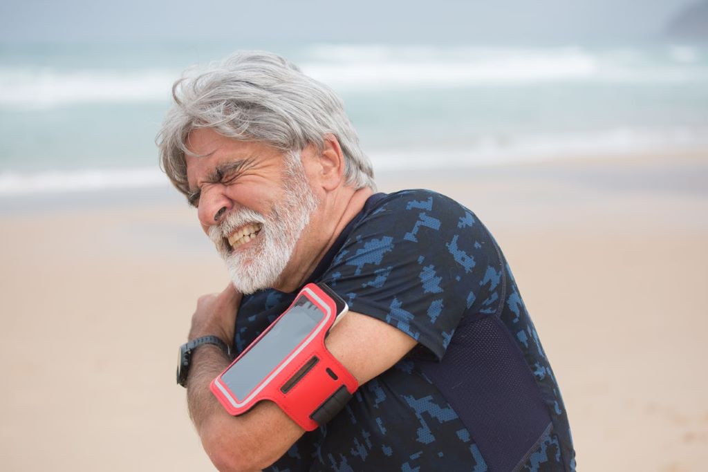
Along with finding ways to prevent or treat fibrosis, Genin and Huebsch said doctors can look for ways to apply this new knowledge about the importance of mechanical stress to treatment of injuries or burns. The findings could help address the high fail rate for treatments of elderly patients with injuries that require reattaching tendon to bone or skin to skin.

For instance, in rotator cuff injuries, there is compelling evidence that patients must start moving their arm to recover function, but equally compelling evidence that patients should immobilise the arm for better recovery. The answer might depend on the amount of collagen a patient produces and the stress fields at play at the recovery site.

By understanding the multidirectional stress fields’ impact on the cell structure, doctors may be able to look at specific patients’ repair and determine a personalised treatment plan.

For instance, a patient who has biaxial stress coming from two directions at the site of injury will potentially need to exercise more to trigger cell repair, Genin said. However, another patient showing signs of uniaxial stress, meaning stress is pulling only one direction, any movement could over-activate cells, so in that case, the patient should keep the injury immobilised. All that and more is still to be worked out and confirmed, but Genin is excited to begin.

“The next generation of disease we’re going to be conquering are diseases of mechanics,” Genin said.

Source: Washington University in St. Louis

New AI Tool for TBI Investigations in Forensics and Law Enforcement

Photo by Tom Jur on Unsplash

A study led by University of Oxford researchers has developed an advanced physics-based AI-driven tool to aid traumatic brain injury (TBI) investigations in forensics and law enforcement. The findings have been published in Communications Engineering.

TBI is a critical public health issue, with severe and long-term neurological consequences. In forensic investigations, determining whether an impact could have caused a reported injury is crucial for legal proceedings, yet there is currently no standardised, quantifiable approach to do this. The new study demonstrates how machine learning tools informed by mechanistic simulations could provide evidence-based injury predictions. This would help police and forensic teams accurately predict TBI outcomes based on documented assault scenarios.

The study’s AI framework, trained on real, anonymised police reports and forensic data, achieved remarkable prediction accuracy for TBI-related injuries:

  • 94% accuracy for skull fractures
  • 79% accuracy for loss of consciousness
  • 79% accuracy for intracranial haemorrhage

In each case, the model showed high specificity and high sensitivity (a low rate of false positive and false negative results).

This research represents a significant step forward in forensic biomechanics. By leveraging AI and physics-based simulations, we can provide law enforcement with an unprecedented tool to assess TBIs objectively.

The framework uses a general computational mechanistic model of the head and neck, designed to simulate how different types of impacts—such as punches, slaps, or strikes against a flat surface—affect various regions. This provides a basic prediction of whether an impact is likely to cause tissue deformation or stress. However, it does not predict on its own any risk of injury. This is done by an upper AI layer which incorporates this information with any additional relevant metadata, such as the victim’s age and height, before providing a prediction for a given injury.

Lead researcher Antoine Jérusalem, Professor of Mechanical Engineering in the Department of Engineering Science, University of Oxford

The researchers trained the overall framework on 53 anonymised real police reports of assault cases. Each report included information about a range of factors which could affect the blow’s severity (e.g., age, sex, body build of the victim/offender). This resulted in a model capable of integrating mechanical biophysical data with forensic details to predict the likelihood of different injuries occurring.

When the researchers assessed which factors had the most influence on the predictive value for each type of injury, the results were remarkably consistent with medical findings. For instance, when predicting the likelihood of skull fracture, the most important factor was the highest amount of stress experienced by the scalp and skull during an impact. Similarly, the strongest predictor of loss of consciousness was the stress metrics for the brainstem.

Understanding brain injuries using innovative technology to support a police investigation, previously reliant on limited information, will greatly enhance the interpretation required from a medical perspective to support prosecutions.

Ms Sonya Baylis, Senior Manager at the National Crime Agency

The research team insists that the model is not intended to replace the involvement of human forensic and clinical experts in investigating assault cases. Rather, the intention is to provide an objective estimate of the probability that a documented assault was the true cause of a reported injury. The model could also be used as a tool to identify high-risk situations, improve risk assessments, and develop preventive strategies to reduce the occurrence and severity of head injuries.

Lead researcher Antoine Jérusalem, Professor of Mechanical Engineering in the Department of Engineering Science at the University of Oxford said: ‘Our framework will never be able to identify without doubt the culprit who caused an injury. All it can do is tell you whether the information provided to it is correlated with a certain outcome. Since the quality of the output depends on the quality of the information fed into the model, having detailed witness statements is still crucial.’

Dr Michael Jones, Researcher at Cardiff University, and Forensics Consultant, said: ‘An “Achilles heel” of forensic medicine is the assessment of whether a witnessed or inferred mechanism of injury, often the force, matches the observed injuries. With the application of machine learning, each additional case contributes to the overall understanding of the association between the mechanism of cause, primary injury, pathophysiology and outcome.’

The study ‘A mechanics-informed machine learning framework for traumatic brain injury prediction in police and forensic investigations’ has been published in Communications Engineering. It was conducted by an interdisciplinary team of engineers, forensic specialists, and medical professionals from the University of Oxford, Thames Valley Police, the National Crime Agency, Cardiff University, Lurtis Ltd., the John Radcliffe Hospital and other partner institutions.

Source: University of Oxford

Time of Injury Matters: Circadian Rhythms Affect Muscle Repair

Photo by Mat Napo on Unsplash

Circadian rhythms doesn’t just dictate when we sleep — it also determines how quickly our muscles heal. A new Northwestern Medicine study in mice, published in Science Advances, suggests that muscle injuries heal faster when they occur during the body’s natural waking hours.

The findings could have implications for shift workers and may also prove useful in understanding the effects of aging and obesity, said senior author Clara Peek, assistant professor of biochemistry and molecular genetics at Northwestern University Feinberg School of Medicine.

The study also may help explain how disruptions like jetlag and daylight saving time changes impact circadian rhythms and muscle recovery.

“In each of our cells, we have genes that form the molecular circadian clock,” Peek said. “These clock genes encode a set of transcription factors that regulate many processes throughout the body and align them with the appropriate time of day. Things like sleep/wake behaviour, metabolism, body temperature and hormones — all these are circadian.”

How the study was conducted

Previous research from the Peek laboratory found that mice regenerated muscle tissues faster when the damage occurred during their normal waking hours. When mice experienced muscle damage during their usual sleeping hours, healing was slowed.

In the current study, Peek and her collaborators sought to better understand how circadian clocks within muscle stem cells govern regeneration depending on the time of day.

For the study, Peek and her collaborators performed single-cell sequencing of injured and uninjured muscles in mice at different times of the day. They found that the time of day influenced inflammatory response levels in stem cells, which signal to neutrophils — the “first responder” innate immune cells in muscle regeneration.

“We discovered that the cells’ signalling to each other was much stronger right after injury when mice were injured during their wake period,” Peek said. “That was an exciting finding and is further evidence that the circadian regulation of muscle regeneration is dictated by this stem cell-immune cell crosstalk.”

The scientists found that the muscle stem cell clock also affected the post-injury production of NAD+, a coenzyme found in all cells that is essential to creating energy in the body and is involved in hundreds of metabolic processes.

Next, using a genetically manipulated mouse model, which boosted NAD+ production specifically in muscle stem cells, the team of scientists found that NAD+ induces inflammatory responses and neutrophil recruitment, promoting muscle regeneration.  

Why it matters

The findings may be especially relevant to understanding the circadian rhythm disruptions that occur in aging and obesity, Peek said.

“Circadian disruptions linked to aging and metabolic syndromes like obesity and diabetes are also associated with diminished muscle regeneration,” Peek said. “Now, we are able to ask: do these circadian disruptions contribute to poorer muscle regeneration capacity in these conditions? How does that interact with the immune system?”

What’s next

Moving forward, Peek and her collaborators hope to identify exactly how NAD+ induces immune responses and how these responses are altered in disease.

“A lot of circadian biology focuses on molecular clocks in individual cell types and in the absence of stress,” Peek said. “We haven’t had the technology to sufficiently look at cell-cell interactions until recently. Trying to understand how different circadian clocks interact in conditions of stress and regeneration, is really an exciting new frontier.”

Source: Northwestern University

Engineered Cartilage from Nasal Septum Cells helps Treat Complex Knee Injuries

Researchers grow cartilage replacements from cells of the nasal septum to repair cartilage injuries in the knee. (Photo: University of Basel, Christian Flierl)

An unlucky fall while skiing or playing football can spell the end of sports activities. Damage to articular cartilage does not heal by itself and increases the risk of osteoarthritis. Researchers at the University of Basel and the University Hospital Basel have now shown that even complex cartilage injuries can be repaired with replacement cartilage engineered from cells taken from the nasal septum.

A team at the Department of Biomedicine led by Professor Ivan Martin, Dr Marcus Mumme and Professor Andrea Barbero has been developing this method for several years. It involves extracting the cells from a tiny piece of the patient’s nasal septum cartilage and then allowing them to multiply in the laboratory on a scaffold made of soft fibres. Finally, the newly grown cartilage is cut into the required shape and implanted into the knee joint.

Earlier studies have already shown promising results. “Nasal septum cartilage cells have particular characteristics that are ideally suited to cartilage regeneration,” explains Professor Martin. For example, it has emerged that these cells can counteract inflammation in the joints.

More mature cartilage shows better results

In a clinical trial involving 98 participants at clinics in four countries, the researchers compared two experimental approaches. One group received cartilage grafts that had matured in the lab for only two days before implantation – similar to other cartilage replacement products. For the other group, the grafts were allowed to mature for two weeks. During this time, the tissue acquires characteristics similar to native cartilage.

For 24 months after the procedure, the participants self-assessed their well-being and the functionality of the treated knee through questionnaires. The results, published in the scientific journal Science Translational Medicine, showed a clear improvement in both groups. However, patients who received more mature engineered cartilage continued to improve even in the second year following the procedure, overtaking the group with less mature cartilage grafts.

Magnetic resonance imaging (MRI) further revealed that the more mature cartilage grafts resulted in better tissue composition at the site of the implant, and even of the neighbouring cartilage. “The longer period of prior maturation is worthwhile,” emphasizes Anke Wixmerten, co-lead author of the study. The additional maturation time of the implant, she points out, only requires a slight increase in effort and manufacturing costs, and gives much better results.

Particularly suited to larger and more complex cartilage injuries

“It is noteworthy that patients with larger injuries benefit from cartilage grafts with longer prior maturation periods,” says Professor Barbero. This also applies, he says, to cases in which previous cartilage treatments with other techniques have been unsuccessful.

“Our study did not include a direct comparison with current treatments,” admits Professor Martin. “However, if we look at the results from standard questionnaires, patients treated with our approach achieved far higher long-term scores in joint functionality and quality of life.”

Based on these and earlier findings, the researchers now plan to test this method for treating osteoarthritis – an inflammatory disease that causes joint cartilage degeneration, resulting in chronic pain and disability.

Two large-scale clinical studies, funded by the Swiss National Science Foundation and the EU research framework programme Horizon Europe, are about to begin. These studies will explore the technique’s effectiveness in treating a specific form of osteoarthritis affecting the kneecaps (ie, patellofemoral osteoarthritis). The activities will further develop in Basel the field of cellular therapies, strategically defined as a priority area for research and innovation at the University of Basel and University Hospital Basel.

Source: University of Basel

Promising Findings in Testing Nasal Spray for TBI Treatment

Source: CC0

A new study led by researchers at Mass General Brigham suggests a nasal spray developed to target neuroinflammation could one day be an effective treatment for traumatic brain injury (TBI). By studying the effects of the nasal anti-CD3 in a mouse model of TBI, researchers found the spray could reduce damage to the central nervous system and behavioural deficits, suggesting a potential therapeutic approach for TBI and other acute forms of brain injury. The results are published in Nature Neuroscience.

“Traumatic brain injury is a leading cause of death and disability – including cognitive decline – and chronic inflammation is one of the key reasons,” said lead author Saef Izzy, MD, FNCS, FAAN, a neurologist and head of the Immunology of Brain Injury Program at Brigham and Women’s Hospital, a founding member of the Mass General Brigham healthcare system. “Currently, there is no treatment to prevent the long-term effects of traumatic brain injury.”

The study examines the monoclonal antibody Foralumab, made by Tiziana, which has been tested in clinical trials for patients with multiple sclerosisAlzheimer’s disease, and other conditions.

“This opens up a whole new area of research and treatment in traumatic brain injury, something that’s almost impossible to treat,” said senior author Howard Weiner, MD, co-director of the Ann Romney Center for Neurologic Diseases at Brigham and Women’s Hospital. “It also means this could work in intracerebral hemorrhage and other stroke patients with brain injury.”

Multiple experiments were done in mouse models with moderate-to-severe traumatic brain injury to explore the communication between regulatory cells induced by the nasal treatment and the microglial immune cells in the brain. Over time, researchers were able to identify how they modulate immune response.

“Modulating the neuroinflammatory response correlated with improved neurological outcomes, including less anxiety, cognitive decline, and improved motor skills,” Izzy said.

In addition to assessing the effects of the treatment, the research team was able to learn about immune response over time and compare the immune responses and effects of TBI in the mice.

The next step in the research is to translate the findings from preclinical models to human patients.

“Our patients with traumatic brain injury still don’t have an effective therapeutic to improve their outcomes, so this is a very promising and exciting time to move forward with something that’s backed up with solid science and get it to patients’ bedsides,” said Izzy.

Once in the clinical setting, Weiner said the hope is this treatment could be used on a variety of traumatic brain injury patients, including football players with repetitive concussions. 

“We envision giving a nasal spray right there on the sidelines,” said Weiner. “It isn’t something we can do yet, but we see the potential.”

Source: Mass General Brigham

What are the Best Methods to Treat Rotator-cuff Tears?

Photo by Kampus Production

Rotator-cuff disorders are the most common cause of shoulder symptoms. Tears of the rotator cuff can result from a substantial traumatic injury or can occur slowly over time. Most degenerative tears occur in the dominant arm of adults over the age of 40, and their prevalence increases with as you age. There are a variety of treatments.

In a recent publication in the New England Journal of Medicine, University of Michigan Health professor and chair of Physical Medicine and Rehabilitation, Nitin Jain, MD, MSPH, talks through the different treatments available for rotator-cuff tears to help bring together a better understanding of how to treat the issue for patients and providers.

Nonoperative treatment, such physical therapy, is the typical approach to treating rotator-cuff tears. However, surgery is considered in certain patients whose rotator-cuff tears don’t resolve with nonoperative treatments. Topical treatments also exist, such as glyceryl trinitrate (nitro-glycerine) gel which appears to have the greatest success.

Symptoms of rotator-cuff tears

“Patients with nontraumatic or degenerative rotator-cuff tears typically experience an onset of shoulder pain that seems to have no cause,” said Jain. 

“However, it is not uncommon for tears to be asymptomatic and become slowly painful over time, or even cause no pain at all.”

Jain says there are some activities that make the injury more painful as the tear worsens. This can include sleeping on your shoulder, overhead activities and/or lifting items above your shoulder level.

“Rotator-cuff tears may also grow over time, but there’s a lack of correlation between patient symptoms and the size and thickness of the tear,” explained Jain.

Your active range of motion and arm strength are usually affected by the tear, which gets assessed by using certain protocols when providers are searching for a diagnosis.

Rehabilitation and physical therapy for rotator-cuff tears 

This is the most common form of treatment for rotator-cuff tears, says Jain: “It is recommended that as the first line of specialist referral, patients seek care from a physical medicine and rehabilitation doctor (physiatrist) or sports medicine doctor.

“Rehabilitation and physical therapy routines address areas such as periscapular muscle weakness, correcting scapular posture and improve rotator cuff muscle strength and endurance.”

In observational studies, more than 80% of patients who received supervised physical therapy reported reduced pain and improved function between 6 months to a year. However, the trial populations consisted of patients with various types of rotator-cuff injuries and had no requirement for advanced imaging to confirm their diagnosis.

“One of the biggest factors in a successful rehabilitation was trust from patients that their physical therapy routine would improve their rotator-cuff condition,” said Jain. “The more patients leaned into the physical therapy routine, the better their outcomes were.”

Other nonpharmacologic therapies for rotator-cuff tears

Evidence suggests that psychosocial distress and depression are associated with shoulder pain and reduced function in patients with rotator-cuff tears.

“Despite this, though, there isn’t much data supporting psychosocial interventions in the treatment of rotator-cuff disorders, even though they show benefit in the treatment of other musculoskeletal disorders such as lower back pain,” said Jain.

In addition to the lack of data for psychological interventions for rotator-cuff repairs, there’s also a lack of high quality trials supporting the use of manual therapy, massage therapy, acupuncture, therapeutic ultrasonography, transcutaneous electrical nerve stimulation, shock-wave therapy or pulsed-electromagnetic-field therapy.

Topical and oral medications and injections for rotator-cuff tears

There isn’t a lot of evidence supporting the use of topical medications in treating rotator-cuff disorders. The topical treatment with the best outcomes so far has been glyceryl trinitrate.

In a small, randomised trial it showed short term benefits in the treatment of rotator-cuff disorders, but it also found there was a considerably high bias towards this treatment from participants in the study.

Topical nonsteroidal anti-inflammatory drugs such as diclofenac and ketoprofen have also been effective in providing pain relief in chronic musculoskeletal pain and tendinitis and have a better safety profile than oral, nonsteroidal anti-inflammatory drugs.

“But high quality evidence supporting their use in rotator-cuff disorders is still lacking,” explained Jain.

For oral medications, randomised research trials have shown that oral nonsteroidal anti-inflammatory drugs (NSAIDs) reduced pain, although modestly, in patients with rotator-cuff disorders. 

“Opioid drugs are generally not recommended due to risks associated with their use and lack of evidence of superiority to nonopioid therapy in a variety of musculoskeletal conditions,” said Jain.

Jain says acetaminophen hasn’t been studied specifically in rotator-cuff disorders, but what has been studied has shown little or no benefit regarding pain or function.

“Rigorous evidence is lacking to inform the use of pain-modulating drugs such as gabapentin, duloxetine, and pregabalin, specifically regarding the nonoperative treatment of rotator-cuff disorders,” said Jain.

Injection of a glucocorticoid, together with a local anaesthetic, has been reported to provide symptomatic pain relief in patients with rotator-cuff disorders.

Small trials have shown short term benefit, about four weeks long, of pain relief through using this method. The injections are performed in the subacromial space of the rotator-cuff for those with subacromial impingement syndrome. Some centres use ultrasound guidance to administer this treatment, which can reduce the risk of an inadvertent injection into the tendon.

Surgical interventions for rotator-cuff injuries

“Surgical interventions are not the initial recommendation when it comes to rotator-cuff repairs. However, they may be considered in some patients whose condition does not improve with conservative treatment,” said Jain.

Observational data supports that surgery is associated with better function and reduced pain in patients who are under 65 years of age and have smaller tears.

Surgical repairs are mostly performed arthroscopically, involving the repair of the torn tendon and resecuring it to the humerus to allow for tendon-to-bone healing as well as a low incidence of complications, explains Jain.

The hypothesis that surgical intervention can reduce the progression of muscle degradation has led some experts to recommend early surgical intervention, but data is still lacking on outcomes of early surgery compared to surgery later.

Source: Michigan Medicine – University of Michigan

Men More Than Three Times as Likely to Die From a Brain Injury, New Study Shows

Photo by Anna Shvets

A new analysis of mortality data reveals the disproportionate impact of traumatic brain injuries (TBI) on older adults, males and certain racial and ethnic groups. The study, published in the peer-reviewed journal Brain Injury, provides a comprehensive analysis of TBI-related deaths across different population groups across the US in 2021.

The findings indicate that suicides remain the most common cause of TBI-related deaths, followed by unintentional falls, and specific groups are disproportionately affected by these tragedies.

Men, in particular, were found to be most likely to die from a TBI – more than three times the rate of women (30.5 versus 9.4). The reasons observed were multifactorial and could reflect differences in injury severity following a fall or motor vehicle crash, to the interaction of sex and age – with TBI outcomes in men worsening with age, while postmenopausal women fare better than men of similar age.

“While anyone is at risk for getting a TBI, some groups have a higher chance than others of dying from one. We identified specific populations who are most affected. In addition to men, older adults are especially at risk, with unintentional falls being a major cause of TBI-related death. American Indian or Alaska Native people also have higher rates of these fatal injuries,” says lead author Alexis Peterson PhD, of the National Center for Injury Prevention and Control at the Centers for Disease Control and Prevention.

“These findings highlight the importance of tailored prevention strategies to reach groups who may be at higher risk and the role healthcare providers can play in reducing TBI-related deaths through early intervention and culturally sensitive care.”

TBI remains a leading cause of injury-related death in the US In 2020, TBIs were associated with around a quarter of all injury-related deaths.

Using data from the National Vital Statistics System, the new analysis identified 69 473 TBI-related deaths among US residents during 2021. The age-adjusted TBI-related mortality rate was 19.5 per 100 000, representing an 8.8% increase from 2020.

Through statistical modeling, the researchers examined the simultaneous effect of multiple factors such as geographic region, sex, race and ethnicity, and age, on TBI-related mortality.

Key findings include:

  • Older adults (75+) had the highest rates of TBI-related deaths, with unintentional falls being the most common cause in this age group.
  • Non-Hispanic American Indian/Alaska Native individuals experienced the highest TBI-related death rate (31.5) compared to other racial and ethnic groups.
  • There were 37,635 TBI-related deaths categorised as unintentional injuries (ie, motor vehicle crashes, unintentional falls, unintentionally struck by or against an object, other).
  • 30,801 were categorized as intentional injuries (ie, all mechanisms of suicide and homicide).
  • Children aged from birth to 17 years accounted for around 4% of TBI-related deaths (2,977).

The authors emphasise the critical role of healthcare providers in preventing TBI-related deaths, particularly with groups at higher risk. “By assessing patients who may be at higher risk for TBI, especially due to falls or mental health challenges, healthcare providers can make timely referrals and recommend culturally tailored interventions to prevent further injury or death,” says Dr Peterson.

Public health efforts should focus on addressing the underlying causes of TBI-related deaths, such as unintentional falls and mental health crises, to help prevent further loss of life. “TBIs remain a significant public health concern, especially among older adults, men, and certain racial and ethnic groups,” says Peterson.  “CDC has proven resources that healthcare providers can use to not only reduce health disparities that increase the risk for TBI but also improve care for anyone affected by a TBI.”

The authors note the COVID-19 pandemic could have influenced TBI-related death trends in 2021. They also acknowledge several limitations of this analysis, including potential misclassification or incomplete documentation of causes on death certificates, which may lead to inaccuracies in estimating TBI-related deaths.

Source: Taylor & Francis Group

Diagnosing and Managing Blast Injuries

Photo by Jeff Kingma on Unsplash

The prevalence of armed conflicts, terrorist attacks and industrial accidents necessitates clinician understanding of blast injuries in both civilian and military settings. Blast injuries are a complex form of trauma, resulting from the explosive release of energy. The severity and types of injury depend on the proximity to the blast, blast pressure and the presence of other elements like fragments and heat.

In a new video published in the New England Journal of Medicine‘s “Video in Clinical Medicine” section, authors from Boston University Chobanian & Avedisian School of Medicine and Boston Medical Center (BMC), in collaboration with the department of anaesthesiology at Walter Reed National Military Medical Center, focus on the mechanisms and classifications of blast injuries and present essential knowledge for initial diagnosis and management.

“Blast injuries present a unique challenge in trauma medicine due to their complex mechanisms and varied presentations. A comprehensive understanding and approach to managing these injuries is essential to improve patient outcomes,” explains corresponding author Rafael Ortega, MD, FASA, chair & professor of anaesthesiology at the school.

The video provides a review on blast injury types including: primary injuries to the lungs, ears and bowels due to the blast wave; secondary injuries caused by trauma from propelled debris; tertiary injuries due to blast wind impacts on the body; quaternary injuries like burns, asphyxiation and exposure to toxic substances; and quinary injuries, clinical repercussions of chemical, radiologic or biologic contaminants occurring post-detonation. Also considered are the types of explosives, such as dynamite and Molotov cocktails.

The authors point out that explosions can inflict injuries on many organ systems and that the diagnosis of blast injuries requires a high index of suspicion to identify silent blunt injuries. They suggest initial assessment should follow advanced trauma life support (ATLS) protocols including imaging methods, such as radiographs, CT scans, and ultrasonography, which are critical for detecting internal injuries. Injuries to the pulmonary, gastrointestinal system, along with neurological, cardiovascular, facial and auditory, musculoskeletal systems are also reviewed.

In terms of diagnosis, the authors indicate that ATLS guidelines offer a structured approach to trauma care after explosions. “However, their application should be tailored to the specific situation and patient needs, with the order of interventions potentially varying based on clinical judgment and immediate life threats,” says Ortega who also is chief of anaesthesiology at BMC.

According to the authors, blast injuries should be managed using a multidisciplinary approach tailored to the individual patient’s injuries. “When possible, involve different specialties such as emergency medicine, trauma surgery, neurosurgery, orthopaedic surgery, maxillofacial surgery, otolaryngology and anaesthesiology to ensure the best possible patient outcomes,” he adds.

In terms of preventive strategies, the authors recommend public education, improved safety regulations for handling explosives, and the use of personal protective equipment which can help reduce the impact of blast injuries and designing public spaces to minimise the impact of blasts.

Source: Boston University School of Medicine

Key Molecule in Wound Healing Identified

Photo by Diana Polekhina on Unsplash

A new study from Karolinska Institutet and the Chinese Academy of Medical Sciences has identified an RNA molecule that is important for skin wound healing. The research, published in Nature Communications, may have implications for the treatment of hard-to-heal wounds.

The study focuses on the molecular processes in wound healing that regulate the transition from inflammation to a proliferative phase, where new cells form to repair damaged tissue. Researchers have now mapped lncRNA (long non-coding RNA molecules) in human skin wounds in tissue samples from Karolinska University Hospital, identifying a key regulator in wound healing.

“Our study reveals that the lncRNA molecule SNHG26 plays a pivotal role in guiding skin cells through the stages of wound healing, from an inflammatory stage to a healing phase,” explains Ning Xu Landén, docent at the Department of Medicine, Solna, Karolinska Institutet.

The researchers also used mouse models to uncover how this molecule interacts with genes involved in inflammation and tissue regeneration. In mice lacking SNHG26, wound healing was delayed, emphasising the molecule’s importance in the balance between inflammation and tissue repair. The discovery paves the way for new therapeutic approaches for acute and chronic wounds.

“By targeting SNHG26, we may be able to accelerate healing and reduce complications, particularly in chronic wounds where prolonged inflammation is a major problem,” says Ning Xu Landén.

Source: Karolinska Institutet

Concussions in Amateur Sport not Linked to Long-term Cognitive Effects

Photo by Olga Guryanova on Unsplash

The impact of concussion while playing sport is different in those who don’t play professionally, says new research.

Sports-related concussions (SRC) may not be associated with long-term cognitive risks for non-professional athletes, a study led by a UNSW medical researcher suggests. In fact, study participants who had experienced an SRC had better cognitive performance in some areas than those who had never suffered a concussion, pointing to potential protective effects of sports participation.

Published in the Journal of Neurology, Neurosurgery and Psychiatry, the research reveals that individuals who reported experiencing any SRC during their lifetime had a marginally better cognitive performance than those who reported no concussions.

The study, a collaboration between researchers at UNSW Sydney, the University of Oxford, the University of Exeter and Harvard University, analysed data from more than 15 000 participants from the UK-based PROTECT study of 50- to 90-year-olds. This ongoing research aims to understand brain ageing and cognitive decline.

“Our findings suggest that there is something about playing sport, even though a person may experience concussion, that may be beneficial for long-term cognitive outcomes,” says lead author Dr Matt Lennon MD, PhD, at UNSW Medicine & Health.

“While it may be that those who play sports have had access to better education and more resources, we controlled for these factors in the analysis, so that doesn’t explain the result. We hypothesise that there may be physical, social and long-term behavioural effects of sport that may make for healthier adults in late-life,” said Dr Lennon.

Largest study of long-term effects of sports concussions

The study is the largest to date examining the long-term cognitive effects of SRC. Researchers collected lifetime concussion histories from 15 214 participants using the Brain Injury Screening Questionnaire. Among them, 6227 (39.5%) reported at least one concussion and 510 (3.2%) at least one moderate-severe concussion. On average, participants reported suffering their last head injury an average of 29 years prior to the study and their first head injury an average of 39 years earlier.

Researchers then compared cognitive function among individuals with 0, 1, 2 and 3+ SRCs and 0, 1, 2 and 3+ non-sports-related concussions (nSRCs)  (i.e. from falls, car accidents, assaults and other causes). The SRC group showed 4.5 percentile rank better working memory than those who hadn’t experienced an SRC, and 7.9% better reasoning capacity than those without concussions.

Those with one SRC also had better verbal reasoning and attention compared to those with no SRC.

Conversely, participants with 3+ nSRCs – so things like accident and assaults – had worse processing speed and attention, and a declining trajectory of verbal reasoning with age.

“This study suggests that there could be long term benefits from sport which could outweigh any negative effects of concussions, which could have important implications for policy decisions around contact sport participation. It may also be that non-sports related head injuries lead to greater brain damage than sports-related concussions,” said senior author Professor Vanessa Raymont from the University of Oxford and Oxford Health NHS Foundation Trust.

The researchers say the study had some limitations.

“The retrospective design of the study, with elderly participants often recalling details of events over three decades in the past, may have affected the reporting of head injuries, even though we used a well-validated head injury screening tool,” said Prof. Raymont.

Study implications

The study looked at mid-to-late-life people who experienced SRC years earlier, whereas most other studies on SRC focus on younger athletes in the immediate period after their head injuries, where cognitive effects are most salient.

“While these results do not indicate the safety of any sport in particular, they do indicate that overall sports may have greater beneficial effects for long-term cognitive health than the damage it causes, even in those who have experienced concussion,” said Dr Lennon.

“This finding should not be overstated – the beneficial effects were small and in people who had two or more sports-related concussions there was no longer any benefit to concussion. Additionally, this study does not apply to concussions in professional athletes whose head injuries tend to be more frequent, debilitating and severe.”

Anne Corbett, Professor at Exeter University and the lead investigator of the PROTECT study, said: “What we see emerging is a completely different profile of brain health outcomes for people who have concussions as a result of sport compared to those that are not related to sport. Concussions that occur during sport do not lead to brain health concerns whereas other concussion types do, especially when people experience multiple concussions. In fact, people who take part in sport seem to have better brain health regardless of whether they have had a concussion whilst taking part or not.”

Source: University of New South Wales