Category: Implants and Prostheses

A New Pacemaker that Works with Light, not Electricity

Pacemakers regulate the heartbeats of people with chronic heart diseases like atrial fibrillation and other forms of arrhythmia. However, pacemaker implantation is an invasive procedure, and the lifesaving pacing the devices provide can be extremely painful. Pacemakers also can only be used to treat a few specific types of disease.

In Science Advances, researchers describe their new pacemaker design that uses light and optogenetics that could be implanted with a less invasive procedure, also causing less pain in operation. As well as triggering cardiac neurons with light, the new design can also be powered by light, removing the need for a battery which has to be surgically replaced.

The study was helmed by researchers in the Gutruf Lab, led by biomedical engineering assistant professor and Craig M. Berge Faculty Fellow Philipp Gutruf.

Currently available pacemakers work by implanting one or two leads, or points of contact, into the heart with hooks or screws. If the sensors on these leads detect a dangerous irregularity, they send an electrical shock through the heart to reset the beat.

“All of the cells inside the heart get hit at one time, including the pain receptors, and that’s what makes pacing or defibrillation painful,” Gutruf said. “It affects the heart muscle as a whole.”

The device Gutruf’s team has developed, yet to be tested in humans, would use a digitally created mesh that would send much more targeted signals.

Modifying cardiac neurons to respond to light

Optogenetics modifies cells, usually neurons, to make them responsive to light. This technique only targets cardiomyocytes, the cells of the muscle that trigger contraction and make up the beat of the heart. This precision will not only reduce pain for pacemaker patients by bypassing the heart’s pain receptors, it will also allow the pacemaker to respond to different kinds of irregularities in more appropriate ways. For example, during atrial fibrillation, the upper and lower chambers of the heart beat asynchronously, and a pacemaker’s role is to get the two parts back in line.

“Whereas right now, we have to shock the whole heart to do this, these new devices can do much more precise targeting, making defibrillation both more effective and less painful,” said Igor Efimov, professor of biomedical engineering and medicine at Northwestern University, where the devices were lab-tested. “This technology could make life easier for patients all over the world, while also helping scientists and physicians learn more about how to monitor and treat the disease.”

To ensure the light signals can reach many different parts of the heart, the team created a design that involves encompassing the organ, rather than implanting leads that provide limited points of contact.

The new pacemaker model consists of four petallike structures made of thin, flexible film, which contain light sources and a recording electrode. The petals, specially designed to accommodate the way the heart changes shape as it beats, fold up around the sides of the organ to envelop it, like a flower closing up at night.

“Current pacemakers record basically a simple threshold, and they will tell you, ‘This is going into arrhythmia, now shock!'” Gutruf said. “But this device has a computer on board where you can input different algorithms that allow you to pace in a more sophisticated way. It’s made for research.”

Because the system uses light to affect the heart, rather than electrical signals, the device can continue recording information even when the pacemaker needs to defibrillate. In current pacemakers, the electrical signal from the defibrillation can interfere with recording capabilities, leaving physicians with an incomplete picture of cardiac episodes. Additionally, the device does not require a battery, which could save pacemaker patients from needing to replace the battery in their device every five to seven years, as is currently the norm.

Gutruf’s team collaborated with researchers at Northwestern University on the project. While the current version of the device has been successfully demonstrated in animal models, the researchers look forward to furthering their work, which could improve the quality of life for millions of people.

Source: University of Arizona

Silver Lining for Titanium Implants Reduces Infection Risk

Photo by Mehmet Turgut Kirkgoz on Unsplash

A novel method of coating titanium implants for orthopaedic and trauma surgery promises to reduce infection complications, according to a paper published in Langmuir: The ACS Journal of Fundamental Interface Science.

Infection remains a major complication when implants such as screws and plates are embedded into people during procedures like joint replacement surgery and spinal fusion surgery. Most infections occur because the devices’ titanium implant surfaces have poor antibacterial and osteoinductive properties, despite titanium possessing the highest biocompatibility of all metals.

Assistant Professor Rahim Rahimi at Purdue University has developed a process which immobilises silver onto the implant surfaces of titanium orthopaedic devices to improve their antibacterial properties and cellular integration. The process can be implemented onto many currently utilised metal implant surfaces.

The antibacterial efficacy of laser-nanotextured titanium surfaces with laser-immobilised silver was tested against both gram-positive (Staphylococcus aureus) and gram-negative (Escherichia coli) bacteria. The surfaces retained efficient and stable antimicrobial properties for more than six days. The laser-nanotextured titanium surfaces also provided a 2.5-fold increase in osseointegration properties compared with a pristine titanium implant surface.

“The first step of the two-step process creates a hierarchical nanostructure onto the titanium implant surface to enhance the bone cells’ attachment,” A/Prof Rahimi said. “The second step immobilises silver with antibacterial properties onto the titanium implant surface.

“The technology allows us to not only immobilise antibacterial silver compounds onto the surface of the titanium implants but also provide a unique surface nanotexturing that allows better settle attachment mineralisation.

“These unique characteristics will allow improving implant outcomes, including less risk of infection and fewer complications like device failure.”

A/Prof Rahimi said the traditional method to address infections caused by implanted orthopaedic devices often utilises antibiotics or other surface modifications that have their own associated complications.

“Long-term antibacterial protection is not possible with these traditional drug coatings because a large portion of the loaded drug is released in a short time,” Rahimi said. “There also is often a mixture of microbes that are found in implant-associated infection; it is essential to choose a bactericidal agent that covers a broad spectrum.”

Source: Purdue University

A Soft Robotic Design for Diabetic Amputee Pain Relief

Proof-of-concept rendering (left) and photo (right) of the prototype of the new microfluidics-enabled soft robotic prosthesis for lower limb amputees.
Credit: Waterloo Microfluidics Laboratory at University of Waterloo

Diabetic amputations often involve neuropathy, and patients detect damage resulting from an ill-fitting prosthesis, leading to further amputation. To solve this, in Biomicrofluidics, scientists described a new type of prosthetic using microfluidics-enabled soft robotics that reduces skin ulcerations and pain in patients who have had an amputation between the ankle and knee.

More than 80% of lower-limb amputations are due to diabetic foot ulcers, and the lower limb is known to swell at unpredictable times, resulting in volume changes of 10% or more.

Typically, the prosthesis used after amputation includes fabric and silicone liners that can be added or removed to improve fit. The amputee needs to manually change the liners, but neuropathy leading to poor sensation makes this difficult and can lead to more damage to the remaining limb.

“Rather than creating a new type of prosthetic socket, the typical silicon/fabric limb liner is replaced with a single layer of liner with integrated soft fluidic actuators as an interfacing layer,” said author Carolyn Ren, from the University of Waterloo. “These actuators are designed to be inflated to varying pressures based on the anatomy of the residual limb to reduce pain and prevent pressure ulcerations.”

The scientists started off with pneumatic actuators to adjust the pressure of the prosthetic socket, but it was quite heavy.

To reduce weight, the group miniaturised the actuators, designing a microfluidic chip with 10 integrated pneumatic valves to control each actuator. The full system is controlled by a miniature air pump and two solenoid valves that provide air to the microfluidic chip. The control box is small and light enough to be worn as part of the prosthesis.

Prosthetics experts provided a detailed map of desired pressures for the prosthetic socket. The group carried out extensive measurements of the contact pressure provided by each actuator and compared these to the desired pressure for a working prosthesis.

All of the actuators produced the right pressures suggesting the new device will work well in the field, with the next step being a more accurate biological model.

The group plans additional research to integrate pressure sensors directly into the prosthetic liner, perhaps using newly available knitted soft fabric that incorporates pressure sensing material.

Source: American Institute of Physics

Partially Paralysed Man Uses Robotic Arms to Feed Himself

Photo by Tara Winstead on Pexels

Recent advances in neural science, robotics, and software have enabled scientists to develop a robotic system that responds to muscle movement signals from a partially paralysed person relayed through a brain-machine interface. Human and robot act as a team to make performing some tasks a piece of cake.

Two robotic arms – a fork in one hand, a knife in the other – flank a seated man, who sits in front of a table, with a piece of cake on a plate. A computerised voice announces each action: “moving fork to food” and “retracting knife.” Partially paralysed, the man makes subtle motions with his right and left fists at certain prompts, such as “select cut location”, so that the machine slices off a bite-sized piece. Now: “moving food to mouth” and another subtle gesture to align the fork with his mouth.

In less than 90 seconds, a person with very limited upper body mobility who hasn’t been able to use his fingers in about 30 years, just fed himself dessert using his mind and some smart robotic hands.

A team led by researchers at the Johns Hopkins Applied Physics Laboratory (APL), in Laurel, Maryland, and the Department of Physical Medicine and Rehabilitation (PMR) in the Johns Hopkins School of Medicine, published a paper in the journal Frontiers in Neurorobotics that described this latest feat using a brain-machine interface (BMI) and a pair of modular prosthetic limbs.

Also sometimes referred to as a brain-computer interface, BMI systems provide a direct communication link between the brain and a computer, which decodes neural signals and ‘translates’ them to perform various external functions, from moving a cursor on a screen to now enjoying a bite of cake. In this particular experiment, muscle movement signals from the brain helped control the robotic prosthetics.

A new approach

The study built on more than 15 years of research in neural science, robotics, and software, led by APL in collaboration with the Department of PMR, as part of the Revolutionizing Prosthetics program, which was originally sponsored by the US Defense Advanced Research Project Agency (DARPA). The new paper outlines an innovative model for shared control that enables a human to manoeuvre a pair of robotic prostheses with minimal mental input.

“This shared control approach is intended to leverage the intrinsic capabilities of the brain machine interface and the robotic system, creating a ‘best of both worlds’ environment where the user can personalise the behaviour of a smart prosthesis,” said Dr Francesco Tenore, a senior project manager in APL’s Research and Exploratory Development Department. The paper’s senior author, Tenore focuses on neural interface and applied neuroscience research.

“Although our results are preliminary, we are excited about giving users with limited capability a true sense of control over increasingly intelligent assistive machines,” he added.

Helping people with disabilities

One of the most important advances in robotics demonstrated in the paper is combining robot autonomy with limited human input, with the machine doing most of the work while enabling the user to customize robot behavior to their liking, according to Dr David Handelman, the paper’s first author and a senior roboticist in the Intelligent Systems Branch of the Research and Exploratory Development Department at APL.

“In order for robots to perform human-like tasks for people with reduced functionality, they will require human-like dexterity. Human-like dexterity requires complex control of a complex robot skeleton,” he explained. “Our goal is to make it easy for the user to control the few things that matter most for specific tasks.”

Dr Pablo Celnik, project principal investigator in the department of PMR said: “The human-machine interaction demonstrated in this project denotes the potential capabilities that can be developed to help people with disabilities.”

Closing the loop

While the DARPA program officially ended in August 2020, the team at APL and at the Johns Hopkins School of Medicine continues to collaborate with colleagues at other institutions to demonstrate and explore the potential of the technology.

The next iteration of the system may integrate previous research that found providing sensory stimulation to amputees enabled them to not only perceive their phantom limb, but use muscle movement signals from the brain to control a prosthetic. The theory is that the addition of sensory feedback, delivered straight to a person’s brain, may help him or her perform some tasks without requiring the constant visual feedback in the current experiment.

“This research is a great example of this philosophy where we knew we had all the tools to demonstrate this complex bimanual activity of daily living that non-disabled people take for granted,” Tenore said. “Many challenges still lie ahead, including improved task execution, in terms of both accuracy and timing, and closed-loop control without the constant need for visual feedback.”

Celnik added: “Future research will explore the boundaries of these interactions, even beyond basic activities of daily living.”

Source: Frontiers

Deep Nerve Stimulation Controls Blood Pressure

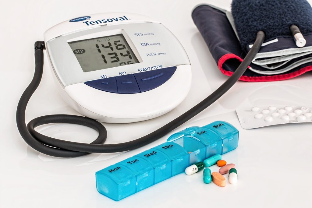
Blood pressure cuff
BP cuff for home monitoring. Source: Pixabay

A study published in Frontiers in Neuroscience demonstrated that blood pressure and renal sympathetic nerve activity (RSNA) can be controlled by bioelectronic treatment. RSNA is often increased in hypertension and renal disease.

Using a custom-wired electrode, Professor Mario Romero-Ortega previously reported that deep peroneal nerve stimulation (DPNS) elicits an acute reduction in blood pressure. The current study, advances that work, focusing on his development of a small implantable wireless neural stimulation system and exploration of different stimulation parameters to achieve a maximum lowered response.

Prof Romero-Ortega integrated a nerve stimulation circuit less than a millimetre in size, with a novel nerve attachment microchannel electrode that can be implanted into small nerves, while enabling external power and DPNS modulation control.

Using this implantable device, his team demonstrated that systolic blood pressure can be lowered 10% in one hour and 16% two hours after nerve stimulation.

“Our results indicate that DPNS consistently induces an immediate and reproducible arterial depressor effect in response to electrical stimulation of the deep peroneal nerve,” reported Prof Romero-Ortega.

While pharmacological treatments are effective, blood pressure remains uncontrolled in 50–60% of resistant hypertensive subjects. Unfortunately, despite the use of multiple antihypertensive drugs in combination, blood pressure remains poorly controlled in 50–60% of the hypertensive population and approximately 12–18% of them develop resistant hypertension, defined as blood pressure greater than 140/90 mmHg despite the use of antihypertensive drugs.

“In this study, DPNS induced an initial increase in RSNA during the first 2–3 seconds, followed by a reduction in renal activity and mean arterial pressure, despite the increase in heart rate,” said Prof Romero-Ortega. “The observed activation of the RSNA during the DPNS was not expected since its activity is associated with hypertension.”

Source: University of Houston

Implant Helps Patient with Neurodegenerative Disease to Walk Again

Patient takes steps with the help of an assistant. Credit: Jmmy Ravier & NeuroRestore

A woman bedridden for over a year due to a debilitating neurodegenerative disease was able to get up and walk again, thanks to an innovative electrical stimulation system which was able to raise her blood pressure on standing and prevent her fainting. The system was developed by a team headed by Professors Jocelyne Bloch and Grégoire Courtine, and was detailed in The New England Journal of Medicine.

Their system includes electronics implanted directly on the spinal cord to reactivate the neurons that regulate blood pressure, thereby preventing the patient from losing consciousness every time she’s in an upright position. This type of implant was already in use for the treatment of low blood pressure in tetraplegic patients.

The female patient in the study suffers from multiple system atrophy-parkinsonian type (MSA-P), a neurodegenerative disease that afflicts several parts of the nervous system, including the sympathetic nervous system. 

MSA-P leads to the loss of sympathetic neurons regulating blood pressure, which results in orthostatic hypotension, a dramatic blood pressure drop when patients are in an upright position, which in some cases results in fainting. This increases fall risks, limits mobility, and can eventually shorten life expectancy. Having to remain in a reclined position to avoid passing out severely impacts patients’ quality of life.

The implant consists of a set of electrodes connected to an electrical-impulse generator typically used to treat chronic pain. After implanting their device directly on the patient’s spinal cord, the researchers found an improvement in the body’s capacity to regulate blood pressure, enabling the patient to remain conscious for longer periods in an upright position and to begin physical therapy to walk again. After being bedridden for 18 months, the patient is now able to walk as far as 250 metres.

For Jocelyne Bloch, this marks an important step toward the treatment of degenerative diseases: “We’ve already seen how this type of therapy can be applied to patients with a spinal-cord injury. But now, we can explore applications in treating deficiencies resulting from neurodegeneration. This is the first time we’ve been able to improve blood-pressure regulation in people suffering from MSA.”

Grégoire Courtine added that “this technology was initially intended for pain relief, not for this kind of application. Going forward, we and our company Onward Medical plan to develop a system targeted specifically to orthostatic hypotension that can help people around the world struggling with this disorder.”

Source: Ecole Polytechnique Federale de Lausanne

Tiny Implantable Electrodes to Treat Drug-resistant Neuropathic Pain

Woman holding her wrist in pain
Credit: Pixabay CC0

Using a rice-grain sized wireless implant to stimulate peripheral nerves from within blood vessels could potentially treat drug-resistant neuropathic pain, according to a study published in Nature Biomedical Engineering.

After receiving a grant, a team set out to create implantable, wirelessly powered nerve stimulators that can be used in place of opioids for pain management. The 1-millimetre large implants are small enough to be placed on stents and delivered within blood vessels adjacent to specific areas of the central and peripheral nervous system.

Co-principal investigator of the study, Sunil A. Sheth, MD, explained: “We’re getting more and more data showing that neuromodulation, or technology that acts directly upon nerves, is effective for a huge range of disorders—depression, migraine, Parkinson’s disease, epilepsy, dementia, etc. – but there’s a barrier to using these techniques because of the risks associated with doing surgery to implant the device, such as the risk of infection. If you can lower that bar and dramatically reduce those risks by using a wireless, endovascular method, there are a lot of people who could benefit from neuromodulation.”

Neuropathic pain can be a disabling disorder that accounts for nearly 40% of chronic pain sufferers, often leading to anxiety, depression, and opioid addiction. Previous studies showed that electrical stimulation is an effective treatment for reducing pain when doctors target the spinal cord and dorsal root ganglia (DRG), a bundle of nerves that carry sensory information to the spinal cord. However, existing DRG stimulators require invasive surgery to implant a battery pack and pulse generator.

According to the researchers, this new type of technology offers a way to perform minimally invasive bioelectronic therapy that helps with more precise placement of the implant and more predictable outcomes. The team are hoping to move forward with regulatory approval, which Dr Sheth estimates may take a few years.

Source: The University of Texas Health Science Center

‘An Alien Knee’: Discomfort with Total Knee Replacement

Photo by Kampus Production from Pexels

A study interviewing patients who received total knee replacement for osteoarthritis find that, despite the welcome pain relief, some also experience less pleasant psychological impacts.

For their study published in Arthritis Care & Research, UK researchers sought to bridge a knowledge gap of where people were dissatisfied with their total knee replacements even though they reported less pain and better function.

Using semi-structured interviews, researchers elicited comments from 34 patients, meant to explore patients’ thoughts about their knee implants. They received a lot of feedback about non-pain discomfort and feelings of dissonance.

“My leg feels like it’s made of lead,” one patient told researchers.

“It feels like someone is holding your knees, when you move, it’s like someone is … putting pressure there,” said another.

A third said: “I know it’s not my knee. It’s an alien knee in there. I don’t really feel connected to it.”

“Typically, the assessment of patient-reported outcomes after joint replacement focuses on functional outcome and pain relief as the main determinant of satisfaction,” the researchers explained. “This narrow perspective is compounded by poor definitions of satisfaction after surgery, and there is little research on how and why some patients express dissatisfaction with joint replacement and what they are dissatisfied about.”

Citing a study of hand surgery patients in which patients “spoke about their hand as if it were an object separate from their self,” the researchers argued that a psychological concept called embodiment could help explain the patients’ feelings of dissonance.

They wrote: “Embodiment refers to the experience of the body as both subject and object, such that this idea impacts the way in which a person sees and interacts with the world, and vice versa. Embodiment provides a way of understanding how one experiences limits of possible action, a sense of control, and empowerment over physical action.”

Initially, the researchers weren’t planning to focus on embodiment, but, they explained, “by the third interview we noted that some participants described sensations of discomfort such as heaviness or numbness when discussing pain and some described their knee as ‘alien,’ ‘foreign,’ or ‘not part of’ themselves. In response to these findings, the interviewer sought to elicit views about any such sensations in subsequent interviews, if this topic was not broached first by the participant.”

Their study emerged from an earlier one focusing on reasons for avoiding healthcare encounters post-surgery and involved the same participants, who had lingering pain and discomfort. The semi-structured interview covered pain as well as how patients managed it. After a third interview, patients who reported feelings of alienation from their implant were asked about it in more detail.

Participants reflected the general knee-replacement population – most in their 60 and 70s, and just over half were women. Of the 34 patients, 24 were between 2 and 4 years out from their surgery.

Physical types of non-pain discomfort were commonly reported, such as feelings of numbness and/or heaviness, as well as sensations of pressure applied externally. One man said it felt like the skin over his knee was stretched tight. Separate from these sensations were reports that the limb no longer felt like a part of them but something foreign, like an external prosthesis. Some patients complained of a lack of control. “That knee just wouldn’t do what it’s told to do,” one told the interviewer.

Others said they hadn’t regained trust in the knee, with one man still using a cane for fear of falling.

Overall, the researchers found that the patients’ experience were quite similar to those of amputees getting used to their prosthetic limbs. This could be partly explained by often experiencing years of pain and loss of function before the joint replacement.

“Presurgical chronic pain, instability, and untrustworthiness might continue to influence [mental] incorporation of the prosthesis afterwards,” the researchers suggested.

And there is a potential clinical implication for the findings: “Our study suggests that the interest for rehabilitation becomes not only strengthening the joint and promoting full recovery to tasks, but also modifying a person’s relationship with the new joint to achieve full incorporation or re-embodiment.”

The researchers proposed that other programmes developed for other conditions could be helpful, such as external prosthesis use as well as complex regional pain syndromes.

“Our focus should not be on the absence or loss of embodiment,” the researchers added, “but on employing a multidisciplinary approach to using the concept to guide the development of pre-rehabilitative strategies and appropriate outcome measures.”

Source: MedPage Today

Recipients of Bionic Eyes Blindsided by Obsolescence

Source: Daniil Kuzelev on Unsplash

After the manufacturer of a bionic eye ended support, hundreds of recipients of the vision-improving implants have been left without support – “literally in the dark”, as one of them put it.

IEEE Spectrum, which first broke the story, reported that Second Sight discontinued its retinal implants in 2019. The retinal implants serve as the source of artificial vision for the users.

The publication wrote that the firm’s focus is currently on developing a brain implant known as the Orion, which also provides artificial vision. However, it only offers very limited support to the 350 or so who have the now-obsolete Argus II implants.

The system consists of a camera mounted on glasses worn by the user, which transmits video to a video processing unit (VPU), which then encodes the images into arrays of black and white pixels. The VPU then relays the pixel to an electrode array behind the retina, which creates flashes of light corresponding to the white pixels. The technology has had a long and costly road from experiment to product, starting with a lab experiment in the 1990s where stimulation of a single electrode in the retina was discovered to create a visible flash of light perceived by a blind patient. It is hugely expensive, with an estimated cost of $150,000 (R2.25 million) even before the surgery and post-surgery training. 

Implantation surgery typically takes a few hours, followed by training to help users interpret the new optical input from their implants. It is not a replacement for sight; rather it is more like a new sense. Users of the system see fleeting changes of grey which some can then use to assist with basic locomotion. However, the technology is still crude and not all benefit to the same degree. While some can make out the stripes on a pedestrian crossing, others never achieve that level of ability.

The technology also comes with some risk. In the postapproval period, 17% experienced adverse events, though this was an improvement over the 40% in the preapproval period. Since the implant can interfere with MRI scans, some have had to consider removal.

IEEE Spectrum contacted a number of patients, who voiced concern over their future. One patient, Ross Doer, said he was delighted when Second Sight told him in 2020 he was eligible for software upgrades. Yet, he heard troubling rumours. When he called his Second Sight vision-rehab therapist, “She said, ‘Well, funny you should call. We all just got laid off,’ ” he recalled. “She said, ‘By the way, you’re not getting your upgrades.’ ”

“Those of us with this implant are figuratively and literally in the dark,” he said.

Second Sight, when contacted by the publication, said that it had to reduce its workforce because of financial difficulties, and though it attempted to provide “virtual support” was unable to assist with repairs or replacements.
Benjamin Spencer, one of the six patients to receive the new Orion implant, said that it was “amazing” and he was able to see his wife for the first time. But knowing what he does now about Second Sight makes him apprehensive, and plans to have his implant removed at the end of the study period.

Speaking to the BBC, Elizabeth M. Renieris, professor of technology ethics at the University of Notre Dame, in the US, described the development as a cautionary tale.

“This is a prime example of our increasing vulnerability in the face of high-tech, smart and connected devices which are proliferating in the healthcare and biomedical sectors,” she said.

“These are not like off-the-shelf products or services that we can actually own or control. Instead we are dependent on software upgrades, proprietary methods and parts, and the commercial drivers and success or failure of for-profit ventures.”

She added that in future, ethical considerations concerning such technology should include “autonomy, dignity, and accountability”.

Source: IEEE Spectrum

Implant Enables Man With Severed Spinal Cord to Walk

Michel Rocatti walking using the spinal stimulation system. ©NeuroRestore-Jimmy Ravier

In a world first, Michel Roccati, a man with a completely severed spinal cord was able to walk again outside the lab with the help of a portable electrical stimulation system that causes his legs to take a step in conjunction with his intention to move and a walker to steady him.

This was a further development of a technology that in 2018 helped David M’zee, who had been left paralysed by a partial spinal cord injury suffered in a sports accident, to walk again. A research team led by Professors Grégoire Courtine, at École Polytechnique Fédérale de Lausanne (EPFL) and Jocelyne Bloch, at Lausanne University Hospital (CHUV) had developed an electrical stimulation system to help people with spinal cord injuries walk again.

They had wanted to see if electrodes could stimulate movement in the parts of the spine damaged so badly that signals no longer reach the nervous system from the brain. That pioneering study was detailed in Nature and Nature NeuroscienceThanks to the electrodes making up for the weakness of the signals in his damaged spinal cord, M’zee was able to voluntarily move his legs and could walk several hundred metres at a time, sometimes without the aid of the rails on the treadmill.

Now a new milestone has just been reached with the technology, and the research team enhanced their system with more sophisticated implants controlled by advanced software. These implants can stimulate the region of the spinal cord that activates the trunk and leg muscles. Thanks to this new technology, three patients with complete spinal cord injury were able to walk again outside the lab. “Our stimulation algorithms are still based on imitating nature,” said Prof Courtine. “And our new, soft implanted leads are designed to be placed underneath the vertebrae, directly on the spinal cord. They can modulate the neurons regulating specific muscle groups. By controlling these implants, we can activate the spinal cord like the brain would do naturally to have the patient stand, walk, swim or ride a bike, for example.”

On a cold, snowy day last December, Michel Roccati – an Italian man who became paralysed after a motorcycle accident four years earlier – braved the icy wind to try out the system outdoors, in central Lausanne. He had recently undergone the surgical procedure in which Prof Bloch placed the new, implanted lead on his spinal cord.

Scientists attached two small remote controls to Michel’s walker and connected them wirelessly to a tablet that forwards the signals to a pacemaker in Michel’s abdomen. The pacemaker in turn relays the signals to the implanted spinal lead that stimulates specific neurons, causing Michel to move. Grasping the walker, Michel pressed a button corresponding to either the left or right leg with the firm intention of taking a step forward, and his feet rose and fell in short steps.

“The first few steps were incredible – a dream come true!” he says. “I’ve been through some pretty intense training in the past few months, and I’ve set myself a series of goals. For instance, I can now go up and down stairs, and I hope to be able to walk one kilometre by this spring.”

Two other patients have also successfully tested the new system, which is described in Nature Medicine. “Our breakthrough here is the longer, wider implanted leads with electrodes arranged in a way that corresponds exactly to the spinal nerve roots,” said Bloch. “That gives us precise control over the neurons regulating specific muscles.” Ultimately, it allows for greater selectivity and accuracy in controlling the motor sequences for a given activity.

While extensive training is necessary for patients to get comfortable using the device, the speed and scope of rehabilitation is amazing. “All three patients were able to stand, walk, pedal, swim and control their torso movements in just one day, after their implants were activated!” said Prof Courtine. “That’s thanks to the specific stimulation programs we wrote for each type of activity. Patients can select the desired activity on the tablet, and the corresponding protocols are relayed to the pacemaker in the abdomen.”

While the progress achievable in a single day is astonishing, the gains after several months are even more impressive. The three patients followed a training regimen based on the stimulation programs and were able to regain muscle mass, move around more independently, and take part in social activities like having a drink standing at a bar. What’s more, because the technology is miniaturized, the patients can perform their training exercises outdoors and not only inside a lab.

Presently there is one key limitation, Prof Bloch said: “We need at least six centimetres of healthy spinal cord under the lesion. That’s where we implant our electrodes.”

As for Roccati, after nine months of Lausanne-based rehab, he now lives independently in Italy. “I continued rehab at home, working alone, with all the devices,” he said. “And I see improvements every day.”

Source: École Polytechnique Fédérale de Lausanne