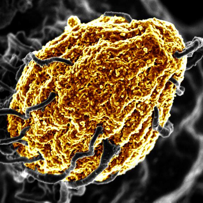
Scanning Electron Micrograph image of a human T cell. Credit: NIH/NIAID
A new study published in Nature has found that certain food proteins can cause T-helper cells in gut-associated lymphoid tissue to become dysfunctional in order for the immune system not to attack that particular food. Understanding how the process could be restarted could aid the development of food allergy treatments.
Led by Marc Jenkins, director of the University of Minnesota Medical School’s Center for Immunology, the research focused on why the immune system does not attack food in the way that it attacks other foreign entities like microbes.
“This study helps explain why our immune systems do not attack our food even though it is foreign to our bodies,” said Jenkins. “We found that ingested food proteins stimulate specific lymphocytes in a negative way. The cells become dysfunctional and eventually acquire the capacity to suppress other cells of the immune system.”
The gut associated-lymphoid tissue is a suppressive environment where lymphocytes that would normally generate inflammation undergo arrested development. This abortive response usually prevents dangerous immune reactions to food.
The research found that T-helper cells lack the inflammatory functions needed to cause gut pathology and yet the cells have the potential to produce regulatory T-cells that may suppress it. This means when people develop an intolerance or allergic reaction to certain foods, there may be a future capability to suppress that reaction by reintroducing dysfunctional lymphocytes.
Further research is needed to identify the mechanisms whereby food-specific lymphocytes become dysfunctional, knowledge which could be used to fight food allergies.
The gut microbiome has been linked to aspects ranging from the immune system to mental health. However, pinpointing how and which bacteria specifically affect which biological process has eluded researchers, until now – a new study figured it out the link between immunity and a major gut bacterium.
Such knowledge is essential for learning how to manipulate gut bacteria to treat or prevent illness.
“Microbiome studies need to move from making associations to determining function and causation,” said Professor Ramnik Xavier, co-author of the study which appears in Nature.
“The real significance of this work was connecting a bacterium, the molecule it makes, the pathway it operates through, and the biological outcome,” said Professor at HMS and co-senior author of the study with Prof Xavier. “That’s very rare.”
Profs Clardy and Xavier and colleagues focused on Akkermansia muciniphila, a species that makes up 3% of the gut microbiome. It gets its name from the intestinal mucus it breaks down.
Studies had suggested that A. muciniphila has a key role in maintaining healthy immune processes, seeming to protect against diseases such as type 2 diabetes and inflammatory bowel disease and make cancer cells more responsive to immune checkpoint therapies. Until now, no research could confirm how that connection worked.
Start to finish
The researchers show that the links begin with a lipid in A. muciniphila‘s cell membrane.
“That discovery was quite surprising. The usual suspects for triggering an immune response would be a protein or a sugar,” said Prof Clardy.
After discovering the molecular structure of the lipid, the team found that it communicates toll-like receptors (TLR) 2 and TLR 1. Found on many immune cells, they detect bacteria and identify hostile ones for the immune system. In this case, versions of TLR2 and TLR1 bound together in a way scientists hadn’t seen before.
The researchers showed in cell cultures that the fat’s activation of TLR2-TLR1 can trigger the release of certain cytokines — immune proteins involved in inflammation — while leaving other cytokines alone.
They also confirmed that the lipid helps maintain immune homeostasis. They found that low doses of the lipid act like a leash, preventing the immune system from reacting to a potentially harmful molecule until that molecule reaches significant levels. On the other end, they saw that high doses of the lipid don’t stimulate an immune response much more than low or medium doses, keeping a healthy ceiling on inflammation.
New doors open
The work introduces new possibilities for developing drugs that piggyback on A. muciniphila‘s ability to manipulate the immune system and fight disease. Members of Prof Clardy’s lab have made that job easier by revealing the molecular structure of the lipid and figuring out how scientists can make it and similar ones easily in the lab.
The study also serves as a model for pinpointing how other members of the gut microbiome act on the body.
“You can change the bacterium and apply the same set of tests,” said Prof Clardy.
Also, contrary to the expectations of many people in the field, such work doesn’t require fancy techniques. The researchers used traditional methods, spectroscopic analysis and chemical synthesis, to find and understand the lipid molecule.
In fact, despite its “remarkable activity,” the lipid has a “generic structure” that would have flown under the radar of more advanced genomic or metabolomic analyses, Prof Clardy said.
“The gut microbiome and the immune system are very complicated, which makes you expect that answers will be complicated too,” he said. “But sometimes complicated things are just lots of simple things.”
Infected cell covered with SARS-CoV-2 viruses. Source: NIAID
Researchers have investigated how antiviral proteins called interferons interact with SARS-CoV-2. The study, published in PNAS, focuses on how the innate immune system defends against this coronavirus, which appears to be adapting to evade this interferon response.
The study was the result of a collaborative effort, including the laboratories of Mario Santiago, PhD, associate professor of medicine and Eric Poeschla, MD, professor of medicine, both at the University of Colorado School of Medicine.
While the adaptive arm of the immune system robustly deals with infection by generating antibodies and T cells, the innate arm forms an earlier, first line of defence by recognising conserved molecular patterns in pathogens.
“SARS-CoV-2 just recently crossed the species barrier into humans and continues to adapt to its new host,” said Prof Poeschla. “Much attention has deservedly focused on the virus’s serial evasions of neutralising antibodies. The virus seems to be adapting to evade innate responses as well.”
The type I Interferon system is a major player in antiviral defence against all kinds of viruses. Virus-infected cells release type I interferons (IFN-α/β), which warn the body of the intrusion. Secreted interferons cause susceptible cells to express powerful antiviral mechanisms to limit viral growth and spread. The interferon pathway could significantly reduce the levels of virus initially produced by an infected individual.
“They are clinically viable therapeutic agents that have been studied for viruses like HIV-1 for years,” explained Prof Santiago. “Here we looked at up to 17 different human interferons and found that some interferons, such as IFNalpha8, more strongly inhibited SARS-CoV-2. Importantly, later variants of the virus have developed significant resistance to their antiviral effects. For example, substantially more interferon would be needed to inhibit the omicron variant than the strains isolated during the earliest days of the pandemic.”
The data suggests that COVID clinical trials on interferons, dozens of which are listed in clinicaltrials.gov, may need to be interpreted based on which variants were circulating when the study was conducted. Researchers say that future work to decipher which of SARS-CoV-2’s multitude of proteins might be evolving to confer interferon resistance may contribute in that direction.
A macrophage digesting a yeast cell (yellow). Credit: NIH
Certain pathogens such as Salmonella have developed strategies to protect themselves from the macrophages’ digesting attempts, causing severe Typhoid infections and inflammations. Scientists report in Nature Metabolism how the inter-organellar crosstalk between phago-lysosomes and mitochondria restricts the growth of such bacteria inside macrophages.
Signals from the digestion cell organelle
As scavenger cells, macrophages have a very prominent digestion organelle, the phago-lysosome, where engulfed microorganisms are commonly degraded into pieces and become inactivated. “It has long been known that the molecule TFEB (Transcription factor EB) is important for the regulation of the phago-lysosomal system. More recent evidence also suggested that TFEB supports the defense against bacteria,” said Max Planck Institute group leader Angelika Rambold.
She and her team wanted to understand how exactly TFEB mediates its anti-bacterial role in macrophages. They confirmed earlier findings showing that a broad range of microbes, bacterial and inflammatory stimuli activate TFEB and thus the phago-lysosomal system.
“It made sense that pathogen signals trigger TFEB as macrophages need a more active digestion system quickly after they devour a meal of bacteria. But, interestingly, the experiments also revealed an additional strong effect of TFEB activation on another intracellular organelle system — mitochondria. This was completely unexpected and novel to us,” said Angelika Rambold.
Instructing mitochondria to increase anti-microbial activity
Composed of inner and outer membranes, mitochondria are the primary sites of cellular respiration and release energy from nutrients. Moreover, the mitochondria in immune cells were recently identified as sources of anti-microbial metabolites.
By using a broad experimental tool set, the investigators identified the pathway controlling an unexpected crosstalk between lysosomes and mitochondria. “Macrophages make use of extensive inter-organellar communication: the lysosome activates TFEB, which shuttles into the nucleus where it controls the transcription of a protein called IRG1. This protein is imported into mitochondria, where it acts as a major enzyme to produce the anti-microbial metabolite itaconate,” explained Angelika Rambold.
Exploiting organelle communication to control bacterial infections
The researchers investigated whether they could make use of this newly identified pathway to control bacterial growth. “We speculated that activating this pathway could be used to target certain bacterial species, such as Salmonella,” said Angelika Rambold. “Salmonella can escape the degradation by the phago-lysosomal system. They manage to grow inside macrophages, which can lead to the spreading of these bacteria to several organs in an infected body,” explained Alexander Westermann, collaborating scientist from the University of Würzburg.
When the researchers activated TFEB in infected macrophages in mice, the TFEB-Irg1-itaconate pathway inhibited the growth of Salmonella inside the cells. These data show that the lysosome-to-mitochondria interplay represents an antibacterial defense mechanism to protect the macrophage from being exploited as a bacterial growth niche.
In light of the increasing emergence of multi-drug resistant bacteria, with more than 10 million expected deaths per year by 2050 according to the various expert groups, it becomes important to identify new strategies to control bacterial infections that escape immune mechanisms. A promising path could be to use the TFEB-Irg1-itaconate pathway or itaconate itself to treat infections caused by itaconate-sensitive bacteria. According to the researchers from more work is needed to assess whether these new intervention points can be successfully applied to humans.
The oral fungal pathogen Candida albicans (red) produce hyphae that allow attachment of another fungus Candida glabrata (green). Yeast cells of Candida glabrata (green) adhere to Candida albicans hyphae (red) both in static culture (left, scanning electron microscopy) and under biofilm conditions of flow (right, confocal fluorescence microscopy). Credit: Edgerton Lab, State University of New York at Buffalo
Candida, a distant cousin of baker’s yeast is notorious for causing various types of thrush that can be a major nuisance, but it can also lead to an invasive and occasionally fatal infection. In a study in the journal Nature Immunology, a research team uncovered a previously unknown defense mechanism employed by the immune system in fighting Candida infections.
In healthy individuals, Candida is present in the microbiome in the gut and on the skin. Normally, Candida is held in check by the immune system, but it can occasionally grow excessively, invading the lining of the mouth, the vagina, the skin or other parts of the body. In severe cases, it can spread to the bloodstream and from there to the kidneys. Such life-threating infections may occur in weakened immune systems. Antibiotics can also unleash local or invasive Candida eruptions by wiping out competing, beneficial bacteria.
Until now, the immune cells that got most of the credit for defending the body against Candida were the small, round lymphocytes of the T cell type, called TH17. These cells were also the ones to take the blame when this defence failed.
In the new study, postdoctoral fellow Dr Jan Dobeš, working together with colleagues in Abramson’s lab, discovered that a powerful commando unit of TH17 cells capable of fighting Candida cannot be generated without crucial early support from an entirely different contingent: a subset of rare lymphoid cells known as type-3 innate lymphoid cells, or ILC3, that express a gene called the autoimmune regulator, or Aire
The two groups of cells belong to the two different arms of the immune system, which, like normal soldiers and specialised ‘commando’ units, join forces against a common enemy. The Aire-ILC3s – part of the more ancient, innate arm – spring into action almost immediately upon encountering a threat – in this case, a Candida infection. The TH17s belong to the immune system’s more recent, adaptive arm, which takes several days or even weeks to respond, but which launches a much more targeted and potent attack than the innate one.
The scientists found that as soon as Candida starts infecting tissues, the Aire-ILC3s engulf the yeast whole, chop them up and display some of the yeast pieces on their surfaces. In this way, the bits are presented to the TH17s, a few of which are generally on call in the lymph nodes, ready for an infection alert. This kind of presentation instructs the specialised T cells to start dividing rapidly, soaring in number from a few lone commandos to several hundred or even thousands of Candida-specific fighters, capable of destroying the yeast at the sites of infection.
“We have identified a previously unrecognised immune system weapon that is indispensable for orchestrating an effective response against the fungal infection,” Abramson said.
Abramson became intrigued by Candida because it commonly leads to severe, chronic infections in people with a rare autoimmune syndrome caused by defects in the Aire gene. Abramson’s lab had conducted extensive studies of this gene, helping to clarify its role in preventing autoimmune disorders. That research, as well as studies by other scientists, had shown that Aire-expressing cells in the thymus instruct developing T cells to refrain from attacking the body’s own tissues. When Aire is defective, T cells fail to receive proper instructions, consequently causing widespread autoimmunity that wreaks havoc in multiple body organs. But one puzzle remained: Why would Aire-deficient patients suffering from a devastating autoimmune syndrome also develop chronic Candida infections?
While trying to complete the Aire puzzle, Dobeš and colleagues found that outside the thymus, Aire is also expressed in a small subset of ILC3s in the lymph nodes. The researchers then genetically engineered two groups of mice: One lacked Aire in the thymus, and the other group lacked it in the ILC3s in the lymph nodes. The first group developed autoimmunity but was able to successfully fight off Candida. In contrast, those in the second group, the ones lacking Aire in ILC3s, were without autoimmunity, but were unable to generate numerous Candida-specific TH17s. Consequently, they failed to effectively eliminate Candida infections. In other words, without Aire-expressing ILC3s, the specialised T cells needed for fighting Candida were not produced in sufficient numbers.
“We found an entirely new role for Aire, one that it plays in the lymph nodes – turning on a mechanism that increases the numbers of Candida-fighting T cells,” Dr Dobeš explained.
An Aire-ILC3 cell (green) “kisses” a Candida-fighting TH17 cell (red), telling it to start dividing (top row), but it ignores other T cells that do not specialize in fighting Candida (bottom row)
These findings open up new directions of research that in the future may help develop new treatments for severe Candida, and possibly for other fungal infections. The newly discovered mechanism might, for example, help produce large numbers of Candida-fighting T cells to be used in cell therapy. If scientists one day decode the signals by which Aire-ILC3s boost T cell proliferation, they might serve as the basis for new therapies.
Fourth-generation e-cigarettes, such as Juul devices, are associated with unique changes in markers of immune responses inside our airways, according to a new peer-reviewed paper in the American Journal of Respiratory and Critical Care Medicine.
Third-generation e-cigarettes include vape pens and box mods. Fourth generation include nicotine-salt-containing e-cigarettes, such as Juul products, and disposable e-cigarettes, which have become increasingly popular, especially after the recent FDA ban on the sale of Juul products.
Researchers in the lab of Professor Ilona Jaspers at the UNC School of Medicine found that users of fourth-generation nicotine-salt-containing devices display a unique mix of cellular biomarkers indicative of immune suppression.
“Our work demonstrates the importance of considering device type in future clinical, epidemiological, and mechanistic studies on the health effects of e-cigarettes,” said Prof Jasper, who led the study. “We also think this research can help regulators determine which products cause the most severe types of biological changes in airway cells important for maintaining proper health.”
E-cigarettes have become popular in the last decade. Some started used them to quit smoking, believing vaping was a safer alternative, both in the short-term and long-term. Also, because electronic cigarettes don’t contain tar, consumers assumed vaping decreased their risk of cancer down the road.
“It’s impossible to know if vaping decreases cancer risk or many other long-term conditions,” Prof Jaspers said. “It took 60 years of research to show that smoking causes cancer.” In contrast, e-cigarettes have only been around for about 15 years. “Still, the research from our lab and many others has shown many of the same acute biological effects in the airways that we have documented in smokers,” she said. “And we’ve seen some changes to cells and immune defences in people who vape that, frankly, we’ve never seen before, which is very concerning.”
A major concern for researchers, doctors, and public health officials is the fact that teenagers who would not have otherwise tried cigarettes began using e-cigarettes, which contain nicotine – with its attendant health implications – and thousands of chemicals, many of which are FDA-approved for eating but not inhaling.
Several studies have documented that inhaling chemical-laden nicotine aerosols suppresses the immune responses in the respiratory tracts of smokers and e-cigarette users. Some studies, including some at UNC, have detailed how different chemicals in various e-cigarettes, including chemicals that make up thousands of different flavours, have adverse effects on airway cells. The Jaspers lab, which has been at the forefront of such research, set out to study the effects of different varieties of e-cigarette devices. For this study, her team collected central airway (sputum) samples from non-smokers, smokers, and users of both third-generation and fourth-generation e-cigarette devices.
They found that users of fourth-generation e-cigarettes had significantly more bronchial epithelial cells in their sputum, suggestive of airway injury because normally, bronchial epithelial cells make up an intact barrier in the airways and are not found in sputum samples. Levels of two proteins, sICAM1 and sVCAM1, were significantly lower in fourth-generation e-cigarette users compared to all other groups. These proteins are important in fighting infections and other disease.
In addition, proteins are important for overall immune defence were significantly lower in fourth versus third generation e-cigarette users. Lower of these proteins – CRP, IFN-g, MCP-1, uteroglobin, MMP-2, and VEGF – indications immune system depression. “Another key finding of the study was that, when examining the mixture of immune markers overall rather than one by one, fourth generation e-cigarette users had the most distinguishable changes out of all of the groups, indicating a shift away from immune homeostasis,” said the study’s lead author, Elise Hickman, PhD.
The study did not demonstrate that e-cigarettes cause cancer, emphysema, COPD, or other long-term diseases associated with long-term cigarette smoking. But researchers think that altering immune responses in the respiratory tract over the course of many years, especially for teens, could play a major role in the development of long-term health conditions and in susceptibility to inhaled pathogens.
In an unexpected finding in studying alopecia, scientists have uncovered an unexpected link between T cells and hair growth, which could potentially be used to treat the condition. The findings, published in Nature Immunology, describe how regulatory T cells interact with skin cells using a hormone as a messenger to generate new hair follicles and hair growth.
Alopecia is an autoimmune condition where the immune system attacks the hair follicles, resulting in hair loss.
“For the longest time, regulatory T cells have been studied for how they decrease excessive immune reactions in autoimmune diseases,” explained Ye Zheng, associate professor at the Salk Institute and the paper’s corresponding author. “Now we’ve identified the upstream hormonal signal and downstream growth factor that actually promote hair growth and regeneration completely separate from suppressing immune response.”
Initially, the researchers were investigating the roles of regulatory T (Treg) cells and glucocorticoid hormones in autoimmune diseases. (Glucocorticoid hormones are cholesterol-derived steroid hormones produced by the adrenal gland and other tissues.) They first investigated how these immune components functioned in multiple sclerosis, Crohn’s disease and asthma.
They found that glucocorticoids and Treg cells did not function together to play a significant role in any of these conditions. So, they thought they’d have more luck looking at environments where Treg cells expressed particularly high levels of glucocorticoid receptors (which respond to glucocorticoid hormones), such as in skin tissue. The scientists induced hair loss in normal mice and mice lacking glucocorticoid receptors in their Treg cells.
“After two weeks, we saw a noticeable difference between the mice — the normal mice grew back their hair, but the mice without glucocorticoid receptors barely could,” says first author Zhi Liu, a postdoctoral fellow in Associate Prof Zheng’s lab. “It was very striking, and it showed us the right direction for moving forward.”
The findings suggested that some sort of communication must be occurring between Treg cells and hair follicle stem cells to allow for hair regeneration.
The scientists then investigated how the regulatory T cells and glucocorticoid receptors behaved in skin tissue samples, and found that glucocorticoids instruct the Treg cells to activate hair follicle stem cells, leading to hair growth. This crosstalk between the T cells and the stem cells depends on a mechanism whereby glucocorticoid receptors induce production of the protein TGF-beta3, all within the regulatory T cells. TGF-beta3 then activates the hair follicle stem cells to differentiate into new hair follicles, promoting hair growth. Additional analysis confirmed that this pathway was completely independent of regulatory T cells’ ability to maintain immune balance.
However, Treg cells don’t normally produce TGF-beta3, as they did here. A database search revaled that this phenomenon occurs in injured muscle and heart tissue, similar to how hair removal simulated a skin tissue injury in this study.
“In acute cases of alopecia, immune cells attack the skin tissue, causing hair loss. The usual remedy is to use glucocorticoids to inhibit the immune reaction in the skin, so they don’t keep attacking the hair follicles,” said Associate Prof Zheng. “Applying glucocorticoids has the double benefit of triggering the regulatory T cells in the skin to produce TGF-beta3, stimulating the activation of the hair follicle stem cells.”
This study revealed that Treg cells and glucocorticoid hormones are not just immunosuppressants but also have a regenerative function. Next, the scientists will look at other injury models and isolate Treg cells from injured tissues to monitor increased levels of TGF-beta3 and other growth factors.
Schizophrenia, which affects how people interact with reality, is difficult to treat because it has many possible causes. In a study published in Cell Reports Medicine, Japanese researchers have identified an autoantibody – an antibody which attacks the body’s own tissues – in some patients with schizophrenia.
Notably, they also found that this autoantibody caused schizophrenia-like behaviours and changes in the brain when they injected it into mice.
When considering possible autoantibodies that might cause schizophrenia, researchers from Tokyo Medical and Dental University (TMDU) had a specific protein in mind. Previous research has suggested that neural cell adhesion molecule (NCAM1), which helps facilitate synaptic connections, may have a role in the development of schizophrenia.
“We decided to look for autoantibodies against NCAM1 in around 200 healthy controls and 200 patients with schizophrenia,” explained lead author of the study Hiroki Shiwaku. “We only found these autoantibodies in 12 patients, suggesting that they may be associated with the disorder in just a small subset of schizophrenia cases.”
The research went on to find out whether these autoantibodies could cause any changes that commonly occur in schizophrenia, so they purified autoantibodies from some of the patients and injected them into the brains of mice.
“The results were impressive,” said the study’s senior author, Hidehiko Takahashi. “Even though the mice only had these autoantibodies in their brains for a short time, they had changes in their behaviour and synapses that were similar to what is seen in humans with schizophrenia.”
The mice given the patient autoantibodies had cognitive impairment and changes in their regulation of the startle reflex, which are both seen in other animal models of schizophrenia. They also had fewer synapses and dendritic spines, which are structures that are important for the connections between brain cells, and are also affected in schizophrenia.
The study findings hold promise for the diagnosis and treatment of schizophrenia can present very differently among patients and is often resistant to treatment. If schizophrenia is indeed caused by autoantibodies against NCAM1 in some patients, this will lead to important improvements in their diagnosis and treatment.
Scanning Electron Micrograph image of a human T cell. Credit: NIH/NIAID
While T cells are the body’s warriors against infection, without rest and maintenance T cells can die, leaving their hosts more susceptible to pathogens, researchers reported in the journal Science.
“We may have to change how we teach T cell biology,” said Professor Lieping Chen, who is the senior author of the study.
T cells remain in a quiescent state until pathogens are detected, but the molecular mechanisms of this state were previously unknown.
In the new study, researchers showed that a protein known as CD8a – which is found in a subset of T cells called CD8 cells – is crucial to keeping the cells in this dormant state. When scientists deleted this protein in mice, the protective CD8 cells were unable to enter a quiescent state and died, leaving the host vulnerable to infections.
Further, they identified another protein, PILRa, that provides a biochemical signal to CD8a. By disrupting this protein pair, both “memory” CD8 cells – previously been exposed to pathogens – and naïve cells died because they lacked the ability to stay in a quiescent state.
The researchers hope that understanding why this resting state is crucial to maintenance and survival of T cells can lead to improved immune system function.
Chen noted that as people age they tend to lose both naïve and memory T cells, making older individuals more susceptible to infections. It is possible that the inability of T cells to remain in a quiescent state could lead to people becoming more susceptible to infections and cancer, the authors suggest.
New research from scientists at La Jolla Institute for Immunology (LJI), shows that hypoxia can activate the same group of immune cells that cause inflammation during asthma attacks. During hypoxia, these cells flood the airways with lung-damaging molecules.
Hypoxia is a known trigger for developing and worsening lung conditions such as severe asthma, chronic obstructive pulmonary disease (COPD), and fibrosis. To treat and prevent these diseases, researchers need to understand why a lack of oxygen would affect the immune system.
“We show how lack of oxygen can be part of a feedback loop that can contribute to even worse inflammation,” says LJI Professor and Chief Scientific Officer Mitchell Kronenberg, PhD, a member of the LJI Center for Autoimmunity and Inflammation. “This work gives us insight into the causes of fibrosis of the lung and severe asthma.”
Prof Kronenberg and colleagues worked with a genetically altered mouse model to mimic the signals of hypoxia in the airway’s epithelial cells, which line the paths to the lungs. They discovered that combining the hypoxia signals with inflammatory signals stimulated the “innate,” or rapidly responding immunity, and an immune cell type called an ILC2.
An ILC2’s job is to make signaling molecules – cytokines – that quickly alert other immune cells to react to a pathogen. Unfortunately, ILC2s sometimes over-react and respond to harmless environmental allergens. In these cases, ILC2s churn out cytokines that drive mucus production and inflammation in the lungs. All this swelling and mucus leads to hypoxia.
As they report in Journal of Experimental Medicine, ILC2s respond to hypoxia as well, adding to the lung damage already caused during an asthma attack.
“That hypoxia may then contribute further to inflammation,” said Prof Kronenberg.
The next step was to figure out exactly how epithelial cells activate ILC2 during hypoxia. LJI Postdoctoral Fellow Jihye Han, PhD, led the work to uncover an unexpected culprit: adrenomedullin (ADM). ADM is known for its role in helping blood vessels dilate, but until now it had no known role in immune function.
Prof Kronenberg was surprised to see ADM involved — but not shocked. “We’re finding that many molecules with no previously known role in the immune system can also be important for immune function,” said Prof Kronenberg. “We need to understand that more generally.”
The researchers showed that human lung epithelial cells exposed to hypoxia also produced ADM. This means ADM or its receptor could be targets for treating inflammatory and allergic lung diseases.
The challenge is to find a balance between dampening the harmful immune response without leaving the body vulnerable to infections. Prof Kronenberg points out that the epithelial cell-ADM-ILC2 connection protected mice from hookworm infections, which damage the lungs and gut.
“ADM is a new target for lung diseases and has been implicated in bacterial pneumonia as well,” said Prof Kronenberg. “But blocking it would have to be done carefully.”