Category: Immune System

Salt Cuts off Regulatory T Cells’ Energy Supply

Spilled salt shaker
Source: Pixabay CC0

Regulatory T cells ensure that immune responses happen in a controlled way. But eating too much salt weakens these cells’ energy supply, thus rendering them temporarily dysfunctional. This salt-induced ‘load shedding’ may have implications for autoimmunity, researchers report in Cell Metabolism.

Excessive salt consumption not only causes cardiovascular problems, it could also adversely impact the immune system. The study found that salt can disrupt regulatory T cells by impairing their energy metabolism. The findings may provide new avenues for exploring the development of autoimmune and cardiovascular diseases.

A few years ago, research by teams led by Professor Dominik Müller and Professor Markus Kleinewietfeld revealed that excess salt in the diet can negatively affect the metabolism and energy balance in certain types of innate immune cells called monocytes and macrophages and stop them from working properly. They further showed that salt triggers malfunctions in the mitochondria. Inspired by these findings, the research groups wondered whether excessive salt intake might also create a similar problem in adaptive immune cells like regulatory T cells.

Important immune regulators

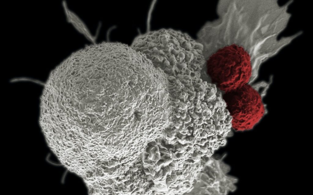
Regulatory T cells, also known as Tregs, are an essential part of the adaptive immune system. They are responsible for maintaining the balance between normal function and unwanted excessive inflammation.

Scientists believe that the deregulation of Tregs is linked to the development of autoimmune diseases like multiple sclerosis. Recent research has identified problems in mitochondrial function of Tregs from patients with autoimmunity, yet the contributing factors remain elusive.

“Considering our previous findings of salt affecting mitochondrial function of monocytes and macrophages as well as the new observations on mitochondria in Tregs from autoimmune patients, we were wondering if sodium might elicit similar issues in Tregs of healthy volunteers,” says Müller, who co-heads the Hypertension-Mediated End-Organ Damage Lab at the Max Delbrück Center and the ECRC.

Previous research has also shown that excess salt could impact Treg function by inducing an autoimmune-like phenotype. In other words, too much salt makes the Treg cells look like those involved in autoimmune conditions. However, exactly how sodium impairs Treg function had not yet been uncovered.

Salt interferes with mitochondrial function of Tregs

The new international study led by Kleinewietfeld and Müller has now discovered that sodium disrupts Treg function by altering cellular metabolism through interference with mitochondrial energy generation. This mitochondrial problem seems to be the initial step in how salt modifies Treg function, leading to changes in gene expression that showed similarities to those of dysfunctional Tregs in autoimmune conditions.

Even a short-term disruption of mitochondrial function had long-lasting consequences for the fitness and immune-regulating capacity of Tregs in various experimental models. The new findings suggest that sodium may be a factor that could contribute to Treg dysfunction, potentially playing a role in different diseases, although this needs to be confirmed in further studies.

“The better understanding of factors and underlying molecular mechanisms contributing to Treg dysfunction in autoimmunity is an important question in the field. Since Tregs also play a role in diseases such as cancer or cardiovascular disease, the further exploration of such sodium-elicited effects may offer novel strategies for altering Treg function in different types of diseases,” says Kleinewietfeld, who heads the VIB Laboratory for Translational Immunomodulation. “However, future studies are needed to understand the molecular mechanisms in more detail and to clarify their potential relationship to disease.”

Source: Max Delbrück Center for Molecular Medicine in the Helmholtz Association

Training Cells to Fight Both Chronic Inflammatory and Infectious Diseases

T cell
Scanning Electron Micrograph image of a human T cell. Credit: NIH/NIAID

Researchers from the University of Queensland have identified a pathway in cells that could be used to reprogram the body’s immune system to fight back against both chronic inflammatory and infectious diseases such as E. Coli.

Reporting their findings in the open-access journal PNAS, Dr Kaustav Das Gupta and Professor Matt Sweet found that a glucose-derived molecule in immune cells can both stop bacteria growing and dampen inflammatory responses.

According to Dr Das Gupta, the discovery is a critical step towards future therapeutics that train immune cells.

“The effects of this molecule called ribulose-5-phosphate on bacteria are striking – it can cooperate with other immune factors to stop disease-causing strains of the E. coli bacteria from growing,” Dr Das Gupta said.

“It also reprograms the immune system to switch off destructive inflammation, which contributes to both life-threatening infectious diseases such as sepsis as well as chronic inflammatory diseases like respiratory diseases, chronic liver disease, inflammatory bowel disease, rheumatoid arthritis, heart disease, stroke, diabetes and dementia.”

The research was carried out on a strain of E. coli bacteria, responsible for 80% of urinary tract infections and also a common cause of sepsis. Pre-clinical trials confirmed the role of this pathway in controlling bacterial infections.

Professor Sweet said that human cells were also used to demonstrate that ribulose-5-phosphate reduces the production of molecules that drive chronic inflammatory diseases.

“Host-directed therapies which train our immune systems to fight infections, will become increasingly important as more types of bacteria become resistant to known antibiotics,” Professor Sweet said.

“A bonus is that this strategy also switches off destructive inflammation, which gives it the potential to combat chronic disease.

“By boosting the immune pathway that generates ribulose-5-phosphate, we may be able to give the body the power to fight back against inflammatory and infectious diseases – not one, but two of the major global challenges for human health.”

Many current anti-inflammatory therapies target proteins on the outside of cells but because this pathway occurs inside cells, the researchers devised a new approach to target the pathway using mRNA technology.

Source: University of Queensland

A New Understanding of Graft-versus-host Disease

T cell
Scanning Electron Micrograph image of a human T cell. Credit: NIH/NIAID

New research published in the journal Immunity challenges the prevailing hypothesis for how donor stem cell grafts cause graft-versus-host disease (GVHD) and offers an alternative model that could guide development of novel therapies.

The study showed in a mouse model that GVHD, which often affects the skin, gut and liver, is maintained by donor T cells that seed those tissues soon after transplant and not by the continual recruitment of T cells from the blood as previously thought.

“This study changes the paradigm of how people think about GVHD,” said co-senior author Warren Shlomchik, MD, professor of medicine and immunology at the University of Pittsburgh School of Medicine. “It provides important mechanistic detail about what’s going on in the tissues affected by GVHD, which could ultimately inform the development of better therapeutics and lead to better outcomes for stem cell recipients.”

Allogeneic stem cell transplantation involves infusion of stem cells from a healthy donor’s blood or bone marrow to a recipient. While often lifesaving for patients with leukaemia and other blood disorders, the treatment also comes with a risk of developing GVHD, a life-threatening disease that occurs when donor alloreactive T cells attack the recipient’s healthy tissues.

According to a widely held theory, GVHD is maintained by T cells that continually migrate from secondary lymphoid organs throughout the body, including the spleen and lymph nodes, to affected tissues via the blood.

However, a different model posits that the disease is maintained locally by T cells in the tissues with little input from the blood. In the new study, Shlomchik, lead author Faruk Sacirbegovic, PhD, research assistant professor of surgery at Pitt, and their team investigated the two hypotheses for how GVHD is sustained in tissues.

The researchers developed a system to track alloreactive T cells in a mouse model of GVHD by labelling individual cells with unique tags to create different T cell “flavours.” By measuring the tags over time, they monitored where the T cells travelled and replicated.

The analysis showed that each tissue affected by GVHD had unique T cell populations with varying frequencies of each T cell flavour.

“This finding is strong evidence that the disease is locally maintained by T cells in each of the tissues,” explained Shlomchik. “If tissues were constantly getting T cells from circulating blood, then the frequencies of T cell flavors in each tissue should become more and more alike over time — but we didn’t see that.”

The team used mathematical models to predict that progenitor T cells seed out into recipient tissues early after transplant, differentiating there into disease-causing cells.

Next a series of experiments was conducted to confirm this prediction and identified these progenitors as T cells expressing a gene called Tcf7.

“We think that progenitor T cells are long-lived in target tissues and are critical for maintaining GVHD,” said co-senior author Thomas Höfer, PhD, professor of theoretical systems biology at the University of Heidelberg. “After the initial seeding phase, the disease is mostly sustained within the tissue itself without a lot of input from new T cells in the blood.”

Stem cell recipients are typically treated with immunosuppressants to prevent and treat GVHD. As these powerful drugs act systemically to suppress the immune system, they also lower immunity to infections and have other side effects.

According to the researchers, the study’s insights could eventually lead to new, targeted therapies for GVHD.

“Now that we know the identity of progenitor cells, we might be able to prevent them forming early post-transplant or target them directly after they’ve formed,” said Shlomchik. “The findings also suggest that treating GVHD in the tissues themselves would be effective – although targeting tissues beyond the skin remains a challenge.”

With better ways to minimise the risk of GVHD after stem cell transplantation, the procedure could become more widely used to treat a broader range of diseases, including blood disorders such as sickle cell anaemia and autoimmune diseases such as lupus and multiple sclerosis.

Source: University of Pittsburgh

Scientists Solve Epstein-Barr Virus Mystery

Photo by National Cancer Institute on Unsplash

Medical science has not yet been able to explain why the Epstein-Barr virus triggers infectious mononucleosis (IM) in some people with initial infections and not in others. But now researchers have identified a unusual T cell response to the virus as the cause, and as a potential target for the development of vaccines. The findings were recently published in the journal Blood.

T cells normally fight the proliferation of the Epstein-Barr virus (EBV) in humans as part of an antiviral immune response. In this response, certain EBV components (peptides) are presented to the T cells by a specific molecule (HLA-E), which is found on the surface of cells infected with EBV. This triggers a non-classical T-cell response that leads to the destruction of the infected cells. Due to a genetic variation (HLA-E*0103/0103), about one third of the population naturally has more HLA-E molecules on EBV-infected cells.

A recently published study has shown that the risk of developing IM following first-time infection with the Epstein-Barr virus depends strongly on this EBV-specific immune response.

“Our research revealed that people with the HLA-E*0103/0103 genetic variation have a lower risk of developing infectious mononucleosis than those who do not have the variation. Our experiments in the lab showed that this gene variation is associated with a highly pronounced EBV-specific -non-classical — immune response,” explained Hannes Vietzen from MedUni Vienna’s Center for Virology, the first author of the study.

Preventive and diagnostic possibilities

EBV is one of the most common viral infections in humans. On initial infection, the virus causes IM in some children and young adults; this disease is characterised by non-specific symptoms, such as fever, as well as exhaustion that in some cases can last for several months. Until now, it was unclear why a first-time EBV infection only leads to IM in a minority of people, while most do not present any symptoms whatsoever. The immune response that the researchers identified could also be a target for research into preventive measures: “This immune response was still measurable years after the initial EBV infection and generally provides long-lasting protection against reinfection with Epstein-Barr, so it might be worth focusing our attention on this mechanism with a view to developing new vaccines in future,” said Hannes Vietzen, looking ahead.

Another finding from the study could also open up new diagnostic options: “The combination of the unfavourable HLA-E genetic variation with certain EBV peptides also appears to play an important role in the development of EBV-associated lymphomas in transplant recipients,” Hannes Vietzen commented. “Analysis of the EBV strains found in these patients could be helpful in identifying high-risk patients at an early stage and treating them in good time.”

Source: Medical University of Vienna

Complex Surface Features on B Cells may Hold Immune Secrets

Image credit: Albert Ludwigs University of Freiburg

Using new microscopic methods in combination with machine learning-based image analysis, researchers from Albert Ludwigs University of Freiburg have discovered new structures on the surface of living B cells that affect the distribution and possibly the function of their antigen receptors. The researchers’ study has been published in The EMBO Journal.

B cells recognise pathogens through specialised receptors on their surface. For the first time, scientists were able to observe how these receptors are distributed on the surface of living and moving cells. They found that the B cell surface is shaped into a characteristic landscape of interconnected ridges and protrusions. On this landscape, the IgM-class B cell antigen receptors (IgM-BCR) accumulate in specific areas. The precision of the receptors’ localisation and their clustering into larger units likely constitute a mechanism that controls receptor signalling and facilitates antigen sensing and thereby the activation of B cells.

In most immunological textbooks, lymphocytes are depicted as round, ball-like cells whose smooth surface carries randomly distributed receptors. The notion of a smooth unstructured B cell surface has already been challenged by electron micrographs of fixed and frozen lymphocytes, revealing thin membrane protrusions called microvilli on the cells’ surface. These tentacle-like structures help immune cells to search for molecular markers of pathogens, so-called antigens. B lymphocytes recognise such antigens through different classes of their B cell antigen receptors (BCR). These antigen receptors are complex molecular machines that, when activated, interact with other molecules to initiate a signalling cascade, leading to the differentiation of B cells into plasma cells and the production of protective antibodies.

The research group of Prof. Dr. Michael Reth collaborated with other imaging specialists to analyse how the IgM-BCR is distributed across the 3D surface of living B cells. For this, they used a technique called lattice light sheet microscopy, LLSM for short. “This method can capture volumetric images of living cells at a very high speed,” explains Dr. Deniz Saltukoglu from Freiburg University, the first author of the study. “In other types of high-resolution microscopy, cells need to be attached to a flat surface, which completely alters the B cells’ outer structures. LLSM allowed us to observe the cells in an environment that mimics biological tissues, meaning that the structures and movements that we saw were largely undisturbed,” she says.

The researchers then developed custom image analysis tools to quantify and objectively characterise the microscopic data. “We needed to segment the images and isolate morphological features,” says Saltukoglu. “So far this had only been done with two-dimensional data, so we had to develop new computational tools for volumetric, time course data.” For this, the researchers drew inspiration from algorithms that are used to map geographical data for archaeological surveys. With this approach, they found that the B cell surface carries a network of elevated ridges, with microvilli growing from the intersections of the network. Within this “cellular landscape”, the IgM-BCRs form clusters that concentrate along the ridges, in close proximity to the bases of the microvilli. The position of these clusters was linked with the dynamic movement of the ridges on the cells’ surface.

“We think that the 3-D location of the antigen receptors controls their activity,” says Reth. “Localisation at the microvilli base may prevent their unwanted activation. Once B cells receive a danger signal, they extent their microvilli and we assume that the IgM-BCR clusters then get recruited to the tip where they are localized in an optimal position for antigen sensing.”

This hypothesis is in line with other findings from Reth’s group, which suggest that the IgM-BCRs are regulated via lateral interactions with regulatory coreceptors. This means that the position and distribution of antigen receptors likely represent additional control mechanisms that affect signalling and activation of cells of the immune system.

Source: Albert Ludwigs University of Freiburg

The Immune System Attacks Tumours at Dawn

Shown here is a pseudo-colored scanning electron micrograph of an oral squamous cancer cell (white) being attacked by two cytotoxic T cells (red), part of a natural immune response. Photo by National Cancer Institute on Unsplash

Researchers have demonstrated that the daily rhythms of the immune system – and in particular that of dendritic cells, its key sentinels – have a hitherto unsuspected impact on tumour growth. These results, published in Nature, indicate that simply changing the time of administration of a treatment could boost its effectiveness.

Like pathogens, cancer cells can be identified and targeted by a specific immune response, which can be enhanced by immunotherapy treatments.

In previous studies, the team from the University of Geneva (UNIGE) and the Ludwig-Maximilians University of Munich (LMU) had shown that the activation of the immune system is modulated according to the time of day, indicating a peak of efficiency early in the morning in humans.

The immune system is no exception to the circadian rhythm. ‘‘By studying the migration of dendritic cells in the lymphatic system, one of the most essential elements of the immune response, we had highlighted the fact that immune activation oscillates throughout the day, with a peak in the late behavioural resting phase,” summarises study leader Christoph Scheiermann, professor at the University of Geneva. In the current study, the group focussed on cancer to assess how this temporal modulation affected tumours.

Temporal profiling of dendritic cells

The scientists injected groups of mice with melanoma cells at six different times of the day and then monitored tumour growth for a fortnight. ‘‘By varying only the time of injection, we observed very surprising results: tumours implanted in the afternoon grew little, while those implanted at night grew much faster, in accordance with the rhythm of activation of the mice’s immune system’’, said Chen Wang, a researcher in Christoph Scheiermann’s lab and first author of this study. The research team then reproduced the experiment with mice that had no immune system. ‘‘There was no longer any difference related to the time of day, thus confirming that the latter is indeed induced by the immune response: the first immune cells activated are the dendritic cells of the skin, which are found 24 hours later in the lymph node. The T cells are then activated and attack the tumour.” Suppressing the internal clocks of the dendritic cells causes the rhythm of activation of the immune system to disappear, confirming their key role.

Lastly, the researchers administered an immunotherapy treatment at different times of the day to mice whose tumour implantation had taken place at the same time. ‘‘This therapeutic vaccine consisted of a tumour-specific antigen, very similar to what is used to treat patients. When administered in the afternoon, the beneficial effect was again increased.’’

Findings in humans

In order to find out whether these results were repeated in humans, the scientists re-examined the data of patients treated with cancer vaccines for melanoma. Melanoma-specific T cells in these patients responded better to treatments administered early in the morning, which corresponds to the human circadian profile, reversed in comparison with mice, which are nocturnal animals. ‘‘This is very encouraging, but it is only a retrospective study of a small cohort of ten people,’’ Christoph Scheiermann points out.

The researchers now want to confirm and refine these initial findings through clinical studies. However, the very idea that a treatment can become more potent depending on the time of day opens up some surprising possibilities.

Source: University of Geneva

Study Reveals a Possible Secret to Viral Infection Resistance in Humans

Colourised scanning electron microscope image of a natural killer cell. Credit: National Institutes of Health

Studying resistance to viral infections in humans is difficult because it’s virtually impossible to disentangle resisting being infected from simply not being exposed. By studying women who were accidentally exposed to hepatitis C (HCV) over 40 years ago, scientists in Ireland have uncovered a secret that may explain why some people are able to resist viral infections.

The extraordinary work, published in Cell Reports Medicine, has wide-ranging implications from improving our fundamental understanding of viral resistance to the potential design of therapies to treat infected people.

From 1977–79, several thousand women in Ireland were exposed to the hepatitis C virus through contaminated anti-D, a medication made using plasma from donated blood and given to Rhesus negative women who are pregnant with a Rhesus positive foetus. The medication prevents the development of antibodies that could be dangerous in subsequent pregnancies. Some of the anti-D used during the 1977–79 period was contaminated with hepatitis C.

Infected women fell into three groups: those who were chronically infected; those who cleared the infection with an antibody response; and those who appeared protected against infection but yet produced no antibodies against hepatitis C.

“We hypothesised that women who seemed to resist HCV infection must have an enhanced innate immune response, which is the ancient part of the immune system that acts as a first line of defence,” said senior author Cliona O’Farrelly, Professor of Comparative Immunology in Trinity’s School of Biochemistry and Immunology.

“To test this we needed to make contact with women exposed to the virus over forty years ago and ask them to help us by allowing us to study their immune systems to hunt for scientific clues that would explain their differing responses.

“After a nationwide campaign over 100 women came forward and we have gained some unique and important insights. That so many women – many of whom have lived with medical complications for a long time – were willing to help is testament to how much people want to engage with science and help pursue research with the potential to make genuine, positive impacts on society. We are deeply grateful to them.”

The scientists ultimately recruited almost 40 women from the resistant group, alongside 90 women who were previously infected.

In collaboration with the Institut Pasteur in Paris they then invited almost 20 women in each group to donate a blood sample that they stimulated with molecules that mimic viral infection and lead to activation of the innate immune system.

“By comparing the response of the resistant women to those who became infected, we found that resistant donors had an enhanced type I interferon response after stimulation,” said first author Jamie Sugrue, PhD Candidate. Type I interferons are a key family of antiviral immune mediators that play an important role in defence against viruses including hepatitis C and SARS-CoV-2, or COVID.

“We think that the increased type I interferon production by our resistant donors, seen now almost 40 years after the original exposure to hepatitis C, is what protected them against infection.

“These findings are important as resistance to infection is very much an overlooked outcome following viral outbreak, primarily because identifying resistant individuals is very difficult – since they do not become sick after viral exposure, they wouldn’t necessarily know that they were exposed. That’s why cohorts like this, though tragic in nature, are so valuable – they provide a unique opportunity to study the response to viral infections in an otherwise healthy population.”

The lab’s efforts are now focused on leveraging these biological findings to unpick the genetics of viral resistance in the HCV donors. Their work on HCV resistance has already helped ignite international interest in resistance to other viral infections such as SARS-CoV-2.

The O’Farrelly lab has expanded its search for virus-resistant individuals by joining in the COVID human genetic effort and by recruiting members of the public who have been heavily exposed to SARS-CoV-2 but never developed an infection.

Source: Trinity College Dublin

Antibodies can Prevent Bacteria from Infiltrating Cells

Adhesion of Bartonella henselae to human cells. B. henselae  (strain Marseille) bacteria (light blue) in an early stage infection process (30 min) to human HeLa-229 cells (red). Adhesion to host cells is mediated by specific interactions between B. henselae  surface proteins and components of the host extracellular matrix including molecules such as fibronectin or collagen. Scale bar: 8 μm.

Using Bartonella henselae bacteria, the cause of cat scratch disease, researchers have demonstrated for the first time that antibodies can prevent certain surface proteins of bacterial pathogens from entering host cells. The findings are important for the development of new drugs against highly resistant infectious agents.

Infections pose a significant threat to human health, especially when pathogens manage to colonise the organism and subsequently cause severe infections. The first step in such an infection always consists of the pathogens attaching themselves to the host cells’ surface. From here, the infections spread, resulting, for example, in infections of deeper tissue layers and organs.

A group of scientists surrounding Prof. Volkhard Kempf from Frankfurt University Hospital’s Institute of Microbiology and Hospital Hygiene has now succeeded in blocking this adhesion mechanism in a bacterium, thereby preventing the infection of host cells. For this purpose, the researchers examined the pathogen Bartonella henselae, usually causing cat scratch disease. Transmitted by cats, the disease mainly affects young children, whose symptoms include swollen and hardened lymph nodes around the site of infection, usually after a scratch or bite injury caused by infected cats.

Bartonella bacteria infect so-called endothelial cells, which line the blood vessels. Via their surface protein Bartonella adhesin A (BadA), they attach themselves to a protein (fibronectin) of the so-called “extracellular matrix,” a network of protein fibers that lie on top of the endothelial cells.

Breaking BadA

To determine which parts of the BadA protein are important in the bacterial adhesion process, the researchers equipped Bartonella bacteria with various genetically modified BadA variants, among others, and then analysed the extent to which these variants were still able to bind fibronectin. Once it was clear which BadA segments were responsible for the binding, the team produced antibodies against them, demonstrating for the first time that such antibodies can prevent infection by such bacteria.

Prof. Volkhard Kempf explains: “Bartonella henselae is not a very dangerous pathogen, and in most cases, cat scratch disease does not require any specific medical treatment. However, for us Bartonella henselae is a very important model organism for far more dangerous pathogens such as Acinetobacter baumannii, a serious pathogen that usually causes wound infection or pneumonia and often shows resistance to several last-choice antibiotics. The BadA protein of Bartonella henselae belongs to the so-called ‘trimeric autotransporter adhesins’, which are also responsible for adhesion to human cells in Acinetobacter and a number of other pathogens. A drug-induced blocking of these adhesins is therefore a promising novel and future approach to combat dangerous bacterial infections.”

The researchers published their findings in Diagnostics.

Source: Goethe University Frankfurt

Iron is Critical for Neutrophils as Well as Red Blood Cells

Red blood cells, platelets and T cells. Source: CC0

In a surprising discovery published in Science Advances, turning off the two proteins that regulate iron uptake results in not only anaemia but also in neutrophil levels plummeting. Iron deficiency, a known defence mechanism against infectious pathogens, can therefore a double-edged sword, as it simultaneously curbs the defensive power of this important arm of the innate immune system.

Iron is an indispensable component, needed for the blood pigment haemoglobin. The iron supply to the cells is controlled by the two proteins IRP-1 and IRP-2. If the cell lacks iron, IRP-1 and IRP-2 crank up the production of the various iron transporter proteins that take iron into the cell. IRP-1 and IRP-2 also ensure that an equally dangerous excess of iron does not occur.

IRP-1 and IRP-2 are essential for survival: mice lacking both control proteins during embryonic development die while still in the womb. But what happens when IRP-1 and IRP-2 fail in adult mice? A team led by Bruno Galy at the German Cancer Research Center (DKFZ) has now investigated this by shutting down IRP production in mice.

As the researchers had expected, the most striking change after IRPs were switched off was a pronounced decrease in red blood cells. Due to the lack of haemoglobin, these erythrocytes reached only a minimal size.

However, the researchers were surprised to see that white blood cells also decreased, mainly due to a deficiency of neutrophils, which account for up to two-thirds of white blood cells in humans.

The neutrophil decline was not caused by a mass die-off but a developmental blockade in the haematopoietic system: the precursor cells in the bone marrow no longer develop into mature neutrophils – an iron-dependent process. Other types of white blood cells, such as monocytes, were unaffected by the IRP-dependent developmental blockade.

Iron limitation is a double-edged sword

“This strong iron dependence of neutrophils was previously unknown. It possibly affects the immune defence against bacterial pathogens,” said senior author Bruno Galy. Yet iron deficiency is one of the body’s defence strategies in bacterial infections since many pathogens are dependent on iron. The body hoards the metal in certain cells to cut off access for pathogens, limiting their ability to replicate.

Galy is involved with another study also in Science Advances, which shows that iron deficiency in blood serum, as typically occurs with infections, leads to a decrease in neutrophils in mice and limits the ability of these immune cells to fight bacteria. “Iron deficiency apparently modulates the innate immune system. It suppresses the maturation of neutrophils and also throttles their defensive power,” commented Bruno Galy. “The limitation of available iron is apparently a double-edged sword: On the one hand, the body thereby prevents bacteria from spreading. On the other hand, the function of an important arm of the innate immune system suffers.”

Inflammation often leads to anaemia, as can be experienced by cancer patients. The researchers next want to investigate whether iron deficiency in chronic inflammation also impairs immune function.

Source: German Cancer Research Center (Deutsches Krebsforschungszentrum, DKFZ)

A Theory Behind Autoimmunity in Type 1 Diabetes

A 3D map of the islets in the human pancreas. Source: Wikimedia

The autoimmune destruction of the pancreatic beta-cells in type 1 diabetes (T1D) has been studied extensively, yet the mystery of what causes autoimmunity is unknown. In a new study, researchers present a testable hypothesis to explain the initiation of autoimmunity – which, if validated, this would allow early detection and possible prevention of T1D in susceptible individuals. This hypothesis is discussed in the journal Diabetes.

“Previous studies have focused on the triggers, genes and proteins that differentiate individuals with T1D from those without diabetes with a focus on the b-cell (b-cells create antibodies) as a target of immune destruction and blood glucose as the main abnormality. Our focus is on metabolic communication as an early instigator with the b-cell as an active participant together with the immune cells,” explained corresponding author Barbara Corkey, PhD, professor at Boston University School of Medicine.

Prof Corkey’s research led her to hypothesise that autoimmunity induction results from one or more major inflammatory events in individuals with susceptible human leukocyte antigens phenotypes plus elevated sensitivity to cytokines and free fatty acids (FFA).

“Illnesses or environmental agents that dramatically increase cytokine production and/or elevate FFA initiate autoimmune destruction in individuals with specific genetic features. Thus, early prevention should be aimed at decreasing elevated lipids and diminishing excessive simultaneous elevation of cytokines or cytokine- and lipid-induced immune cell proliferation,” she said.

Prof Corkey believes that the characteristics that make individuals susceptible to autoimmune destruction could also apply to other autoimmune diseases such as toxic shock syndrome and possibly long COVID.

Source: Boston University School of Medicine