Category: Immune System

An Entirely New COVID-related Syndrome

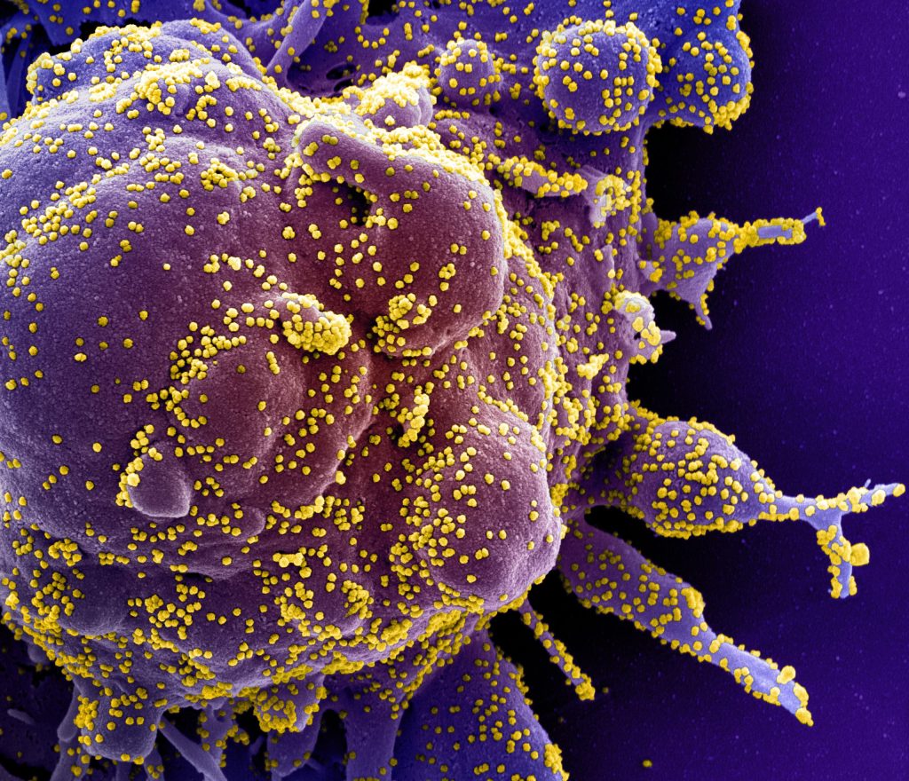
SARS-CoV-2 infecting a human cell. Credit: NIH

A rare autoimmune disease has been newly described as a COVID-related syndrome, following an investigation by the University of California San Diego School of Medicine and Leeds University.

It started when Pradipta Ghosh, MD, a professor in the Departments of Medicine and Cellular and Molecular Medicine at UC San Diego School of Medicine, received an email from Dennis McGonagle, PhD, professor of investigative rheumatology at the University of Leeds in the UK. This was the beginning of an international collaboration, one that uncovered a previously overlooked COVID-related syndrome and resulted in a paper in eBioMedicine, a journal published by The Lancet.

McGonagle asked if she was interested in collaborating on a COVID-related mystery. “He told me they were seeing mild COVID cases,” Ghosh said. “They had vaccinated around 90 percent of the Yorkshire population, but now they were seeing this very rare autoimmune disease called MDA5 – autoantibody associated dermatomyositis (DM) in patients who may or may not have contracted COVID, or even remember if they were exposed to it.”

McGonagle told of patients with severe lung scarring, some of whom presented rheumatologic symptoms – rashes, arthritis, muscle pain – that often accompany interstitial lung disease. He was curious to know if there was a connection between MDA5-positive dermatomyositis and COVID.

“DM is more common in individuals of Asian descent, particularly Japanese and Chinese,” Ghosh said. “However, Dr McGonagle was noting this explosive trend of cases in Caucasians.”

“But that’s the least of the problem,” Ghosh said. “Because he said, ‘Oh, and by the way, some of these patients are progressing rapidly to death.'”

Ghosh is the founding director of the Institute for Network Medicine at UC San Diego School of Medicine, home to the Center for Precision Computational Systems Network (PreCSN – the computational pillar within the Institute for Network Medicine). PreCSN’s signature asset is BoNE – the Boolean Network Explorer, a powerful computational framework for extracting actionable insights from any form of big-data.

“BoNE is designed to ignore factors that differentiate patients in a group while selectively identifying what is common (shared) across everybody in the group,” Ghosh explained. Previous applications of BoNE allowed Ghosh and her team to identify other COVID-related lung and heart-afflicting syndromes in adults and children, respectively.

As a rheumatologist, McGonagle specialises in inflammatory and autoimmune conditions. Ghosh said that McGonagle’s roster of patients, all within the UK’s National Health System (NHS), helped to facilitate the investigation.

“The NHS has a centralised health care database with comprehensive medical records for a large population, making it easier to access and analyse health data for research purposes,” Ghosh explained.

Ghosh and McGonagle put together a team to probe what they found was indeed an entirely new syndrome.

The study began with McGonagle lab’s detection of autoantibodies to MDA5 – an RNA-sensing enzyme whose functions include detecting COVID and other RNA viruses. A total of 25 patients from the group of 60 developed lung scarring, also known as interstitial lung disease. Ghosh noted that the lung scarring was bad enough to cause eight people in the group to die due to progressive fibrosis. She said that there are established clinical profiles of MDA5 autoimmune diseases.

“But this was different,” Ghosh said. “It was different in behaviour and rate of progression – and in the number of deaths.”

Ghosh and the UC San Diego team explored McGonagle’s data with BoNE. They found that the patients who showed the highest level of MDA5 response also showed high levels of interleukin-15.

“Interleukin-15 is a cytokine that can cause two major immune cell types,” she explained. “These can push cells to the brink of exhaustion and create an immunologic phenotype that is very, very often seen as a hallmark of progressive interstitial lung disease, or fibrosis of the lung.”

BoNE allowed the team to establish the cause of the Yorkshire syndrome – and pinpoint a specific single nucleotide polymorphism that is protective. By right of discovery, the group was able to give the condition a name: MDA5-autoimmunity and Interstitial Pneumonitis Contemporaneous with COVID. It’s MIP-C for short, “Pronounced ‘mipsy,'” Ghosh said, adding that the name was coined to make a connection with MIS-C, a separate COVID-related condition of children.

Ghosh said that it’s extremely unlikely that MIP-C is confined to the United Kingdom. Reports of MIP-C symptoms are coming from all over the world. She said she hopes the team’s identification of interleukin-15 as a causative link will jump start research into treatment.

Source: University of California – San Diego

Interferons Drive Lupus Symptoms and Affect Treatment

A woman with Systemic Lupus Erythematosus. Source: Wikimedia CC0

In a new study, researchers from Johns Hopkins Medicine say they have uncovered insights as to why lupus symptoms and severity present differently in individuals with the autoimmune condition. The team says this is a crucial step forward in understanding biological mechanisms behind lupus, and may also lead to shifts in how clinicians treat patients with the condition.

The full report, published in Cell Reports Medicine, concludes that specific combinations and elevated levels of immune system proteins, known as interferons, are associated with certain lupus symptoms such as skin rashes, kidney inflammation and joint pain. Interferons normally help to fight infection or disease, but are overactive in lupus, causing widespread inflammation and damage. The study also shows that other common lupus-related symptoms cannot be explained by increased interferon levels.

“For years, we have accumulated knowledge that interferons play a role in lupus,” says corresponding author and rheumatologist Felipe Andrade, MD, PhD, associate professor of medicine at the Johns Hopkins University School of Medicine. He says this research began with questions about why certain lupus treatments were ineffective for some patients. “We have seen instances where the patient surprisingly didn’t improve – we wondered if certain interferon groups were involved.”

Some lupus treatments are designed to suppress a specific group of interferons, known as interferon I. In clinical trials for these treatments, the team observed some patients failing to improve, despite genetic tests showing high interferon I levels before treatment, or what experts call a high interferon signature. The team believed that two other interferon groups, interferon II and interferon III, may be to blame for these poor treatment responses.

To investigate, the team looked at how different combinations of interferon I, II or III, and their overactivity, may present in people with lupus. Researchers took 341 samples from 191 participants to determine the activity of the three interferon groups, and used human cell lines engineered to react to the presence of each specific interferon group to analyse the samples. Through this process, researchers determined that the majority of participants fell into four categories: those only with increased interferon I; those with a combination of increased interferons I, II and III; those with a combination of increased interferons II and III; or those with normal interferon levels.

Researchers were able to use these findings to also make several associations between these interferon combinations and lupus symptoms. In those with elevated interferon I, lupus was mainly associated with symptoms affecting the skin, such as rashes or sores. Participants with elevated levels of interferon I, II and III exhibited the most severe presentations of lupus, often with significant damage to organ systems, such as the kidneys.

Not every symptom found in lupus was associated with elevated interferons, though. The formation of blood clots and low platelet counts, which also affect clotting, did not have an association with increased levels of interferon groups I, II or III. Researchers say this indicates that both interferon-dependent and other biological mechanisms are involved in this complex disease. The study also found that genetic testing of genes associated with these interferon groups, or the interferon signature, did not always indicate elevated interferon levels. They plan to investigate this in future studies.

“What we’ve seen in our study is that these interferon groups are not isolated; they work as a team in lupus and can give patients different presentations of the disease,” says rheumatologist Eduardo Gómez-Bañuelos, MD, PhD, assistant professor of medicine at Johns Hopkins and the study’s first and additional corresponding author. Evaluating a patient’s elevated interferon combinations allows for a better understanding of how they may react to treatments, and would allow clinicians to group them into clinical subtypes of lupus, Gómez-Bañuelos explains.

Source: Johns Hopkins Medicine

Experimental Type 1 Diabetes Drug Shields Pancreas Cells from Immune System Attack

A 3D map of the islet density routes throughout the healthy human pancreas. Source: Wikimedia CC0

An experimental monoclonal antibody drug called mAb43 appears to prevent and reverse the onset of clinical type 1 diabetes in mice, in some cases lengthening the animals’ lifespan, report scientists at Johns Hopkins Medicine.

The drug is unique, according to the researchers, because it targets insulin-making beta cells in the pancreas directly and is designed to shield those cells from attacks by the body’s own immune system cells. The drug’s specificity for such cells may enable long-term use in humans with few side effects, say the researchers. Monoclonal antibodies are made by cloning, or making identical replicas of, an animal (including human) cell line.

The findings, published in Diabetes, raise the possibility of a new drug for type 1 diabetes, an autoimmune condition which has no cure or means of prevention. Unlike type 2 diabetes, in which the pancreas makes too little insulin, in type 1 diabetes, the pancreas makes no insulin because the immune system attacks the pancreatic cells that make it.

The lack of insulin interferes with the body’s ability to regulate blood sugar levels.

According to Dax Fu, PhD, associate professor of physiology at the Johns Hopkins University School of Medicine and leader of the research team, mAb43 binds to a small protein on the surface of beta cells, which dwell in clusters called islets. The drug was designed to provide a kind of shield or cloak to hide beta cells from immune system cells that attack them as “invaders.” The researchers used a mouse version of the monoclonal antibody, and will need to develop a humanised version for studies in people.

For the current study, the researchers gave 64 non-obese mice bred to develop type 1 diabetes a weekly dose of mAb43 via intravenous injection when they were 10 weeks old. After 35 weeks, all mice were non-diabetic. One of the mice developed diabetes for a period of time, but it recovered at 35 weeks, and that mouse had early signs of diabetes before the antibody was administered.

In five of the same type of diabetes-prone mice, the researchers held off giving weekly mAb43 doses until they were 14 weeks old, and then continued dosages and monitoring for up to 75 weeks. One of the five in the group developed diabetes, but no adverse events were found, say the researchers.

In the experiments in which mAb43 was given early on, the mice lived for the duration of the monitoring period of 75 weeks, compared with the control group of mice that did not receive the drug and lived about 18-40 weeks.

Next, the researchers, including postdoctoral fellows Devi Kasinathan and Zheng Guo, looked more closely at the mice that received mAb43 and used a biological marker called Ki67 to see if beta cells were multiplying in the pancreas. They said, after treatment with the antibody, immune cells retreated from beta cells, reducing the amount of inflammation in the area. In addition, beta cells slowly began reproducing.

“mAb43 in combination with insulin therapy may have the potential to gradually reduce insulin use while beta cells regenerate, ultimately eliminating the need to use insulin supplementation for glycaemic control,” says Kasinathan.

The research team found that mAb43 specifically bound to beta cells, which make up about 1% or 2% of pancreas cells.

Another monoclonal antibody drug, teplizumab, received US Food and Drug Administration approval in 2022. Teplizumab binds to T cells, making them less harmful to insulin-producing beta cells. The drug has been shown to delay the onset of clinical (stage 3) type 1 diabetes by about two years, giving young children who get the disease time to mature and learn to manage lifelong insulin injections and dietary restrictions.

“It’s possible that mAb43 could be used for longer than teplizumab and delay diabetes onset for a much longer time, potentially for as long as it’s administered,” says Fu.

Source: John Hopkins Medicine

Scientists Discover Immune Key for Chronic Viral Infections

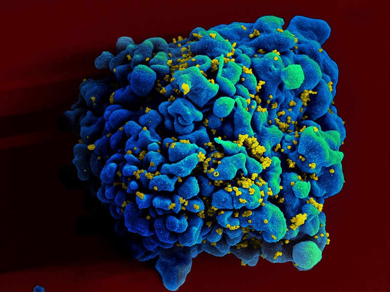
Colourised scanning electron micrograph of HIV (yellow) infecting a human T9 cell (blue). Credit: NIH

Australian researchers have discovered a previously unknown rogue immune cell that can cause poor antibody responses in chronic viral infections. The finding, published in the journal, Immunity, may lead to earlier intervention and possibly prevention of some types of viral infections such as HIV or hepatitis.

One of the remaining mysteries of the human immune system is why ‘memory’ B cells often only have a weak capacity to protect us from persistent infections.

In an answer to this, researchers from the Monash University Biomedicine Discovery Institute have now discovered that chronic viral infection induces a previously unknown immune B memory cell that does not produce high levels of antibodies.

Importantly the research team, led by Professor Kim Good-Jacobson and Dr Lucy Cooper, also determined the most effective time during the immune response for therapeutics such as anti-viral and anti-cancer drugs to better boost immune memory cell development.

“What we discovered was a previously unknown cell that is produced by chronic viral infection. We also determined that early intervention with therapeutics was the most effective to stop this type of memory cell being formed, whereas late intervention could not,” Professor Good-Jacobson said.

According to Dr Cooper, chronic viral infections have been known to alter our ability to form effective long-term protective antibody responses, but how that happens is unknown.

“In the future, this research may result in new therapeutic targets, with the aim to reduce the devastating effect of chronic infectious diseases on global health, specifically those that are not currently preventable by vaccines,” she said.

“Revealing this new immune memory cell type, and what genes it expresses, allows us to determine how we can target it therapeutically and whether that will lead to better antibody responses.”

The research team are also looking to see whether this population is a feature of long COVID, which results in some people having a reduced capacity to fight off the symptoms of COVID infection long after the virus has dissipated.

Source: Monash University

How Glucocorticoids Reprogram Immune Cells to Slow them Down

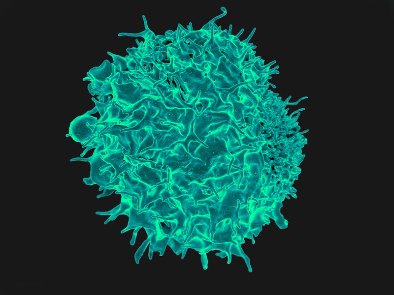
Scanning electron micrograph of a T cell lymphocyte. Credit: NIH / NIAID

Cortisone and other related glucocorticoids are extremely effective at curbing excessive immune reactions. But previously, astonishingly little was known about how they exactly do that. A team of researchers have now explored the molecular mechanism of action in greater detail. As the researchers report in the journal Nature, glucocorticoids reprogram the metabolism of immune cells, activating the body’s natural “brakes” on inflammation. These findings lay the groundwork for development of anti-inflammatory agents with fewer and less severe side effects.

The glucocorticoid cortisone is naturally present in the body as the stress hormone cortisol, which is released to improve the body’s responses to stress. Cortisol intervenes in sugar and fat metabolism and affects other parameters, including blood pressure and respiratory and heart rate. At higher doses, it also inhibits immune system activity, making it it useful for medical purposes. Due to their excellent efficacy, synthetic glucocorticoid derivates that inhibit inflammation even more strongly are used to treat a wide range of immune-mediated inflammatory diseases.

Glucocorticoids affect not only genes, but also cellular energy sources

Glucocorticoid-based medications come with side effects, especially at higher doses and when administered for longer periods. These side effects are related to the other effects of the body’s own cortisol, and include hypertension, osteoporosis, diabetes, and weight gain. With the aim of developing anti-inflammatory agents with fewer and less severe side effects, a team of researchers from from Charité – Universitätsmedizin Berlin, Uniklinikum Erlangen and Ulm University has now conducted a closer study of how the immunosuppressive effects of glucocorticoids exactly works.

Lead researcher Prof Gerhard Krönke, director of the Department of Rheumatology and Clinical Immunology at Charité, explains: “It was previously known that glucocorticoids activate a number of genes in different cells of the body. But through this mechanism, they mainly activate the resources present in the body. This does not adequately explains its strong immunosuppressive effect. In our study, we have now been able to show that glucocorticoids affect more than just the gene expression in immune cells. It also affects the cell´s powerhouses, the mitochondria. And that this effect on cell metabolism is in turn crucial to the anti-inflammatory effects exerted by glucocorticoids.”

Swords to plowshares

For the study, the research team focused on macrophages, a type of immune cell responsible for eliminating intruders such as viruses and bacteria. These cells can also play a role in the emergence of immune-mediated inflammatory diseases. In a mouse model, the researchers studied how these immune cells responded to inflammatory stimuli in a laboratory setting and what effects additional administration of a glucocorticoid had. The researchers observed that in addition to its effect on gene expression, glucocorticoids had a major effect in reversing changes in the cell metabolism that had been initiated by the inflammatory stimuli.

“When macrophages are put into ‘fight’ mode, they redirect their cellular energy into arming for a fight. Instead of supplying energy, their mitochondria produce the components needed to fight intruders,” Krönke says, describing the processes involved. “Glucocorticoids reverse the process, switching the ‘fight’ mode back off and turning swords into plowshares, so to speak. A tiny molecule called itaconate plays an especially important role in this.”

Itaconate mediates anti-inflammatory effect of glucocorticoids

Itaconate is an anti-inflammatory substance that the body naturally produces inside its mitochondria. Macrophages produce it early on when they are activated so that the inflammatory reaction will subside after a certain period. Generation of this natural immune “brake,” however, requires sufficient fuel. When the cell´s powerhouses are arming up for a fight, that is no longer the case, so itaconate production dwindles to a halt after a while. With normal, short-term inflammation, this timing is effective because the immune response has also subsided in the meantime.

“With a persistent inflammatory stimulus, the drop-off in itaconate production is an issue because there is then no immune ‘brake’ even though the immune system is still running on all cylinders, eventually contributing to chronic inflammation,” explains Dr Jean-Philippe Auger, a scientist at the Department of Medicine 3 – Rheumatology and Immunology at Uniklinikum Erlangen and the first author of the study. “This is where glucocorticoids intervenes. By reprogramming the mitochondrial function, they ramp up the formation of itaconate in the macrophages, restoring its anti-inflammatory effect.”

The search for new active substances

Using animal models for asthma and rheumatoid arthritis, the researchers showed how much the anti-inflammatory effect of glucocorticoids depends on itaconate. Glucocorticoids had no effect in animals unable to produce itaconate. So, if itaconate mediates the immunosuppressant effect of cortisone, what about administering itaconate directly, instead of glucocorticoids?

“Unfortunately, itaconate isn’t a particularly good candidate as an anti-inflammatory drug, because it’s unstable, and due to its high reactivity, it could cause side effects if administered systemically,” Krönke explains. “Aside from that, we assume the processes in humans to be a bit more complex than those in mice. So our plan is to look for new synthetic compounds that are just as effective as glucocorticoids at reprogramming the mitochondrial metabolism inside immune cells, but have fewer and less severe side effects.”

Source: Charité – Universitätsmedizin Berlin

Liver Immune System Quickly ‘Eats up’ LDL Cholesterol

Colourised electron micrograph image of a macrophage. Credit: NIH

A new study reveals that immune cells in the liver react to high cholesterol levels and eat up excess cholesterol that can otherwise cause damage to arteries. The findings, published in Nature Cardiovascular Research, suggest that the response to the onset of atherosclerosis begins in the liver.

Immediate response from the liver

In the current study, researchers from Karolinska Institutet wanted to understand how different tissues in the body react to high levels of LDL, commonly called ‘bad cholesterol’, in the blood.

To test this, they created a system where they could quickly increase the cholesterol in the blood of mice.

“Essentially, we wanted to detonate a cholesterol bomb and see what happened next,” says Stephen Malin, lead author of the study and principal researcher at the Department of Medicine, Solna, Karolinska Institutet.

“We found that the liver responded almost immediately and removed some of the excess cholesterol.”

However, it wasn’t the typical liver cells that responded, but a type of immune cell called Kupffer cells that are known for recognising foreign or harmful substances and eating them up. The discovery made in mice was also validated in human tissue samples.

“We were surprised to see that the liver seems to be the first line of defence against excess cholesterol and that the Kupffer cells were the ones doing the job,” says Stephen Malin.

“This shows that the liver immune system is an active player in regulating cholesterol levels, and suggests that atherosclerosis is a systemic disease that affects multiple organs and not just the arteries.”

Several organs could be involved

The researchers hope that by understanding how the liver and other tissues communicate with each other after being exposed to high cholesterol, they can find new ways to prevent or treat cardiovascular and liver diseases.

“Our next step is to look at how other organs respond to excess cholesterol, and how they interact with the liver and the blood vessels in atherosclerosis,” says Stephen Malin. “This could help us develop more holistic and effective strategies to combat this common and deadly disease.”

Source: Karolinska Institutet

Scientists Peer into a Transporter Protein for Inflammatory Signals

In the human body, a protein carrier called SPNS2 transports S1P molecules from endothelial cells to rally immune cell response in infected organs and tissues, resulting in inflammation. By enlarging the entire SPNS2 structure using nanoparticles, the S1P molecules contained within can be viewed via cryogenic electron microscopy. Using this information, small molecules can be developed to inhibit this signalling pathway and treat inflammatory diseases.

Scientists at the National University of Singapore and colleagues in China have analysed the structure of the SPNS2 protein at an atomic level that could provide greater insights into how S1P signalling molecules are released to communicate with the immune cells to regulate inflammatory responses. Their findings are published in Cell Research.

“Seeing is believing. This work shows that SPNS2 is directly exporting S1P for signalling and it is possible to inhibit its transport function with small molecules. This work provides the foundation for understanding of how S1P is released by SPNS2 and how this protein function is inhibited by small molecules for treatment of inflammatory diseases,” said team leader Dr Nguyen Nam Long.

The SPNS2 protein allows the binding of the S1P signalling molecules to trigger the immune cells to leave the lymph nodes and induce inflammation in different parts of the body when needed.

Made up of amino acids, the SPNS2 protein is malleable enough to change its shape and structure to release the S1P signalling molecules through small cavities found within the protein.

Through the discovery of how the SPNS2 protein releases S1P molecules, the SPNS2 structure can be exploited for future drug development.

Similar to discovering how the shape of the lock looks like before the key can be designed, this finding sheds more light into how future drugs can be designed to better target the protein to increase drug efficacy.

This finding builds on previous research which found that deleting SPNS2 protein from a pre-clinical model effectively blocks the S1P signalling pathway so that the S1P signalling molecules are unable to be transported to prompt immune cells to leave the lymph node to induce inflammation.

Both SPNS2 protein and S1P signalling molecule are required for immune cell recruitment to inflammatory organs, which goes towards treating various inflammatory diseases.

“Using pre-clinical models, we have shown that targeting SPNS2 proteins in the body blocks inflammatory responses in disease conditions, such as multiple sclerosis. This work has provided us a possibility to inhibit its transport function with small molecules that will go a long way to treating inflammatory diseases more efficiently and effectively,” said Dr Nguyen.

Source: National University of Singapore, Yong Loo Lin School of Medicine

Smoking Affects the Immune System Many Years after Quitting

Photo by Sara Kurfess on Unsplash

Researchers from Institut Pasteur have discovered that the immune impacts of smoking can last for many years, leaving smokers with effects on some of their bodies’ defence mechanisms acquired while smoking. These findings, which for the first time reveal a long-term memory of the effects of smoking on immunity, are published in the journal Nature.

Individuals’ immune systems vary significantly in terms of how effectively they respond to microbial attacks. But how can this variability be explained? What factors cause these differences? “To answer this key question, we set up the Milieu Intérieur cohort comprising 1000 healthy individuals aged 20 to 70 in 2011,” explains Darragh Duffy, Head of the Translational Immunology Unit at the Institut Pasteur and last author of the study. While certain factors such as age, sex and genetics are known to have a significant impact on the immune system, the aim of this new study was to identify which other factors had the most influence.”

The scientists exposed blood samples taken from individuals in the Milieu Intérieur cohort to a wide variety of microbes and observed their immune response by measuring levels of secreted cytokines(1). Using the large quantities of data gathered for individuals in the cohort, the team then determined which of the 136 investigated variables (body mass index, smoking, number of hours’ sleep, exercise, childhood illnesses, vaccinations, living environment, etc) had the most influence on the immune responses studied. Three variables stood out: smoking, latent cytomegalovirus infection(2) and body mass index. “The influence of these three factors on certain immune responses could be equal to that of age, sex or genetics,” points out Darragh Duffy.

As regards smoking, an analysis of the data showed that the inflammatory response, which is immediately triggered by infection with a pathogen, was heightened in smokers, and moreover, the activity of certain cells involved in immune memory was impaired. In other words, this study shows that smoking disrupts not only innate immune mechanisms, but also some adaptive immune mechanisms. “A comparison of immune responses in smokers and ex-smokers revealed that the inflammatory response returned to normal levels quickly after smoking cessation, while the impact on adaptive immunity persisted for 10 to 15 years,” observes Darragh Duffy. “This is the first time it has been possible to demonstrate the long-term influence of smoking on immune responses.”

Basically, the immune system appears to have something resembling a long-term memory of the effects of smoking. But how? “When we realised that the profiles of smokers and ex-smokers were similar, we immediately suspected that epigenetic processes were at play(3),” says Violaine Saint-André, a bioinformatician in the Institut Pasteur’s Translational Immunology Unit and first author of the study. “We demonstrated that the long-term effects of smoking on immune responses were linked to differences in DNA methylation(4) – with the potential to modify the expression of genes involved in immune cell metabolism – between smokers, ex-smokers and non-smokers.” It therefore appears that smoking can induce persistent changes to the immune system through epigenetic mechanisms.

“This is a major discovery elucidating the impact of smoking on healthy individuals’ immunity and also, by comparison, on the immunity of individuals suffering from various diseases,” concludes Violaine Saint-André.

Notes:

(1) proteins secreted by a large number of immune cells to communicate among themselves and participate in immune defense.

(2) a virus in the herpes family that is often asymptomatic though dangerous to foetuses.

(3) changes in DNA that affect how genes are expressed, i.e. how they are used by cells.

(4) methylation is a type of chemical modification. Methyl groups position themselves on DNA, changing the way in which the genome is read in the cell.

Source: Institut Pasteur

‘Junk Cells’ Actually Have a Powerful Role against Malaria

Red blood cell Infected with malaria parasites. Colourised scanning electron micrograph of red blood cell infected with malaria parasites (teal). The small bumps on the infected cell show how the parasite remodels its host cell by forming protrusions called ‘knobs’ on the surface, enabling it to avoid destruction and cause inflammation. Uninfected cells (red) have smoother surfaces. Credit: NIAID

Researchers from The Australian National University (ANU) have discovered a previously unknown ability of a group of immune system cells, known as Atypical B cells (ABCs), to fight infectious diseases such as malaria.

The discovery, published in Science Immunology, provides new insight into how the immune system fights infections and brings scientists a step closer to harnessing the body’s natural defences to combat malaria.

The scientists say ABCs could also be key to developing new treatments for chronic autoimmune conditions such as lupus. According to the researchers, ABCs have long been associated with malaria, as malaria patients have more of these cells in their system compared to the general population.

“In this study, we wanted to understand the mechanisms that drive the creation of ABCs in the immune system, but also find out whether these cells are good or bad for us when it comes to fighting infection,” lead author Dr Xin Gao, from ANU, said.

“Although ABCs are known to contribute to chronic inflammatory diseases and autoimmunity, we’ve discovered a previously unknown ability of these cells to fight disease. In this sense, ABCs are like a double-edged sword.

“Contrary to past belief, ABCs are not junk cells; they are more important than we thought.

“Our research found that ABCs are also instrumental in developing T follicular helper cells. These helper cells generate powerful antibodies that help the body fight malaria parasites.

“Antibodies can block parasites in the blood as they travel from the site of the infectious mosquito bite to the liver, where the infection is first established.”

In 2022, malaria killed more than 600 000 people worldwide. Although the disease is preventable and curable, scientists face an uphill battle to find long-lasting treatments as malaria parasites continue to find new ways to build resistance to current therapies.

Using gene-editing technology on mice, the ANU researchers discovered a gene called Zeb2 is crucial to the production of ABCs.

“We found that manipulating the Zeb2 gene disrupted the creation of ABCs in the immune system,” study co-author Professor Ian Cockburn, from The ANU John Curtin School of Medical Research, said.

“Importantly, we found that mice without the Zeb2 gene were unable to control malaria infection.

“Therefore, the findings show that ABCs play a crucial role in fighting malaria infections.”

The researchers say targeting ABCs could also pave the way for new treatments for certain autoimmune diseases such as lupus.

“ABCs also appear in large numbers in many autoimmune diseases, including lupus, which can be life-threating in severe cases,” Professor Cockburn said.

“By developing a better understanding of the role of ABCs in the immune system and the cells’ role in fighting disease, it could bring us a step closer to one day developing new and more effective therapies.”

Source: Australian National University

Understanding How T Cells Target Tuberculosis will Enhance Vaccines and Therapies

Tuberculosis bacteria. Credit: CDC

La Jolla Institute for Immunology (LJI) is working to guide the development of new tuberculosis vaccines and drug therapies. Now a team of LJI scientists has uncovered important clues to how human T cells combat Mycobacterium tuberculosis, the bacterium that causes TB. Their findings were published recently in Nature Communications.

“This research gives us a better understanding of T cell responses to different stages in tuberculosis infection and helps us figure out is there are additional diagnostic targets, vaccine targets, or drug candidates to help people with the disease,” says LJI Research Assistant Professor Cecilia Lindestam Arlehamn, PhD, who led the new research in collaboration with LJI Professors Bjoern Peters, PhD, and Alessandro Sette, Dr.Biol.Sci.

The urgent need for TB research

According to the World Health Organization, more than 1.3 million people died of TB in 2022, making it the second-leading infectious cause-of-death after COVID. “TB is a huge problem in many countries,” says Lindestam Arlehamn.

Currently, a vaccine called bacille Calmette-Guerin (BCG) protects against some severe cases of TB. Unfortunately, BCG doesn’t consistently prevent cases of pulmonary TB, which can also be deadly.

Although there are drug treatments for TB, more and more cases around the world have proven drug resistant.

To help stop TB, Lindestam Arlehamn and her colleagues are learning from T cells. Instead of targeting an entire pathogen, T cells look for specific markers, called peptides sequences, that belong to the pathogen.

When a T cell recognises a certain part of a pathogen’s peptide sequence, that area is termed an “epitope.”

Uncovering T cell epitopes gives scientists vital information on how vaccines and drug treatments might take aim at the same epitopes to stop a pathogen.

T cells take aim at a range of TB epitopes

For the new study, the researchers worked with samples from patients who were mid-treatment for active TB. These samples came from study participants in Peru, Sri Lanka, and Moldova.

By looking at T cells in patients from three different continents, the researchers hoped to capture a wide diversity of genetics and environmental factors that can affect immune system activity.

In their analysis, the LJI team uncovered 137 unique T cell epitopes. They found that 16% of these epitopes were targeted by T cells found in two or more patients. The immune system appeared to be working hard to zoom in on these epitopes.

Going forward, Lindestam Arlehamn’s laboratory will investigate which of these epitopes may be promising targets for future TB vaccines and drug therapies.

A step toward better diagnostics

The new study is also a step toward catching TB cases before they turn deadly.

Because Mycobacterium tuberculosis is an airborne bacteria, a person can be exposed without ever realizing it. Once exposed, many people go months or years without any symptoms.

This inactive, or “latent,” TB can turn into active TB if a person’s immune system weakens, for example, during pregnancy or due to an infection such as HIV.

For the new study, the researchers also compared samples from active TB patients with samples from healthy individuals.

The scientists uncovered key differences in T cell reactivity between the two groups.

“For the first time, we could distinguish people with active TB versus those that have been exposed to TB – or unexposed individuals,” says Lindestam Arlehamn.

Lindestam Arlehamn says it may be possible to develop diagnostics that detect this tell-tale T cell reactivity that marks a person’s shift from latent to active TB. “Can we use this peptide pool to look for high-risk individuals and try and follow them over time?” she says.

Source: La Jolla Institute for Immunology