Category: Immune System

Initial Immune Reaction Determines Severity of COVID

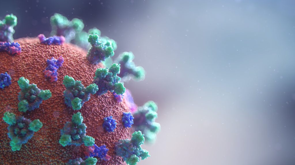
Image source: CDC on Unsplash

Researchers have found that the course of severe COVID could be determined very early on, depending on the body’s initial reaction to the disease in the upper airway as well as inflammatory reactions.

Scientists at the Ragon Institute of MGH, MIT, and Harvard; the Broad Institute of MIT and Harvard; Boston Children’s Hospital (BCH); MIT; and the University of Mississippi Medical Center (UMMC) wondered whether COVID’s path towards severe disease could start much earlier than expected — perhaps even within the initial response created when the virus enters the nose.

To test this, they studied cells taken from nasal swabs of patients at the time of their initial COVID diagnosis, comparing patients who went on to develop mild COVID to those who progressed into more severe disease and eventually required respiratory support. Their results showed that patients who went on to develop severe COVID exhibited a much more muted antiviral response in the cells collected from those early swabs, compared to patients who had a mild course of the disease. The paper appears in Cell.

“We wanted to understand if there were pronounced differences in samples taken early in the course of disease that were associated with different severities of COVID as the disease progressed,” said co-senior author José Ordovás-Montañés, an associate member in the Klarman Cell Observatory at Broad and assistant professor at BCH and Harvard Medical School. “Our findings suggest that the course of severe COVID may be determined by the body’s intrinsic antiviral response to initial infection, opening up new avenues for early interventions that could prevent severe disease.”

To understand the early response to infection, Sarah Glover of the Division of Digestive Diseases at UMMC and her laboratory collected nasal swabs from 58 people, 35 of whom were just recently diagnosed with COVID, representing a variety of disease states from mild to severe. Seventeen swabs came from healthy volunteers and six came from patients with other causes of respiratory failure. The team  sequenced RNA from these samples to find out what kind of proteins the cells were making — a snapshot of the cell’s activity when collected.

By studying a cell’s transcriptome, which is its collection of RNA, can researchers understand how a cell is responding to environmental changes such as a viral infection. It can even be used to see if individual cells are infected by an RNA virus-like SARS-CoV-2.

“Our single-cell sequencing approaches allow us to comprehensively study the body’s response to disease at a specific moment in time,” said co-senior author Alex Shalek, who is also an associate professor at MIT in the Institute for Medical Engineering & Science, the Department of Chemistry, and the Koch Institute for Integrative Cancer Research. “This gives us the ability to systematically explore features that differentiate one course of disease from another as well as cells that are infected from those that are not. We can then leverage this information to guide the development of more effective preventions and cures for COVID and other viral infections.”

Analysing the transcriptome, the team investigated how epithelial and immune cells were responding to early COVID infection from the single-cell transcriptome data. Firstly, in patients who progressed to severe COVID, the initial interferon-driven antiviral response was muted. Second, patients with severe COVID had higher amounts of highly inflammatory macrophages, and high inflammation levels are often seen in severe or fatal COVID.

Since these samples were taken well before COVID had peaked in the patients, both these findings indicate that COVID’s course may be determined by the initial response of the nasal epithelial and immune cells to the virus. The weak initial antiviral response may allow a rapid spread of the virus, making it more likely to move from upper to lower airways, while the recruitment of inflammatory immune cells could help drive the dangerous inflammation in severe disease.

Finally, the team also identified infected host cells and pathways associated with protection against infection — cells and responses unique to patients that went on to develop mild disease. These findings may allow researchers to discover new therapeutic strategies for COVID and other respiratory viral infections.

If the early stages of infection can determine disease, it could enable the development of early interventions that can help prevent the development of severe COVID. Potential markers of severe disease were also identified, genes that were expressed in mild, but not severe COVID.

“Nearly all our severe COVID samples lacked expression of several genes we would typically expect to see in an antiviral response,” said co-first author Carly Ziegler, a graduate student in the Health Science and Technology Program, MIT and Harvard.

“If further studies support our findings, we could use the same nasal swabs we use to diagnose COVID-19 to identity potentially severe cases before severe disease develops, creating an opportunity for effective early intervention,” said Ziegler.

Source: Broad Institute of MIT and Harvard

Journal information: Ziegler, C G K., et al (2021) Impaired local intrinsic immunity to SARS-CoV-2 infection in severe COVID-19. Cell. doi.org/10.1016/j.cell.2021.07.023.

Autoimmune Problems May Cause Fibromyalgia

Photo by Sasun Bughdaryan on Unsplash

New research has shown that many fibromyalgia syndrome (FMS) symptoms are caused by antibodies that increase the activity of pain-sensing nerves throughout the body.

The results show that fibromyalgia is a disease of the immune system, rather than the currently held view that it originates in the brain.

Characterised by widespread muskoleskeletal pain, as well as fatigue and emotional distress, fibromyalgia is estimated to affect1 in 40 people (80% of which are women). It most commonly develops between the ages of 25 and 55, although children can also get it.  

The study by Institute of Psychiatry, Psychology & Neuroscience (IoPPN) at King’s College London, in collaboration with the University of Liverpool and the Karolinska Institute,, demonstrates that the increased pain sensitivity, muscle weakness, reduced movement, and reduced number of small nerve-fibres in the skin that are typical of FMS are all a consequence of patient antibodies.

The researchers injected mice with antibodies from people living with FMS and saw that the mice became more sensitive to pressure and cold, as well as displaying reduced movement grip strength. In contrast, those injected with antibodies from healthy people were unaffected, showing that patient antibodies cause at least part of the disease.

Furthermore, the mice injected with fibromyalgia antibodies recovered after a few weeks, when antibodies had been cleared from their system. This finding strongly suggests that therapies which reduce antibody levels in patients are likely to be effective treatments. Such therapies are already available and are used to treat other disorders that are caused by autoantibodies.

Primary investigator Dr David Andersson, from King’s IoPPN said: “The implications of this study are profound. Establishing that fibromyalgia is an autoimmune disorder will transform how we view the condition and should pave the way for more effective treatments for the millions of people affected. Our work has uncovered a whole new area of therapeutic options and should give real hope to fibromyalgia patients.

“Previous exploration of therapies has been hampered by our limited understanding of the illness. This should now change. Treatment for FMS is focussed on gentle aerobic exercises, as well as drug and psychological therapies designed to manage pain, although these have proven ineffective in most patients and have left behind an enormous unmet clinical need.”

Dr. Andreas Goebel, the study’s principle clinical investigator from the University of Liverpool said, “When I initiated this study in the UK, I expected that some fibromyalgia cases may be autoimmune. But David’s team have discovered pain-causing antibodies in each recruited patient. The results offer amazing hope that the invisible, devastating symptoms of fibromyalgia will become treatable.”

Professor Camilla Svensson, the study’s primary investigator from Karolinska Institute said, “Antibodies from people with FMS living in two different countries, the UK and Sweden, gave similar results, which adds enormous strength to our findings. The next step will be to identify what factors the symptom-inducing antibodies bind to. This will help us not only in terms of developing novel treatment strategies for FMS, but also of blood-based tests for diagnosis, which are missing today.

Dr Craig Bullock, Research Discovery and Innovations Lead at Versus Arthritis said: “This research shows that antibodies found in human blood can cause fibromyalgia-like symptoms in mice, suggesting that these antibodies play a crucial role in the condition. Further research is needed but this offers hope to the millions of people with fibromyalgia that an effective treatment could be found in the relatively near future.”  

Source: Medical Xpress

Journal information: More information: Andreas Goebel et al, Passive transfer of fibromyalgia symptoms from patients to mice, Journal of Clinical Investigation (2021). DOI: 10.1172/JCI144201

Connective Tissue Protein Has Immune Role

Photo by National Cancer Institute on Unsplash

A new study finds that a connective tissue protein also encourages immune responses that fight bacterial infections, while restraining responses that can be deadly in sepsis.

The study focuses on the extracellular matrix (ECM) of connective tissues, once viewed merely as structural material. It is now increasingly recognised as a signaling partner with nearby cells in normal function, as well as being involved in disease. Fibroblasts are important players in the ECM; these cells make tough structural matrix proteins like collagen. The study was published online June 28 in the Proceedings of the National Academy of Sciences.

The new analysis found that lumican, a protein-sugar combination (proteoglycan) secreted by fibroblasts, and known to partner with collagen in connective tissues, also promotes immune system responses in immune cells called macrophages that fight bacterial infections. The study also found that lumican protects tissues by holding back a different type of immune response that reacts to DNA, whether from an invading virus, or released from cell death.

Such inflammatory responses are a transition into healing, but in sepsis they grow out of control, causing damage to the body’s own tissues. Sepsis affects 48.9 million people worldwide, the authors said, but the ECM’s role the condition is largely unknown.

“Lumican may have a dual protective role in ECM tissues, promoting defense against bacteria on the one hand, and on the other, limiting immune overreactions to DNA that cause self-attack, or autoimmunity,” said corresponding study author Shukti Chakravarti, PhD, professor in the Department of Ophthalmology and the Department of Pathology at NYU Langone Health.

The findings suggest that connective tissue, and extracellular matrix proteins like lumican, usually operate outside of cells, but as disease or damage break down ECM, get sucked into and regulate immune cells homing in on the damage.

Lumican  interacts with two proteins on surfaces of immune cells that control the activity of toll-like receptors, which recognise structural patterns common to molecules made by invading microbes, said the researchers. As they are less specific than other parts of the immune system, toll-like receptors can also cause attacks by immune cells on the body’s own tissues if over-activated.

In this study, the researchers found that lumican promotes the ability of toll-like receptor (TLR)-4 on the surfaces of immune cells to recognise bacterial cell-wall toxins called lipopolysaccharides (LPS). Lumican, by attaching to two proteins, CD14 and Caveolin1, probably using collagen-covered regions, stabilises their interactions with TLR4 to increase its ability to react to LPS. This results in production of the signalling protein TNF alpha, which amplifies immune responses.

Along with describing the effect of lumican on the surfaces of immune cells, the new study finds that lumican is taken up from outside cells into membrane-bound pouches, called endosomes, and pulled into cells. Such compartments deliver ingested bacteria to other endosomes that destroy them, heighten inflammation, or produce protective interferon responses. Once pulled inside, the researchers found, lumican bolstered TLR4 activity by slowing down its passage into lysosomes, pockets where such proteins are broken down and recycled.

However, while it encouraged TLR4 activity on cell surfaces, lumican, once inside immune cells, had the opposite effect on toll-like receptor 9 (TLR9), which reacts to DNA instead of bacterial LPS.

Mice with the lumican gene deleted had trouble both fighting off bacterial infections (less cytokine response, slower clearance, greater weight loss), and trouble restraining the immune overreaction to bacteria (sepsis). Elevated lumican levels were also found in human sepsis patients’ blood plasma, and that human immune cells (blood monocytes) treated with lumican had elevated TLR4 activity but suppressed TLR9 responses.

“As an influencer of both processes, lumican-based peptides could be used as a lever, to tweak inflammation related to TNF-alpha, or endosomal interferon responses, to better resolve inflammation and infections,” suggested George Maiti, PhD, a postdoctoral fellow in Dr Chakravarti’s lab.

“Our results argue for a new role for ECM proteins at sites of injury. Taken up by incoming immune cells it shapes immune responses beyond the cell surface by regulating the movement and interaction of endosomal receptors and signaling partners,” said Dr Chakravarti.

Source: NYU Langone Health

Journal information: George Maiti et al., “Matrix lumican endocytosed by immune cells controls receptor ligand trafficking to promote TLR4 and restrict TLR9 in sepsis,” PNAS (2021). www.pnas.org/cgi/doi/10.1073/pnas.2100999118

In the Immune Battle, MRSA Uses Toxins to Fight Dirty

Scanning electron micrograph of methicillin-resistant Staphylococcus aureus and a dead human neutrophil. Credit: NIAID

Researchers have uncovered a novel trick employed by the bacterium Staphylococcus aureus — MRSA uses toxins to ‘fight dirty’ and stifle the immune response. This finding is a step towards one day producing a vaccine against MRSA.

Every year, there are some 700 000 deaths due to the emerging global threat of antimicrobial resistance (AMR). Turning the tables against AMR requires immediate action, and the development of novel vaccines to prevent such infections in the first place, are an attractive and potentially very effective option.

Staphylococcus aureus is the causative agent of the infamous MRSA ‘superbug’, one of the chief concerns of AMR. Immunologists from Trinity College Dublin, working with scientists at GSK, discovered the deadly bacteria’s new trick to foil the immune system. They found that the bacterium interferes with the host immune response by causing toxic effects on white blood cells, preventing them from carrying out their infection-fighting jobs.

The study also showed that the toxicity could be lessened following vaccination with a mutated version of a protein specifically engineered to throw a spanner in the MRSA works. This could one day lead to a vaccine for humans.

Rachel McLoughlin, Professor in Immunology in Trinity’s School of Biochemistry and Immunology and the Trinity Biomedical Sciences Institute (TBSI), said: “As a society we are witnessing first-hand the powerful impact that vaccination can have on curbing the spread of infection. However, in the backdrop of the COVID epidemic we must not lose sight of the fact that we are also waging war on a more subtle epidemic of antimicrobial resistant infection, which is potentially equally deadly.

“In this study we have identified a mechanism by which a protein made by the bacterium – known as Staphylococcal Protein A (SpA) – attacks and rapidly kills white blood cells. This protein has been widely studied for its immune evasion capacity and has a well-documented role in rendering antibodies raised against the bacterium non-functional.

“Here we uncover a previously undocumented strategy by which SpA forms immune complexes through its interaction with host antibodies, that in turn exert toxic effects on multiple white blood cell types. This discovery highlights how important it will be for effective vaccines to be capable of disarming the effects of protein A.”

Dr Fabio Bagnoli, Director, Research & Development Project Leader, GSK, said: “Our collaboration with Trinity College Dublin and in particular with Professor Rachel McLoughlin, a worldwide recognised expert on staphylococcal immunology, is critical for increasing our knowledge on protective mechanisms against S. aureus.”

The study documents the latest discovery made by this group at Trinity under an ongoing research agreement with GSK Vaccines (Siena, Italy). Overall, this collaboration aims to increase understanding of the immunology of Staphylococcus aureus infection to advance development of next-generation vaccines to prevent MRSA infections.

Source: Trinity College Dublin

Journal information: Fox, P. G., et al. (2021) Staphylococcal Protein A Induces Leukocyte Necrosis by Complexing with Human Immunoglobulins. Scientific Reports. doi.org/10.1128/mBio.00899-21.

Uncovering Albumin’s Role in Fertility and Inflammation

Source: CC0

Researchers have discovered that albumin (Alb), one of the most abundant proteins in the body, activates a proton channel (hHv1), also widespread in the body, giving sperm the ability to penetrate and fertilise an egg, and also allowing white blood cells to produce inflammatory mediators to fight infection.

The study explored the physiological connection between Alb and human voltage-gated proton channels (hHv1), which are both essential to cell biology in health and diseases. Researchers also demonstrated the mechanism by which Alb binds directly to hHv1 to activate the channel. This research explains how sperm are triggered to fertilise, and neutrophils are stimulated to release mediators in the innate immune response, describing a new role for Alb in physiology that will operate in the many tissues expressing hHv1.

“We found that the interaction of Alb and hHv1 activates sperm when they leave semen and enter the female reproductive tract because Alb is low in semen and high in the reproductive tract. We now understand why albumin supplementation improves IVF,” explained first author Ruiming Zhao, PhD, from the Department of Physiology & Biophysics at UCI School of Medicine. “We also found the same Alb/hHv1 interaction allows the white blood cells called neutrophils to produce and secrete the inflammatory mediators that kill bacteria and fight infection. However, it’s important to note that the inflammatory response itself can lead to disease.”

Alb’s stimulating role in the physiology of sperm and neutrophils via hHv1 pointed to its having other enhancing or deleterious roles in the other tissues, including the central nervous system, heart and lungs, and influencing cancers of the breast and gastrointestinal tract.

“It is exciting to discover that a common protein has the power to activate the proton channel.  This finding suggests new strategies to block or enhance fertility, and to augment or suppress the innate immune response and inflammation,” said senior author Steve A. N. Goldstein, MD, PhD, vice chancellor of Health Affairs at UCI.

hHv1 is involved in many biological processes in addition to the capacitation of sperm and the innate immune responses included in the study.  The channels have notable roles in proliferation of cancer cells, tissue damage during ischaemic stroke, and hypertensive kidney injury. Because Alb’s presence and involvement varies, the potentiation of hHv1 by Alb can be either beneficial or detrimental in different diseases or conditions.

“We have modeled the structural basis for binding of Alb to the channel that leads to activation and changes in cellular function, and we are now conducting in vivo studies of viral and bacterial infections.  Our next steps include studies of the effects of inhibitors of the Alb-hHv1 interaction on infection, inflammation and fertility,” said Goldstein.  

Source: University of California, Irvine

Journal information: Ruiming Zhao et al, Direct activation of the proton channel by albumin leads to human sperm capacitation and sustained release of inflammatory mediators by neutrophils, Nature Communications (2021). DOI: 10.1038/s41467-021-24145-1

Chronic Inflammation Ages the Pituitary Gland in Mice

Photo by Robina Weermeijer on Unsplash

Researchers have discovered that the pituitary gland in mice ages due to an age-related form of chronic inflammation — which raises the possibility of slowing or even partially repairing this process. 

The pituitary gland is a small, globular gland located underneath the brain that plays a major role in the hormonal system, explained Professor Hugo Vankelecom, a stem cell biologist from the Department of Development and Regeneration at KU Leuven. “My research group discovered that the pituitary gland ages as a result of a form of chronic inflammation that affects tissue and even the organism as a whole,” he said. “This natural process usually goes unnoticed and is referred to as ‘inflammaging’ — a contraction of inflammation and ageing. Inflammaging has previously been linked to the ageing of other organs.”

Because of the pituitary’s pivotal role in the body, its ageing may contribute to the reduction of hormonal processes and hormone levels in our body – such as in menopause.

The study also provides significant insight into the stem cells in the ageing pituitary gland. In 2012, Prof Vankelecom and colleagues showed that a prompt reaction of these stem cells to injury in the gland leads to repair of the tissue, even in adult animals.

“As a result of this new study, we now know that stem cells in the pituitary do not lose this regenerative capacity when the organism ages. In fact, the stem cells are only unable to do their job because, over time, the pituitary becomes an ‘inflammatory environment’ as a result of the chronic inflammation. But as soon as the stem cells are taken out of this environment, they show the same properties as stem cells from a young pituitary.”

Could damage be repaired?

This insight opens up a number of potential therapeutic avenues: would it be possible to reactivate the pituitary? This wouldn’t just involve slowing down hormonal ageing processes, but also repairing the damage caused by a tumour in the pituitary, for example. 

“No fewer than one in every 1000 people is faced with this kind of tumour — which causes damage to the surrounding tissue — at some point.

“The quality of life of many of these patients would be drastically improved if we could repair this damage. We may be able to do so by activating the stem cells already present — for which our present study also provides new indications — or even by transplanting cells. That said, these new treatment options are not quite around the corner just yet, as the step from fundamental research to an actual therapy can take years to complete. For the time being, our study sets out a potential direction for further research.”

The study also brings up another interesting approach: using anti-inflammatory drugs to slow down pituitary ageing or even rejuvenate an ageing pituitary. “Several studies have shown that anti-inflammatory drugs may have a positive impact on some ageing organs. No research has yet been performed on this effect in relation to the pituitary.”

From mice to humans

Since Prof Vankelecom and colleagues studied the pituitary of mice, further research is required to demonstrate whether their findings also apply to humans. Prof Vankelecom cautioned, however: “Mice have a much greater regeneration capacity than humans.

“They can repair damaged teeth, for instance, while humans have lost this ability over the course of their evolution. Regardless, there are plenty of signs suggesting that pituitary processes in mice and humans are similar, and we have recent evidence to hand that gene expression in the pituitaries of humans and mice is very similar. As such, it is highly likely that the insights we gained will equally apply to humans.”

Source: KU Leuven

Journal information: Vennekens, A., et al. (2021) Interleukin-6 is an activator of pituitary stem cells upon local damage, a competence quenched in the aging gland. Proceedings of the National Academy of Sciences. doi.org/10.1073/pnas.2100052118.

Study Reveals Natural Killer Cells’ Fuel Source

Pictured is a false-colour scanning electron micrograph of an oral squamous cancer cell (white) being attacked by two cytotoxic T cells (red), part of a natural immune response. Photo by National Cancer Institute on Unsplash

Scientists have discovered how natural killer (NK) cells fuel their activities when fighting infections, which will in turn help inform the development of immune therapies.

When it comes to dealing with infections and cancer, if T cells are like a team of specialist doctors in an emergency room, then NK cells are the paramedics: They arrive first on the scene and perform damage control until reinforcements arrive. Their existence was revealed in the 1970s when scientists were trying to characterise T cell cytotoxicity.

NK cells belong to our innate immune system, which dispatches these first responders, and they come with a built-in ability to recognise and respond to danger. Learning what powers NK cells is an ongoing area of immunology research, with important clinical implications.

“There’s a lot of interest right now in NK cells as a potential target of immunotherapy,” said Joseph Sun, an immunologist in the Sloan Kettering Institute. “The more we can understand what drives these cells, the better we can program them to fight disease.”

First responders

Previous studies have shown that aerobic glycolysis provides the energy for T cells to carry out their protective activities. But it was not known whether NK cells use this form of metabolism in performing their functions.

Dr Sun and his colleagues studied NK cells in animal models instead of in vitro, in order to find out, in a natural setting, what type of metabolism NK cells use and compare it to T cells. They discovered that NK cells increase aerobic glycolysis about five days before T cells respond with their own glycolytic surge.

“This fits with the idea that NK cells are innate immune cells that are really critical for mounting a rapid response,” said Research Fellow Sam Sheppard.

The findings are relevant to ongoing efforts to use NK cells as immunotherapy in people with cancer and other conditions. These are particularly relevant for procedures that make use of NK cells as a form of cell therapy—when cells are grown outside the body and then introduced back into the patient.

Finding a delicate balance

“If you’re growing these cells in a dish and you push them to divide too rapidly, they may not have as much potential to undergo aerobic glycolysis when you put them into a patient,” Dr Sheppard explained.

For researchers designing clinical trials, the goal is to find a balance between encouraging NK cells to multiply and preserving their stamina. These NK cells are the paramedics of our immune system, so it’s important to keep them speedy and responsive.

The findings were reported June 1, 2021, in the journal Cell Reports.

Source: Eureka Alert

Surprising Mechanism of Action Discovered for Stem Cell Drugs

Two cytotoxic T cells (red) attacking an oral squamous cancer cell. Photo by National Cancer Institute on Unsplash

A new study revealed surprising insights into how specialised drugs that regenerate immune cells lost to chemotherapy actually work. 

In cancer patients following chemotherapy, there is a decrease in immune cells because chemotherapy also impacts the stem cells in bone marrow, which were meant to develop into new immune cells. This means that the immune system is then left short of immune cells to fight new infections.

Certain drugs exist, such as plerixafor, that can stimulate the release of stem cells from the bone marrow into the blood stream, so that they can be harvested and then reintroduced into the patients after treatment. These stem cells develop into new immune cells, bolstering the immune system. However, there was a lack of detailed knowledge of how these drugs actually worked.

Now, a study conducted in mice by researchers at the University of Copenhagen demonstrates how the medicine works at the cell level—and, surprisingly, how plerixafor, one of the two applied and tested drugs, is more effective than the other, despite the fact that the other drug, on paper, appears to be the most effective of the two. This discovery may not just help improve stem cell transplantation; it may also lead to improved drugs in the future.

“We have tested two drugs for stem cell transplantation which appear to have the same effect. What they do is block a receptor, causing the bone marrow to release stem cells into the blood. What the new study shows, though, is that they do not just block the receptor; one of the two drugs also affects other signaling pathways in the cell. And in short, that makes it more effective than the other of the two drugs,” explained PhD student Astrid Sissel Jørgensen from the Department of Biomedical Sciences at the University of Copenhagen.

“We used to believe that all we had to do was block the receptor, and that the two drugs had the same effect. It now appears that there is more to it,” she said.

The drugs tested by the researchers mobilise stem cells by acting as CXCR4 receptor antagonists. There are several drugs that target this receptor, including drugs inhibiting HIV replication.

“The drugs not only block the receptor’s normal signaling. One of the two drugs we have tested also affect some of the other cell pathways and even make the receptor withdraw into the cell and disappear from the surface,” explained corresponding author Professor Mette Rosenkilde. The study results revealed that one of the two drugs makes the bone marrow release more stem cells into the blood.

These findings on how the drugs affect cell pathways differently is also known as biased signalling. Mechanisms like these are what make the one drug more effective in practice than on paper, and they challenge the current view of these drugs.

“The results of our study directly influence our view of drugs used for stem cell transplantation. In the long term, though, it may also affect our view of future drugs, and how new drugs should be designed to have the best possible effect, both in connection with stem cell mobilisation, but also for treating HIV infections, where this particular receptor also plays a main role,” said Prof Rosenkilde.

Source: Medical Xpress

Journal information: Astrid S. Jørgensen et al, Biased action of the CXCR4-targeting drug plerixafor is essential for its superior hematopoietic stem cell mobilization, Communications Biology (2021). DOI: 10.1038/s42003-021-02070-9

Immune Cells Respond to Threats with Six ‘Words’

Scanning electron microscope of a dead yeast cell being engulfed by a macrophage. Credit: National Institute of Allergy and Infectious Diseases (NIAID)

A new study has identified six ‘words’ that certain immune cells use to turn on defence genes, an important step towards discovering how the immune system coordinates itself to handle threats.

In addition, they discovered that using two of these words incorrectly can activate the wrong genes, resulting in the autoimmune disease known as Sjögren’s syndrome.

“Cells have evolved an immune response code, or language,” said senior author Alexander Hoffmann, a professor at UCLA. “We have identified some words in that language, and we know these words are important because of what happens when they are misused. Now we need to understand the meaning of the words, and we are making rapid progress. It’s as exciting as when archeologists discovered the Rosetta stone and could begin to read Egyptian hieroglyphs.”

Listening in on macrophages

Immune cells constantly assess their external environment, and communicate with signalling codons (‘words’) to tell the nucleus which genes to turn on in response to invading pathogens. These codons consist of a sequence of actions by a DNA binding protein that produces a word, like typing letters in sequence on a keyboard.

The researchers focused on words used by macrophages, which clear up harmful substances, pathogens and dead cells. ‘Listening’ to macrophages in healthy mice, they identified six specific codon-words that correlated to immune threats. They repeated this with mice that contained a mutation akin to Sjögren’s syndrome in humans to see if this disease is caused by the defective use of these words.

“Indeed, we found defects in the use of two of these words,” Prof Hoffmann said. “It’s as if instead of saying, ‘Respond to attacker down the street,’ the cells are incorrectly saying, ‘Respond to attacker in the house.'”

According to the researchers, the findings suggest that Sjögren’s doesn’t result from chronic inflammation as it has been thought to, but rather from a codon-word confusion that leads the body to attack itself. New treatments could focus on correcting the miscommunication.

Cracking the code

According to Prof Hoffman, the reason immune cells can mount a specific response to each pathogen is due to ‘signalling pathways’. These link receptor molecules on the immune cells with different defence genes. The transcription factor NFκB is one such pathway, acting as a central regulator of immune cell responses to pathogen threats.

“The macrophage is capable of responding to different types of pathogens and mounting different kinds of defences. The defence units—army, navy, air force, special operations—are mediated by groups of genes,” he said. “For each immune threat, the right groups of genes must be mobilised. That requires precise and reliable communication with those units about the nature of the threat. NFκB dynamics provide the communication code. We identified the words in this code, but we don’t yet fully understand how each defense unit interprets the various combinations of the codon-words.”

Calling up the wrong units can not only be ineffective but destructive as in Sjögren’s.

To crack the language, researchers studied how 12 000 cells communicated in response to 27 immune threat conditions. Based on possible arrangements of NFκB dynamics, they drew up a list of over 900 possible ‘words’, resembling three-letter words.

Then, using a telecommunications industry algorithm developed in the 1940s, they monitored the rate at which each of the potential words came up when macrophages responded to threats. They discovered that six specific dynamical features, or ‘words,’ were most frequently correlated with that response.

This would be like listening to a conversation and finding that certain three-letter words tend to be used, such as “the,” “boy,” “toy,” and “get,” but not “biy” or “bey,” explained lead author Adewunmi Adelaja, who earned his PhD in Hoffmann’s laboratory and is now pursuing his MD at UCLA.

The researchers found that teaching a machine learning algorithm the six words, it was able to recognise the stimulus when simulated cells were ‘talking’. They then explored what would happen if the computer only had five words available. They found that the algorithm made more errors in recognising the stimulus, which led the team to conclude that all six words are required for reliable cellular communication.

The scientists also used calculus to study the biochemical molecular interactions inside the immune cells that produce the words.

Source: UCLA

Journal information: Adewunmi Adelaja et al. Six distinct NFκB signaling codons convey discrete information to distinguish stimuli and enable appropriate macrophage responses, Immunity (2021). DOI: 10.1016/j.immuni.2021.04.011

Sepsis Leaves a Dangerous Imprint in Immune System

E. Coli bacteria. Image by Gerd Altmann from Pixabay

New research suggests that sepsis can cause alterations in the functioning of defence cells that persist even after the patient is discharged from hospital.

This cellular reprogramming creates a disorder the authors term ‘post-sepsis syndrome’, symptoms of which include frequent reinfections, cardiovascular alterations, cognitive disabilities, declining physical functions, and poor quality of life.This explains why so many patients who survive sepsis die sooner after hospital discharge than patients with other diseases or suffer from post-sepsis syndrome, immunosuppression and chronic inflammation.

The article reviews studies done to investigate cases of septic patients who died up to five years after hospital discharge.

Sepsis is one of the main causes of death in intensive care units, sepsis is a life-threatening systemic organ dysfunction triggered by the body’s dysregulated response to a pathogen, usually a bacterium or fungus. While fighting the pathogen, the defence system injures the body’s own tissues and organs.

If not caught and treated in time, the condition can lead to septic shock and multiple organ failure. Patients with severe COVID and other infectious diseases have an increased risk of developing and dying from sepsis.

Worldwide, new sepsis cases are estimated to reach some 49 million per year. Hospital mortality from septic shock exceeds 40% globally, up to 55% in Brazil, according to the Sepsis Prevalence Assessment Database (SPREAD) study, conducted with support from FAPESP.

“The massive infection and the accompanying intense immune response with a cytokine outpouring during sepsis may promote irreversible cell metabolic reprogramming. Cell reprogramming is unlikely to occur in leukocytes or bone marrow only. This might happen in several tissues and cells that prompt systemic organ dysfunctions […] Bacteria can transfer genetic material to host cell DNA as eukaryotic cells develop tools to protect themselves against the microorganism invasion. The latter may induce cell biology and metabolic reprogramming that remains even after the infection’s elimination,” the investigators wrote in the article.

According to Raquel Bragante Gritte, joint first author with Talita Souza-Siqueira, one of the hypotheses was that metabolic reprogramming begins in the bone marrow, whose cells acquire a pro-inflammatory profile.

“Our analysis of blood samples from patients even three years after ICU discharge showed that monocytes [a type of defense cell] were activated and ready for battle. They should have been neutral. Monocytes are normally activated only when they are ‘recruited’ to the tissue,” Gritte told Agência FAPESP. Both Gritte and Souza-Siqueira are researchers at Cruzeiro do Sul University (UNICSUL) in the state of São Paulo, Brazil.

The researchers conducted a follow-up study of 62 patients for three years after discharge from the ICU at USP’s University Hospital, analysing alterations in monocytes, neutrophils and lymphocytes, as well as microRNAs, in order to identify prognostic markers and factors associated with post-sepsis syndrome.

“Our hypothesis is that white blood cells conserve a memory of sepsis, which helps explain why patients remain sick after they leave hospital,” said co-author Rui Curi, Professor at UNICSUL, and Director of Butantan Institute.

The investigators suggest that sepsis may create a specific macrophage phenotype that stays active even after hospital discharge. “Cell metabolism reprogramming is also involved in the functions and even generation of the different lymphocyte subsets. Several stimuli and conditions change lymphocyte metabolism, including microenvironment nutrient availability,” they wrote.

The next stage of research will be bone marrow studies to understand how cells are reprogrammed by sepsis. “We think the key to this alteration is in bone marrow,” she said. “However, another possibility is that activation occurs in the blood. We’ll need to do more in-depth research to find answers.”

Source:
News-Medical.Net