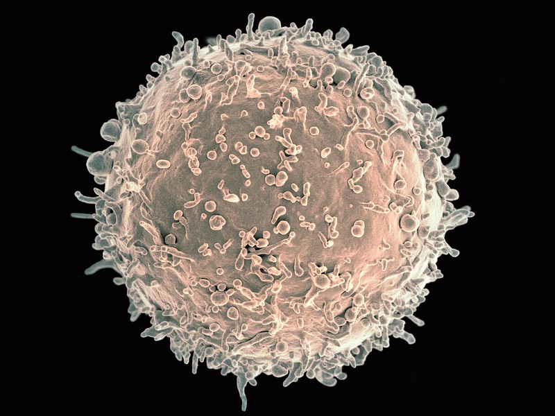
Scanning electron micrograph image of a human B cell. Credit: NIH/NIAID
New research in Nature Immunology has found that B cells play a surprising role in increasing or decreasing the bone marrow’s output of white blood cells. The findings may lead to new treatments for conditions that arise when white blood cell production goes out of balance.
Professor Matthias Nahrendorf, senior author of the study, explained that the nervous system plays a role in controlling blood cell production through neurotransmitters. “This is for instance important in people exposed to stress, where stress hormones — part of the ‘fight-or-flight’ response controlled by the sympathetic nervous system — may increase bone marrow activity and cardiovascular inflammation in response to the neurotransmitter noradrenaline,” he said. The parasympathetic nerves on the other hand, slow down responses and bring about a state of calm to the body, mainly through the neurotransmitter acetylcholine.
Because acetylcholine can have a protective effect against inflammation and heart disease, the researchers studied this neurotransmitter in the bone marrow. “When we looked into how acetylcholine acts on the production of blood cells, we found that it does the expected — it reduces white blood cells, as opposed to noradrenaline, which increases them,” said Prof Nahrendorf. “What was unexpected though was the source of the neurotransmitter acetylcholine.”
In the bone marrow, the typical nerve fibres that are known to release acetylcholine were not found. Instead, it was the antibody-producing B cells supplied the acetylcholine in the bone marrow. “Thus, B cells counter inflammation — even in the heart and the arteries — via dampening white blood cell production in the bone marrow. Surprisingly, they use a neurotransmitter to do so,” said Prof Nahrendorf.
Tapping into this process may help investigators develop strategies to block inflammation in cardiovascular conditions such as atherosclerosis. “Ultimately this may lead to new therapeutics that combat myocardial infarction, stroke, and heart failure,” said Prof Nahrendorf.
A newly published study has identified a key regulatory mechanism in inflammation that may lead to new targets for resolving that inflammation –and the inflammation of patients with sepsis, cancer and COVID.
In the journal PNAS, scientists reported their discovery of a regulatory pathway for immune response after infection or injury, such as burns. Dysregulation of this pathway could differentiate those who are at risk of fatal sepsis or help identify targets to resolve this unregulated inflammation.
“We are very excited about the findings in this paper and the far-reaching impacts it could have on understanding a key regulatory step in the immune response,” said co-lead author Cindy McReynolds, who holds a doctorate in pharmacology and toxicology.
In a rodent model, the research team found that the metabolites of linoleic acid formed by the enzyme, soluble epoxide hydrolase (sEH), drive damaging inflammation after injury. These metabolites, known as lipid mediators, regulate inflammation, blood pressure and pain. Drugs that inhibit the sEH enzyme and reduce inflammation could lead to better outcomes.
“Our previous work identified that these same lipid mediators were up-regulated in severe COVID infections, and we are now finding that these compounds play a role in modulating the immune response so that the body is unable to fight infection or respond properly to trauma without leading to a potentially fatal overreaction,” said Dr McReynolds.
“This dysregulation has fatal consequences in serious diseases such as COVID, cancer, sepsis, burn, where fatality rates can be as high as 40 percent in severe cases,” she said. “An understanding of these pathways can help identify patients at risk of developing serious disease or identify new therapeutic targets for treatment.”
“The immunological disbalance we see in many cases of severe burn injury, trauma and sepsis pose a huge clinical challenge as we lack the understanding of how to diagnose and treat it,” explained co-lead author Dr Christian Bergmann. “With this work, we reveal an important mechanism how immune cells are functionally disabled by sEH-derived metabolites of linoleic acid.”
“The natural compounds we are studying in this paper are metabolites of linoleic acid (LA), an essential fatty acid the body needs in very small amounts to survive and is only available through the diet,” Dr McReynolds elaborated. “At lower concentrations, these metabolites are necessary for regulating thermogenesis and heart health but promote inflammation at higher concentrations. LA is more stable and much cheaper than longer chain polyunsaturated fatty acids, so heavily processed foods have higher LA content to increase shelf-life. Additionally, agricultural practices, such as feeding animals corn-based diets, have increased LA in meats and dairy products.”
“As a result, we are consuming the highest amount of linoleic acid and have the highest recorded concentration of LA in our fatty tissue in human history,” McReynolds said. “As our bodies respond to stress or disease, we metabolise LA into the regulatory metabolites that were monitored in this paper. At higher concentrations, the immune system was unable to properly respond to infection, thereby promoting a sustained immune response. These observations are important in inflammatory-driven diseases, such as sepsis and COVID, but could also be important in understanding many of the increased chronic diseases we are seeing in our population.”
There appears to be a link between eating meat, gut bacteria and multiple sclerosis, according to new research published in EBioMedicine. The study teased out subtle connections that could lead to a better understanding of the causes of the disease.
The autoimmune disease multiple sclerosis (MS) is more prevalent in specific regions, particularly the northern mid-latitudes, suggesting that geography is somehow linked to the disease, perhaps involving diet. However, the exact relationships between diet, immune response, and MS has been a mystery. What exactly triggers the body to attack the myelin sheaths in MS in the first place is unknown.
Growing evidence suggests that bacteria might play a role. Gut bacteria affect the immune system, and diet affects the gut. Researchers studied the gut microbiome, immune systems, diet, and blood metabolites in 25 MS patients and 24 healthy controls to look for any subtle but important correlations.
“We found a number of gut bacteria associated with MS and severity of disability of MS patients,” said Dr Yanjiao Zhou. “We also found increased autoimmune markers and signature metabolites in MS. But what is really interesting is how these systems connect with each other, and how diet is involved in these connections. Using multi-OMICS approaches, we try to close the loop and show the associations between multiple systems.”
Meat eating was the strongest link in their analysis, where higher meat consumption saw a decrease in the population of Bacteroides thetaiotaomicron, a bacteria associated with digesting carbohydrates from vegetables.
Higher meat consumption, seen in the MS patients, was also linked to an increase in T-helper 17 cells in the immune system, and an increase in S-adenosyl-L-methionine (SAM) in their blood.
Meat eating was not a predictor of MS. But the evidence suggested that, in MS, something causes gut bacteria to disassociate with the immune system, leading to heightened T-helper 17 cells and autoimmune attacks on the nervous system. And it tends to be associated with eating meat.
Future research aims to recruit more volunteers, including those with more severe MS. Eventually they hope to understand more of the cause-and-effect between diet, bacterial ecosystems in the gut, and immune response, and potentially help prevent or mitigate MS symptoms in people suffering from the disease.
Scientists have found that four COVID vaccines (Pfizer-BioNTech, Moderna, J&J/Janssen, and Novavax) prompt the body to make effective, long-lasting T cells against SARS-CoV-2. These T cells can recognise SARS-CoV-2 Variants of Concern, including Delta and Omicron.
The new study, published in Cell, showed that the vast majority of T cell responses are also still effective against Omicron, reducing the odds of illness for up to six months, regardless of vaccine.
These data come from adults who were fully vaccinated, but not yet boosted. The researchers are now investigating T cell responses in boosted individuals and people who have experienced “breakthrough” COVID cases.
The study also shows that fully vaccinated people have fewer memory B cells and neutralising antibodies against the Omicron variant. This finding is in line with initial reports of waning immunity from laboratories around the world.
Without enough neutralising antibodies, Omicron is more likely to cause a breakthrough infection, and fewer memory B cells means a slower production of more neutralising antibodies.
Co-first author Camila Coelho, PhD, said: “Our study revealed that the 15 mutations present in Omicron RBD can considerably reduce the binding capacity of memory B cells.”
Neutralising antibodies and memory B cells are only two arms of the body’s adaptive immune response. , T cells do not prevent infection, rather they patrol the body and destroy cells that are already infected, which prevents a virus from multiplying and causing severe disease.
The team believes the “second line of defence” from T cells helps explain Omicron’s reduced severity in vaccinated people. The variant also appears to infect different tissues.
To know whether the vaccine-induced T cells they detected in their study were actually effective against variants such as Delta and Omicron, the scientists took a close look at how the T cells responded to different viral “epitopes.”
Every virus is made up of proteins that form a certain shape or architecture. A viral epitope is a specific landmark on this architecture that T cells have been trained to recognise. Current COVID vaccines were designed to teach the immune system to recognise specific epitopes on the initial variant of SARS-CoV-2, specifically targeting the Spike protein which the virus uses to access human cells. As the virus has mutated, its architecture has changed, and the concern is that immune cells will no longer recognise their targets.
The new study shows that while the architecture of Omicron is different enough to evade some neutralising antibodies and memory B cells, memory T cells still do a good job of recognising their targets, even on the highly mutated Omicron variant. Overall, at least 83 percent of the CD4+ (helper) T cell responses and 85 percent of the CD8+ T cell responses stayed the same, no matter the vaccine or the variant.
The memory B cells that do bind Omicron are likely to also contribute to protection against severe disease, forming multiple lines of defence.
Researchers are now focusing on measuring T cells, B cells and antibody responses after COVID booster shots, and also characterising immune responses after a breakthrough infection.
A new study from the University of East Anglia and Quadram Institute sheds light on how our immune cells make use of body fat to fight infection. The research, published today in Nature Communications, could lead to new approaches to treating people with bacterial infections.
The work could one day help treat infections in vulnerable and older people, the researchers said. The team studied Salmonella bacteria and tracked fatty acid movement and consumption in live stem cells. They then examined the immune response to Salmonella bacterial infection, by analysing liver damage.
They uncovered how blood stem cells respond to infection, by acquiring high energy fatty acids from the body’s fat stores. In the bone marrow where blood stem cells are resident, infection signals drive adipocytesto release their fat stores as fatty acids into the blood.
And they identified that these high energy fatty acids are then taken up by blood stem cells, effectively feeding the stem cells and enabling them to make millions of Salmonella-fighting white blood cells. The researchers also identified the mechanism by which the fatty acids are transferred and discusses the potential impact this new knowledge could have on future treatment of infection.
Dr Stuart Rushworth, from UEA’s Norwich Medical School, said: “Our results provide insight into how the blood and immune system is able to respond to infection.
“Fighting infection takes a lot of energy and fat stores are huge energy deposits, which provide the fuel for the blood stem cells to power up the immune response.
“Working out the mechanism through which this ‘fuel boost’ works gives us new ideas on how to strengthen the body’s fight against infection in the future.”
Dr Naiara Beraza, from the Quadram institute, said: “Our results allow us to understand how our immune system uses fat to fuel the response to infection. Defining these mechanisms will enable us to develop new therapeutics to treat infections in the liver.”
Researchers have uncovered differences in immune pathway activation to influenza infection between individuals of European and African genetic ancestry, according to a study published in Science. Many of the genes that were associated with these immune response differences to influenza are also enriched among genes associated with COVID disease severity.
“The lab has been interested in understanding how individuals from diverse populations respond differently to infectious diseases,” said first author Haley Randolph, a graduate student at the University of Chicago. “In this study, we wanted to look at the differences in how various cell types respond to viral infection.”
The researchers examined gene expression patterns in peripheral mononuclear blood cells, a diverse set of specialised immune cells that play important roles in the body’s response to infection. These cells were gathered from men of European and African ancestry and then exposed the cells to flu in a laboratory setting. This let the team examine the gene signatures of a variety of immune cell types, and observe how the flu virus affected each cell type’s gene expression.
The results showed that individuals of European ancestry showed an increase in type I interferon pathway activity during early influenza infection.
“Interferons are proteins that are critical for fighting viral infections,” said senior author Luis Barreiro, PhD, Associate Professor of Medicine at UChicago. “In COVID-19, for example, the type I interferon response has been associated with differences in the severity of the disease.”
This increased pathway activation hindered the replication of the virus more and limited viral replication later on. “Inducing a strong type I interferon pathway response early upon infection stops the virus from replicating and may therefore have a direct impact on the body’s ability to control the virus,” said Barreiro. “Unexpectedly, this central pathway to our defense against viruses appears to be amongst the most divergent between individuals from African and European ancestry.”
The researchers saw a variety of differences in gene expression across different cell types, suggesting a constellation of cells that work together to fight disease.
Such a difference in immune pathway activation could explain influenza outcome disparities between different racial groups; Non-Hispanic Black Americans are more likely to be hospitalised due to the flu than any other racial group.
However, these results are not evidence for genetic differences in disease susceptibility, the researchers point out. Rather, possible differences in environmental and lifestyle between racial groups could be influencing gene expression, and affecting the immune response.
“There’s a strong relationship between the interferon response and the proportion of the genome that is of African ancestry, which might make you think it’s genetic, but it’s not that simple,” said Barreiro. “Genetic ancestry also correlates with environmental differences. A lot of what we’re capturing could be the result of other disparities in our society, such as systemic racism and healthcare inequities. Although some of the differences we show in the paper can be linked to specific genetic variation, showing that genetics do play some role, such genetic differences are not enough to fully explain the differences in the interferon response.”
These differences in viral susceptibility may not be confined to just influenza. Comparing a list of genes associated with differences in COVID severity, the researchers found that many of the same genes showed significant differences in their expression after flu infection between individuals of African and European ancestry.
“We didn’t study COVID patient samples as part of this study, but the overlap between these gene sets suggests that there may be some underlying biological differences, influenced by genetic ancestry and environmental effects, that might explain the disparities we see in COVID outcomes,” said Barreiro.
As they explore this further, the researchers hope to figure out which factors contribute to the differences in the interferon response, and immune responses more broadly, to better predict individual disease risk.
A key protein called TWEAK damages skin cells in psoriasis patients, according to a new study in mice and with human skin cells, and targeting TWEAK may help control the disease.
Although there are effective treatments for psoriasis, an autoimmune disease that shows up as patches of red, inflamed skin and painful, scaly rashes, not everyone responds to these therapies – and for many, the relief is temporary.
“These therapies don’t reduce disease by 100 percent, and they don’t cure the disease” says La Jolla Institute for Immunology (LJI) Professor Michael Croft, PhD. “And if you take patients off those drugs, the disease almost always comes back.”
“We think TWEAK might be considered a potential target for the treatment of psoriasis,” said first author Rinkesh Gupta, PhD, a postdoctoral fellow at LJI. “It’s good to have this chance to develop a new therapeutic option.”
The findings build on the lab’s earlier research showing that TWEAK can interact with keratinocytes, the most common type of skin cell. By investigating TWEAK-deficient mice, the researchers found that TWEAK is a driver of inflammation in a model of psoriasis. The new study, published in Science Immunology, shows that TWEAK does not work alone; it teams up with two other proteins, tumour necrosis factor (TNF) and interleukin-17 (IL-17), to trigger inflammation. These three seem to control inflammatory molecule production and the expression of additional inflammation-associated proteins in patients with psoriasis.
“The fact that they work together suggests the disease is essentially driven by all three of those particular proteins at the same time,” explained Prof Croft. “The primary implication is that TWEAK will also be a good drug target. as has already been proven for TNF and IL-17.”
The researchers tested this idea with a mouse model of psoriasis to compare how well a TWEAK-inhibitor measured up to therapies inhibiting IL-17 or TNF.
“If you inhibit TWEAK from working on its receptor on keratinocytes, you get the same therapeutic effect as when you inhibit TNF or IL-17,” said Dr Gupta. A particularly encouraging aspect of this finding since TNF and IL-17 are both FDA-approved drug targets for psoriasis.
Prof Croft thinks TWEAK inhibitors have potential as therapies for many types of skin diseases. “We think TWEAK is involved in skin inflammation in general,” he said.
His lab is now investigating the role of TWEAK in atopic dermatitis, and while a distinct disease from psoriasis, they do have a few things in common – and there are not as many good treatments for atopic dermatitis.
“There’s certainly a lot of room for improvement in treatment of atopic dermatitis patients,” he said.
A recent study shows that T helper cells produced by people who received either of the two available messenger RNA (mRNA) vaccines for COVID persist six months after vaccination, at only slightly reduced levels from two weeks after vaccination. They are also at significantly higher levels than in unvaccinated individuals.
In the study, published in Clinical Infectious Diseases, the researchers also found that the T cells they studied recognise and help protect against the highly infectious delta variant of SARS-CoV-2.
“Previous research has suggested that humoral immune response – where the immune system circulates virus-neutralising antibodies – can drop off at six months after vaccination, whereas our study indicates that cellular immunity – where the immune system directly attacks infected cells – remains strong,” said Professor Joel Blankson, MD, PhD, study senior author. “The persistence of these vaccine-elicited T cells, along with the fact that they’re active against the delta variant, has important implications for guiding COVID vaccine development and determining the need for COVID boosters in the future.”
The researchers sampled blood from 15 study participants at three times: prior to vaccination, between seven and 14 days after their second Pfizer/BioNTech or Moderna vaccine dose, and six months after vaccination. The median age of the participants was 41 and none had evidence of prior SARS-CoV-2 infection.
CD4+ T lymphocytes are nicknamed helper T cells because they assist another type of immune system cell, the B lymphocyte (B cell), to respond to antigens on viruses such as SARS-CoV-2. Activated by the CD4+ T cells, immature B cells become either plasma cells that produce antibodies to mark infected cells for disposal from the body or memory cells that ‘remember’ the antigen’s biochemical structure for a faster response to future infections. Therefore, a CD4+ T cell response can serve as a measure of how well the immune system responds to a vaccine and yields humoral immunity.
The researchers found that the number of helper T cells recognising SARS-CoV-2 spike proteins was very low pre-vaccination, with a median of 2.7 spot-forming units (SFUs, the level of which is a measure of T cell frequency) per million peripheral blood mononuclear cells (PBMCs, identified as any blood cell with a round nucleus, including lymphocytes). Between 7 and 14 days after vaccination, the T cell frequency rose to a median of 237 SFUs per million PBMCs. At six months after vaccination, the level dropped slightly to a median of 122 SFUs per million PBMCs – a T cell frequency still significantly higher than before vaccination.
Six months after vaccination, the number of T cells recognising the delta variant spike protein was not significantly different from that of T cells attuned to the original virus strain’s protein.
“The robust expansion of T cells in response to stimulation with spike proteins is certainly indicated, supporting the need for more study to show booster shots do successfully increase the frequency of SARS-CoV-2-specific T cells circulating in the blood,” said Prof Blankson. “The added bonus is finding that this response also is likely strong for the delta variant.”
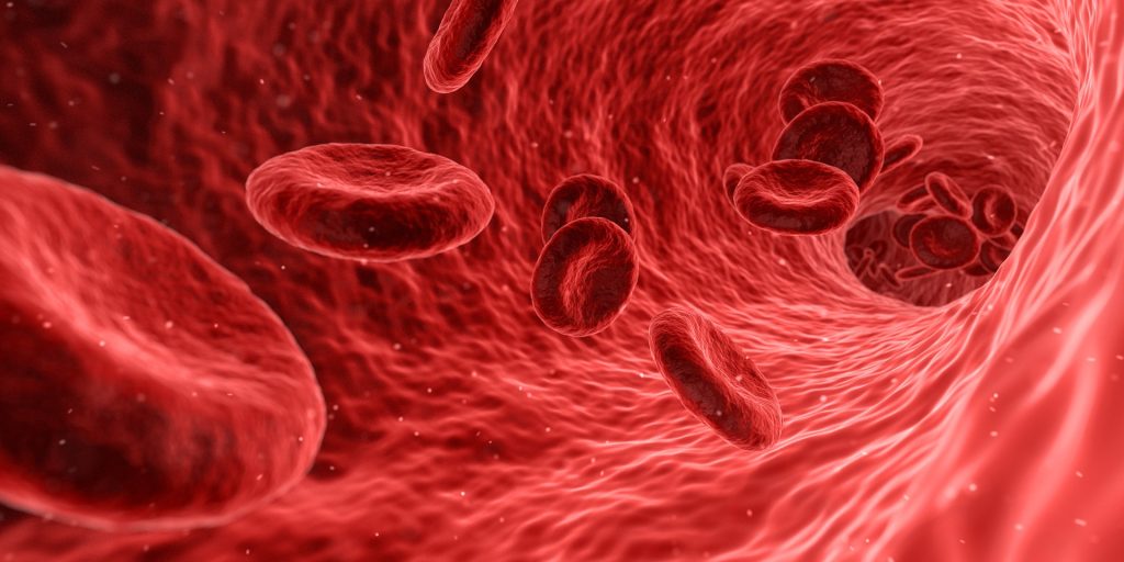
Red blood cells have been discovered to have a critical function as immune sensors by binding cell-free DNA (nucleic acid) present in the body’s circulation during sepsis and COVID.
This DNA-binding capability triggers their removal from circulation, driving inflammation and anaemia during severe illness and playing a much larger role in the immune system than previously thought. Scientists have long known that red blood cells also interacted with the immune system, but not whether they directly altered inflammation, until now. The study appears in Science Translational Medicine.
“Anaemia is common, affecting about a quarter of the world’s population. Acute inflammatory anaemia is often seen early after an infection such as parasitic infections that cause malaria,” said senior author Nilam Mangalmurti, MD, an assistant professor of Medicine at Penn. “For a long time we haven’t known why people, when they are critically ill from sepsis, trauma, COVID, a bacterial infection, or parasite infection, develop an acute anaemia. These findings explain one of the mechanisms for the development of acute inflammatory anaemia for the first time.”
Toll-like receptors (TLRs) play a key role in the immune system by activating immune responses like cytokine production. Analysing the red blood cells of about 50 sepsis patients and 100 COVID patients the study found that, during these illnesses, red blood cells express more TLR9 on their surface.
When the red blood cells bind too much inflammation-causing nucleic acid, they lose their normal structure, causing the body to no longer recognise them, prompting macrophages to engulf them. When this happens, it causes the immune system to become activated in otherwise unaffected organs, creating inflammation. The discovery of this mechanism will allow research on blocking this specific receptor and creating targeted therapies for autoimmune diseases, infectious diseases, and various inflammatory illnesses associated with acute anaemia.
“Right now when patients in the ICU become anaemic, which is almost all of our critically ill patients, the standard is to give them blood transfusions, which has long been known to be accompanied by a host of issues including acute lung injury and increased risk of death,” Prof Mangalmurti said. “Now that we know more about the mechanism of anaemia, it allows us to look at new therapies for treating acute inflammatory anaemia without transfusions, such as blocking TLR9 on the red blood cells. Targeting this TLR9 may also be a way to dampen some of the innate immune activation without blocking this receptor in immune cells, which are very important for the host when fighting a pathogen or injury.”
This DNA-binding discovery could also have implications for research into using red blood cells in diagnostics, Prof Mangalmurti said. For example, a physician might be able to take red blood cells from a patient with pneumonia, sequence the nucleic acid absorbed from the infection, and identify the specific kind of pathogen to better determine what kind of antibiotic to prescribe.
Prof Mangalmurti and colleagues are looking at whether this is a valid option in diagnosing infection in critically ill patients and if this DNA-binding mechanism by red blood cells is a universal mechanism of anaemia in parasitic infections.
A new study recently published in Nature has found that immune protection resulting from COVID protection creates lasting effects in memory B cells.
Unlike circulating antibodies, which peak soon after vaccination or infection only to fade a few months later, memory B cells can remain to ward off severe disease for decades. They also evolve over time, learning to produce successively more potent ‘memory antibodies’ that are more effective at neutralising the virus and with better adaptation to variants.
Though vaccination instils higher levels of circulating antibodies than natural infection, the study suggests that not all memory B cells are created equal. While vaccination gives rise to memory B cells that evolve over a few weeks, natural infection births memory B cells that continue to evolve over several months, producing highly potent antibodies adept at eliminating even viral variants.
Though the findings suggest an advantage from natural infection over vaccination, this does not outweigh the dangers of illness and death from COVID, the researchers warn.
“While a natural infection may induce maturation of antibodies with broader activity than a vaccine does – a natural infection can also kill you,” explained Professor Michel C. Nussenzweig, head of Rockefeller’s Laboratory of Molecular Immunology. “A vaccine won’t do that and, in fact, protects against the risk of serious illness or death from infection.”
When any virus enters the body, immune cells immediately release circulating antibodies, which decay at variable rates depending on the vaccine or infection. They may confer protection for months or years but then dwindle in number, allowing possible reinfection.
Long term protection is provided by memory B cells that produce memory antibodies. Studies suggest that memory B cells for smallpox last at least 60 years after vaccination; those for Spanish flu, nearly a century. And while memory B cells don’t necessarily block reinfection, they can prevent severe disease.
Recent studies have suggested that within five months of receiving a vaccine or recovering from a natural infection, some no longer retain sufficient circulating antibodies to keep the novel coronavirus at bay, but memory B cells remain vigilant. Until now, however, scientists did not know whether the vaccines could be expected to provide the sort of robust memory B cell response seen after natural infection.
Prof Nussenzweig and colleagues resolved to tease out any differences in memory B cell evolution by comparing blood samples from convalescent COVID patients to those from never-infected mRNA-vaccinated individuals.
Vaccination and natural infection elicited similar numbers of memory B cells, which rapidly evolved between the first and second dose of the Pfizer and Moderna vaccines, producing increasingly potent memory antibodies. But after two months, progress stalled. The memory B cells were present in large numbers and expressed potent antibodies, but the antibodies were not getting any stronger. Also, although some of these antibodies were able to neutralize Delta and other variants, there was no overall improvement in breadth.
The researchers found that in convalescent patients, however, memory B cells continued to evolve and improve up to one year after infection. With every memory B cell update, more potent and more broadly neutralising memory antibodies were coming out.
There are several potential reasons that memory B cells produced by natural infection might be expected to outperform those produced by mRNA vaccines, the researchers said.
It is possible that the body responds differently to viruses that enter through the respiratory tract than those that are injected. Or perhaps an intact virus goads the immune system in a way the vaccines’ spike protein antigens simply cannot. It may also be possible that the virus persists in the naturally infected for weeks, giving the body more time to mount a robust response. The vaccine, on the other hand, is flushed out of the body mere days after triggering the desired immune response.
Memory B cells appear to undergo limited bouts of evolution in response to mRNA vaccines, a finding which may have significant implications for booster shots. A booster with the current mRNA vaccine would likely stimulate memory cells to produce antibodies strongly protective against the original virus and somewhat less so against the variants, Prof Nussenzweig said.
“When to administer the booster depends on the object of boosting,” he said. “If the goal is to prevent infection, then boosting will need to be done after 6 to 18 months depending on the immune status of the individual. If the goal is to prevent serious disease, boosting may not be necessary for years.”