As an adult-onset psychiatric disorder, schizophrenia is thought to be triggered by some combination of environmental factors and genetics, although the exact cause remains unclear. In a study published in the journal Cell Genomics, researchers find a correlation between schizophrenia and somatic copy-number variants, a type of mutation that occurs early in development but after genetic material is inherited. This study is one of the first to rigorously describe the relationship between somatic genetic mutations and schizophrenia risk.
“We originally thought of genetics as the study of inheritance. But now we know that genetic mechanisms go way beyond that,” says senior author Chris Walsh, an investigator at the Howard Hughes Medical Institute and chief of genetics and genomics at Boston Children’s Hospital. “We’re looking at mutations that are not inherited from the parents.”
The researchers analysed genotype-marker data from over 20,000 blood samples of people with or without schizophrenia from the Psychiatric Genomics Consortium. They ultimately identified two genes, NRXN1 and ABCB11, that correlated with schizophrenia cases when disrupted in utero. NRXN1, a gene that helps transmit signals throughout the brain, has been associated with schizophrenia before. However, this is the first study to associate somatic, not inherited, NRXN1 mutations with schizophrenia.
Unlike inherited mutations, which are present in all the cells of the body, somatic mutations are only present in a fraction of cells based on when and where a mutation occurred. If a mutation occurs early in development, it is expected that the variant is present throughout the body in a mosaic pattern. On the basis of this principle, researchers can identify somatic mutations that occurred early in development and are present not only in the brain but also in a fraction of cells in the blood.
“If a mutation occurs after fertilisation when there are only two cells, the mutation will be present in half of the cells of the body,” says Walsh. “If it occurs in one of the first four cells, it will be present in about a quarter of the cells of the body, and so on.”
The second gene the researchers identified, ABCB11, is most known to encode a liver protein. “That one came out of nowhere for us,” says Eduardo Maury, a student in Harvard-MIT’s MD-PhD program. “There have been some studies associating mutations in this gene with treatment-resistant schizophrenia, but it hasn’t been strongly implicated in schizophrenia per se.”
When the team investigated further, they found that ABCB11 is also expressed in very specific subsets of neurons that carry dopamine from the brainstem to the cerebral cortex. Most schizophrenia drugs are thought to act on these cells to decrease an individual’s dopamine levels, so this might explain why the gene is associated with treatment resistance.
Next, the team is working towards identifying other acquired mutations that might be associated with schizophrenia. Given that the study analysed blood samples, it will be important to look at more brain-specific mutations that might have been too subtle or recent in a patient’s life for this analysis to detect. In addition, somatic deletions or duplications might be an under-investigated risk factor associated with other disorders.
“With this study, we show that it is possible to find somatic variants in a psychiatric disorder that develops in adulthood,” says Maury. “This opens up questions about what other disorders might be regulated by these kinds of mutations.”
An international team of researchers has developed a new method to deliver drugs into the inner ear, according to a new study in Science Translational Medicine. The discovery was possible by harnessing the natural flow of fluids in the brain and employing a little-understood backdoor into the cochlea. When combined to deliver a gene therapy that repairs inner ear hair cells, the researchers were able to restore hearing in deaf mice.
“These findings demonstrate that cerebrospinal fluid transport comprises an accessible route for gene delivery to the adult inner ear and may represent an important step towards using gene therapy to restore hearing in humans,” says lead author Barbara Canlon, professor at Karolinska Institutet.
The number of people worldwide predicted to have mild to complete hearing loss is expected to grow to around 2.5 billion by mid-century. The primary cause is the death or loss of function of hair cells found in the cochlea – which relay sounds to the brain – due to mutations of critical genes, aging, noise exposure, and other factors.
While hair cells do not naturally regenerate in humans and other mammals, gene therapies have shown promise and in separate studies have successfully repaired the function of hair cells in neo-natal and very young mice.
“However, as both mice and humans age, the cochlea, already a delicate structure, becomes enclosed in the temporal bone. At this point, any effort to reach the cochlea and deliver gene therapy via surgery risks damaging this sensitive area and altering hearing,” says Barbara Canlon.
In the new study, the researchers describe a little-understood passage into the cochlea called the cochlear aqueduct. The cochlear aqueduct is a thin boney channel no larger than several strands of hair.
Channel for spinal fluid
A new study shows that the cochlear aqueduct acts as a conduit between the cerebrospinal fluid found in the inner ear and the rest of the brain.
Scientists are developing a clearer picture of the mechanics of the glymphatic system, the brain’s unique process of removing waste. Because the glymphatic system pumps cerebrospinal fluid deep into brain tissue to wash away toxic proteins, researchers have been eyeing it as a potential new way to deliver drugs into the brain, a major challenge in developing drugs for neurological disorders.
The new study represented an opportunity to put the drug delivery potential of the glymphatic system to the test, while at the same time targeting a previously unreachable part of the auditory system.
Employing several imagining and modeling technologies, the researchers were able to develop a detailed portrait of how fluid from other parts of the brain flows through the cochlear aqueduct and into the inner ear.
The team then injected an adeno-associated virus into the cisterna magna, a large reservoir of cerebrospinal fluid found at the base of the skull.
The virus found its way into the inner ear via the cochlear aqueduct and delivered a gene therapy that expresses a protein called vesicular glutamate transporter-3, which enables the hair cells to transmit signals and rescue hearing in adult deaf mice.
“This new delivery route into the ear may not only serve the advancement of auditory research but also prove useful when translated to humans with progressive genetic-mediated hearing loss,” says Barbara Canlon.
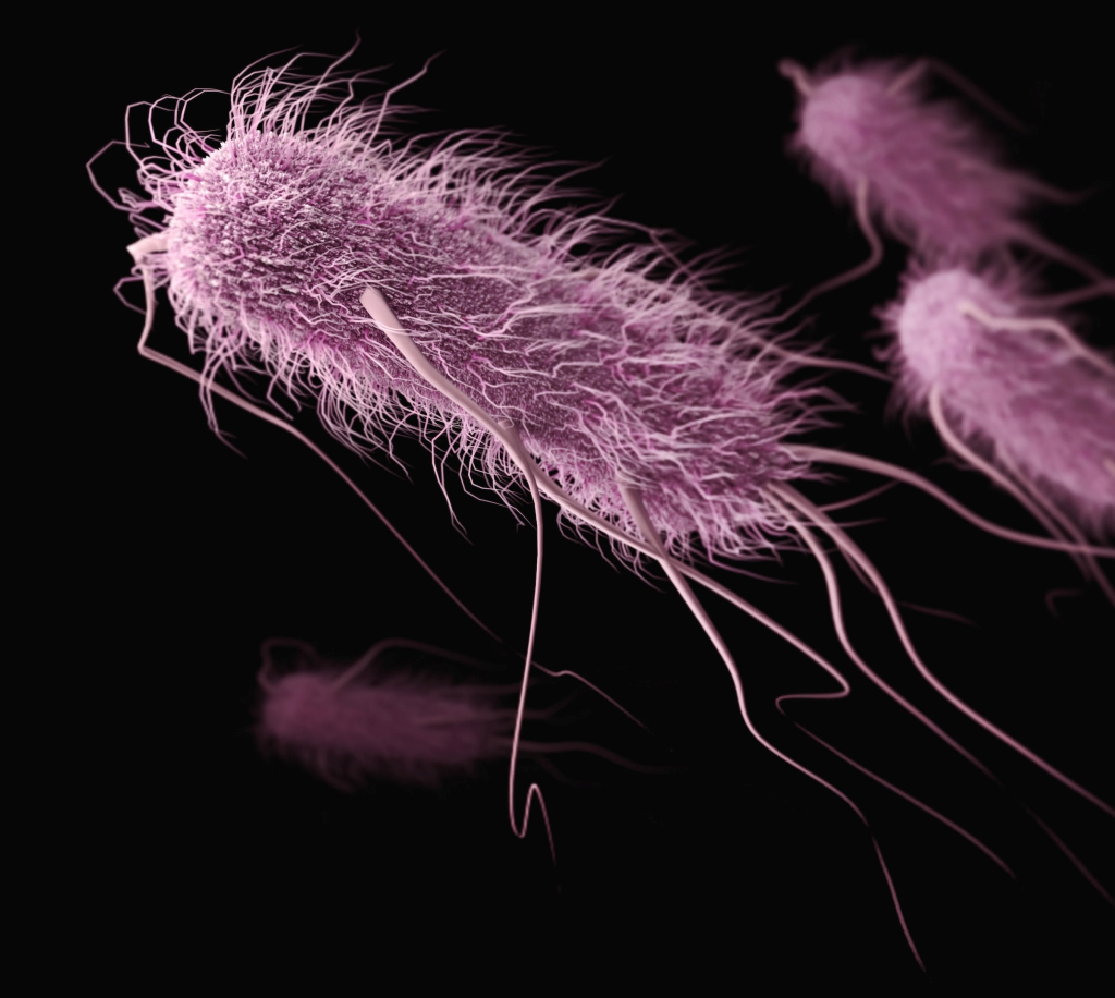
Researchers have identified how pathogenic genes in some Providencia spp., which have gained attention as causes of food poisoning as well as enterohaemorrhagic Escherichia coli. O157 and Salmonella, are transferred within bacterial cells. Their findings are expected to provide new insights into the identification of infection routes of Providencia spp. and the establishment of preventive methods for food poisoning.
Recently, Providencia spp. which have been detected in patients with gastroenteritis, and similar to enterohemorrhagic Escherichia coli. O157 and Salmonella spp., have been attracting attention as causative agents of food poisoning. For children with low immunity, food poisoning can be lethal as it causes severe symptoms such as diarrhoea and dehydration, so clarifying the source of infection and pathogenic factors of Providencia spp., and establishing preventive methods are urgent issues worldwide.
A joint research group led by Professor Shinji Yamasaki, Dr Sharda Prasad Awasthi, a Specially Appointed Lecturer, and graduate student Jayedul Hassan from the Graduate School of Veterinary Science, Osaka Metropolitan University, determined how the pathogenic genes in some Providencia spp. such as Providencia alcalifaciens and Providencia rustigianii are transferred within bacterial cells of genus Providencia. The group has also elucidated that the pathogenic genes of Providencia rustigianii are also transferred to other bacterial cells belonging to Enterobacteriaceae.
Professor Yamasaki concluded, “This achievement is expected to provide new insights into the identification of infection routes of Providencia spp. and the establishment of preventive methods for food poisoning.”
South African scientists – notably, the team headed by Professor Tulio de Oliveira – were thrown into the global spotlight through their pivotal role in detecting and monitoring the emergence of new variants of SARS-CoV-2 – the Beta variant in 2020 and Omicron in 2021. De Oliveira is now at the University of Stellenbosch, but for much of the pandemic headed the KwaZulu-Natal Research Innovation and Sequencing Platform (KRISP).
The country’s advanced genomic sequencing capabilities and proactive surveillance efforts allowed for the early identification of the variants and the discoveries played a crucial role in alerting the global scientific community to the potential for viral mutations and the need for enhanced monitoring.
Now, scientists worldwide believe it is critical to continue investing in genomics to support disease control in public health in South Africa and the broader continent.
What is genomics?
The World Health Organization (WHO) defines genomic surveillance as “the process of constantly monitoring pathogens and analysing their genetic similarities and differences”. It is done through a method known as whole genome sequencing, which determines the entire genetic makeup of specific organisms or cell types. This method is also able to detect changes in areas of genomes, which can help scientists to establish how specific diseases form. The results of genomic sequencing can also be used in diagnosing and treating diseases.
Genomic sequencing enables scientists to read the DNA and RNA of pathogens and understand what they are and how they spread between people – and to develop vaccines and other measures to deal with them.
The US Centers for Disease Control (CDC) explains, “All organisms (bacteria, vegetable, mammal) have a unique genetic code, or genome that is composed of nucleotide bases (A, T, C, and G). If you know the sequence of the bases in an organism, you have identified its unique DNA fingerprint or pattern. Determining the order of bases is called sequencing. Whole genome sequencing is a laboratory procedure that determines the order of bases in the genome of an organism in one process.
“Scientists conduct whole genome sequencing by following these four main steps:
DNA shearing: Scientists begin by using molecular scissors to cut the DNA, which is composed of millions of bases (A’s, C’s, T’s, and G’s), into pieces that are small enough for the sequencing machine to read.
DNA barcoding: Scientists add small pieces of DNA tags, or bar codes, to identify which piece of sheared DNA belongs to which bacteria. This is similar to how a bar code identifies a product at a grocery store.
DNA sequencing: The bar-coded DNA from multiple bacteria is combined and put in a DNA sequencer. The sequencer identifies the A’s, C’s, T’s, and G’s, or bases, that make up each bacterial sequence. The sequencer uses the bar code to keep track of which bases belong to which bacteria.
Data analysis: Scientists use computer analysis tools to compare sequences from multiple bacteria and identify differences. The number of differences can tell the scientists how closely related the bacteria are, and how likely it is that they are part of the same outbreak…”
Time to expand
At a recent conference held at Stellenbosch University’s new state-of-the-art Biomedical Medical Research Institute, de Oliveira stressed that African and other experts should now build on their success in COVID-19 genomics to expand to other pathogens such as influenza, H5N1, and climate-amplified pathogens.
John Sillitoe, the Director of the Genomic Surveillance Unit at the Wellcome Sanger Institute in the United Kingdom, agreed.
“It is important now to focus on endemic diseases so we can improve our understanding and control of endemic diseases. We should also be looking at TB, particularly with the increased prevalence in drug resistance and reduced response to drugs. For other African countries, malaria should be a key focus area. We know that drug resistance now is spreading into Africa from South East Asia and understanding the right combination of drugs to use is something that is easily identifiable through genomic surveillance.”
But surveillance is also about being ready for the next pandemic.
“There’s that classic line that, ‘diseases take no notice of national borders’,” Sillitoe said in an interview. “So, it is really important that we can get as wide a picture of surveillance as possible to identify something new emerging as soon as possible.”
Marco Salemi, Professor of Experimental Pathology at the Department of Pathology, Immunology, and Laboratory Medicine at the University of Florida College of Medicine, said Africa and the world need to be “proactive, rather than reactive” in the battle against future epidemics. He said the world is currently focused on monitoring the COVID-19 pandemic. “But we forget this is this huge reservoir of pathogens out there which we know so little about and which can become more and more of a threat, especially because of climate change – so we need to understand more about all these pathogens in the wild, in animals, and their potential to jump to humans, especially with the rate of globalisation on the planet … Events of zoonotic transmissions will become more and more frequent. We need to face it.”
Building capacity
De Oliveira is of the view that Africa could, in the next few years, potentially, “leapfrog over the rest of the world” in genomic surveillance, thanks to its success in COVID-19 genomics and its experience in using genomics to monitor other pathogens over the past 20 years.
We won’t be starting from scratch.
The use of genomics in infectious diseases started in the mid-eighties during the HIV epidemic, when scientists realised HIV was a complex virus that existed in many different sub-types. Scientists around the world started using genomic tools to sequence the HIV virus, track its origin, and trace the way the virus disseminated.
Genomics has, however, changed dramatically since the 1980s.
“There have been many attempts… to use genomics for public health purposes, but the key factor that was always missing was the ability to generate DNA sequencing in real-time,” said Salemi. “Real-time means there is an epidemic, with cases happening today – and we need to generate sequences within one or two days and then to analyse the genomic data and then to have actionable information that can be immediately transmitted to the public health authorities so that they can act within a few days.”
“Now the technological and computational limitations of the past few years have been overcome, and, as was clearly shown during the COVID-19 pandemic, we have machines that can generate literally thousands of sequences, like coronavirus sequences, in less than one day, or even within a few hours. At the same time, we have high-performance computer clusters, and super calculators that are capable of analysing this data in a very short time,” he said.
These technical advances would, of course, be of little value without people to use them and develop them further.
“Investment has been made on the continent in infectious disease surveillance and genomics surveillance specifically, and so we have lots of experts on the continent who know a lot about infectious diseases and how viruses work, and why it’s important to look at the genomics to trace when there is going to be a new outbreak,” says Professor Zané Lombard, Principal Medical Scientist in the Division of Human Genetics at the University of the Witwatersrand. “South Africa’s role during COVID-19 showcased what can happen quickly and effectively for public health interventions if you have the right experts with the right platform and expertise and infrastructure in place to do that kind of surveillance.”
De Oliveira and his team have worked closely with the Africa Centres for Disease Control and Prevention (Africa CDC) to scale genomic surveillance on the continent and have actively collaborated with other African countries to share expertise, resources, and genetic data in a bid to foster a continent-wide approach to genomic surveillance.
They have also helped set up large genomics facilities in Zimbabwe, Mozambique, and Botswana.
The Africa CDC, through its Pathogen Genomics Initiative (Africa PGI), has, for the past few years, been building a continent-wide genomic disease surveillance network. In 2019, when the PGI started its work, only seven of the African Union’s 55 member states had public health institutions with the equipment and staff to do genetic sequencing. Today, 31 African nations are able to do genetic sequencing for surveillance of COVID, malaria, cholera, Ebola, and other diseases.
De Oliveira said the continent’s experience in genomic surveillance of pathogens in Africa evolved to “unheard-of” levels during COVID. “We’ve been trying to advance genomic surveillance in Africa for the past two decades, and when the pandemic came, we had the right expertise to deal with viruses and respiratory pathogens such as tuberculosis, so we were able to pivot for SARS-CoV-2. In the end, South Africa and Africa became an example to follow for the whole world.
“All the investments we have made in genomic surveillance for COVID can now be leveraged and advanced to other areas of genomics in Africa… including for rare diseases, for cancer diagnostics, and human genomics. Finally, we have the tools and the equipment, as well as the support, to do advanced genomics in Africa, as we have dreamt of doing for the last twenty years.”
What it means in practical terms
Asked what it means, practically, to build capacity for genomics research, Lombard said one aspect is the establishment of strong laboratories. “Historically, if infrastructure was not available locally, researchers would partner with international labs and send their samples to have their sequencing done there. The problem with that was that expertise in using [that] technique was not being built locally,” she said. “It is really important to train the right people who know how to do the laboratory experiments but also to interpret the data correctly.
“It’s not only about building the infrastructure in the labs but also about training the individuals and making sure there are job opportunities locally for them,” she said.
Turning to the machines used in genomics, Lombard said, “The most popular machine these days is called a next-generation sequencer. These can read the whole DNA sequence of a virus.”
Salemi added, “Some of these sequencers are very large and some are even little portable boxes. Some can sequence thousands of samples at a time, while others are capable of sequencing a few dozen samples at a time. The samples, depending on the virus (or pathogen) being tested for, are taken from blood samples, nasal swabs, or sputum from patients, from faeces, urine, or from the skin.
“The BMRI (at Stellenbosch University) – which has the largest sample storage capacity in the southern hemisphere – can store five million samples at minus 80 degrees. If someone wants to build a lab that includes top-of-the-line computational capacity, it will cost anything from $40 million (over 700 million), but to start a small operation to do a few hundred sequences of a virus every week, $100 000 to $200 000 (roughly R17 million to R34 million) is enough, which has been done in many different African countries during the pandemic.”
Training is key
While all the scientists interviewed agreed that laboratories are important in building capacity for genomics research, they stressed that what is really needed is to train more individuals.
“More people need to be trained in genomics but also in bioinformatics, which is a really important component of this work. The technology component is becoming very smart and automated, but the data being generated is becoming more and more complex, with bigger data sets. Dealing with these,” Lombard said, “requires special data analysis skills and bioinformatics skills. The field of bioinformatics will need investment so that we can deal with the deluge of data that will come out.”
She said South African and other African universities are taking this skills need seriously, with many initiatives to offer undergraduate and post-graduate training programmes in these areas.
Salami agreed. “The most important part of building capacity is the human training. I find it naïve and sad when I hear politicians talking about building top-of-the-line laboratories, when, what they really need to do is to start building human capacity. Africa is an amazing reservoir (from which to build these skills) because 50 percent of the continent [are] people who are less than 30 years old. There are about 27 excellent laboratories all over Africa. We need to start creating a strong next generation of scientists.”
In support of this, de Oliveira is trying to raise 100 million dollars to implement real-time genomic research to enable the African continent to respond to new epidemics.
He said during COVID, the Network for Genomics Surveillance was founded and funded by the Department of Science and Innovation and the South African Medical Research Council (SAMRC). This funding was until 2021.
The Centre for Epidemic Response and Innovation, which is led by de Oliveira and forms part of the BMRI, is funded by the Africa CDC, the WHO, the Rockefeller Foundation, and the Elma Foundation. These funders support the work in South Africa and in other African countries, as well as the SA government. The BMRI was mostly funded by Stellenbosch University to the effect of R900 million, while the Department of Higher Education provided about R300 million. CERI occupies one floor of the BMRI.
In de Oliveira’s words, “This truly is the genome era for Africa.”
Diagram comparing the nose shape of a Neanderthal with that of a modern human by Dr Macarena Fuentes-Guajardo.
Neanderthal genes comprise some 1 to 4% of the genome of present-day humans whose ancestors migrated out of Africa, and new research has shown that their lingering presence shapes the immune systems and metabolism of people of non-African ancestry. Some of these genetics changes are detrimental, but are slowly being replaced by human versions.
A multi-institution research team including Cornell University has developed a new suite of computational genetic tools to address the genetic effects of interbreeding between humans of non-African ancestry and Neanderthals that took place some 50 000 years ago. (The study applies only to descendants of those who migrated from Africa before Neanderthals died out, and in particular, those of European ancestry.)
In a study published in eLife, the researchers reported that some Neanderthal genes are responsible for certain traits in modern humans, including several with a significant influence on the immune system. Overall, however, the study shows that modern human genes are winning out over successive generations.
“Interestingly, we found that several of the identified genes involved in modern human immune, metabolic and developmental systems might have influenced human evolution after the ancestors’ migration out of Africa,” said study co-lead author April (Xinzhu) Wei, an assistant professor of computational biology in the College of Arts and Sciences. “We have made our custom software available for free download and use by anyone interested in further research.”
Using a vast dataset from the UK Biobank consisting of genetic and trait information of nearly 300 000 British people of non-African ancestry, the researchers analysed more than 235 000 genetic variants likely to have originated from Neanderthals. They found that 4303 of those differences in DNA are playing a substantial role in modern humans and influencing 47 distinct genetic traits, such as how fast someone can burn calories or a person’s natural immune resistance to certain diseases.
Unlike previous studies that could not fully exclude genes from modern human variants, the new study leveraged more precise statistical methods to focus on the variants attributable to Neanderthal genes.
While the study used a dataset of almost exclusively white individuals living in the United Kingdom, the new computational methods developed by the team could offer a path forward in gleaning evolutionary insights from other large databases to delve deeper into archaic humans’ genetic influences on modern humans.
“For scientists studying human evolution interested in understanding how interbreeding with archaic humans tens of thousands of years ago still shapes the biology of many present-day humans, this study can fill in some of those blanks,” said senior investigator Sriram Sankararaman, an associate professor at the University of California, Los Angeles. “More broadly, our findings can also provide new insights for evolutionary biologists looking at how the echoes of these types of events may have both beneficial and detrimental consequences.”
Genes that make bacteria resistant to antibiotics are much more widespread in our environment than was previously realised. A new study published in Microbiome shows that bacteria in almost all environments carry resistance genes, with a risk of them spreading and aggravating the problem of bacterial infections that are untreatable with antibiotics.
“We have identified new resistance genes in places where they have remained undetected until now. These genes can constitute an overlooked threat to human health,” says Erik Kristiansson, a professor in the Department of Mathematical Sciences.
According to the World Health Organisation (WHO), antibiotic resistance is one of the greatest threats to global health. When bacteria become resistant to antibiotics, it becomes difficult or impossible to treat illnesses such as pneumonia, wound infections, tuberculosis and urinary tract infections. According to the UN Interagency Coordination Group on Antimicrobial Resistance (IACG) 700,000 people die each year from infections caused by antibiotic-resistant bacteria.
Looking for resistance genes in new environments
The genes that make bacteria resistant have long been studied, but the focus has traditionally been on identifying those resistance genes that are already prevalent in pathogenic bacteria. Instead, in the new study from Sweden, researchers have looked at large quantities of DNA sequences from bacteria to analyse new forms of resistance genes in order to understand how common they are. They have traced the genes in thousands of different bacterial samples from different environments, in and on people, in the soil and from sewage treatment plants. The study analysed 630 billion DNA sequences in total.
“The data requires a great deal of processing before information can be obtained. We have used metagenomics, a methodology, that allows vast quantities of data to be analysed,” says Juan Inda Díaz, a doctoral student in the Department of Mathematical Sciences, and the article’s lead author.
The study showed that the new antibiotic resistance genes are present in bacteria in almost all environments. This also includes human microbiomes and, more alarmingly, pathogenic bacteria, which can lead to more infections that are difficult to treat. The researchers found that resistance genes in bacteria that live on and in humans and in the environment were ten times more abundant than those previously known. And of the resistance genes found in bacteria in the human microbiome, 75% were not previously known at all.
The researchers stress the need for more knowledge about the problem of antibiotic resistance.
“Prior to this study, there was no knowledge whatsoever about the incidence of these new resistance genes. Antibiotic resistance is a complex problem, and our study shows that we need to enhance our understanding of the development of resistance in bacteria and of the resistance genes that could constitute a threat in the future,” says Kristiansson.
Preventing bacterial outbreaks in healthcare
The research team is currently working on integrating the new data into the international EMBARK project (Establishing a Monitoring Baseline for Antibiotic Resistance in Key environments). The project is coordinated by Johan Bengtsson-Palme, an assistant professor in the Department of Life Sciences at Chalmers, and aims to take samples from sources such as wastewater, soil and animals to get an idea of the way in which antibiotic resistance is spreading between humans and the environment.
“It is essential for new forms of resistance genes to be taken into account in risk assessments relating to antibiotic resistance. Using the techniques we have developed enables us to monitor these new resistance genes in the environment, in the hope that we can detect them in pathogenic bacteria before they are able to cause outbreaks in a healthcare setting,” says Bengtsson-Palme.
The method used by the researchers is called metagenomics, and is not new, but so far has not been used to analyse new types of antibiotic resistance genes in such large quantities. Metagenomics is a method of studying the metagenome, which is the complete gene set of all different organisms in a given sample or within a given environment. Using the method, it is also possible to study microorganisms that cannot be grown in a lab.
The first gene mapping study on eyebrow thickness in Europeans discovered three previously unreported genetic loci, as reported in the Journal of Investigative Dermatology. The study conducted by the International Visible Trait Genetics (VisiGen) Consortium demonstrates that eyebrow appearance has partly the same and partly different underlying genes in people from different parts of the world.
The appearance of human eyebrows is not just a matter of grooming but is in the genes. Eyebrow thickness, as any other appearance trait, is highly heritable. Thus far, genetic knowledge on eyebrow thickness has been very limited and solely restricted to non-Europeans. This study is the first genome-wide association study (GWAS) on eyebrow thickness in Europeans. By identifying new genes and rediscovering some of the genes previously identified in non-Europeans, the study expands genetic knowledge on human eyebrow variation, which is of broad interest and has implications for dermatology and other fields.
Previous studies were performed among Latin American and Chinese individuals, establishing four eyebrow thickness -associated genetic loci. Because no European eyebrow thickness GWAS had been reported, researchers did not know whether the genetic eyebrow thickness effects described in non-Europeans persist in Europeans, or whether there are European-specific genetic loci involved in eyebrow thickness, or both.
Lead investigator Prof Dr Manfred Kayser, Department of Genetic Identification, Erasmus MC University Medical Center Rotterdam, and co-chair of the VisiGen Consortium responsible for this study, commented, “Despite the immense efforts in mapping genes underlying human complex traits, we still know much more about the genes that make us sick than about those behind our healthy looks. For the first time, we performed a gene mapping study on eyebrow thickness variation in Europeans. Previous genetic knowledge on eyebrow thickness was limited and solely restricted to non-Europeans. We discovered new genes involved in eyebrow variation in Europeans and rediscovered some of the genes previously identified in non-Europeans.”
The study among 9948 individuals from four groups of European ancestry not only discovered three previously unreported genetic loci associated with eyebrow thickness, but also rediscovered two of the four genetic loci previously found in non-Europeans. Two other genetic loci previously reported in non-Europeans had minimal effects in Europeans, due to very low allele frequencies in Europeans.
Prof Dr Kayser concluded, “Our study significantly improves the genetic knowledge of human eyebrow appearance by increasing the number of known genes from four to seven and delivers new targets for future functional studies. By having demonstrated that eyebrow variation is determined by both shared and distinct genetic factors across continental populations, our findings underline the need for studying populations of different ancestries for unveiling the genetic basis of human traits, including, but not restricted to, physical appearance.”
The biology underpinning a rare genetic mutation that allows its carrier to feel almost no pain, heal faster and had reduced anxiety and fear, has been uncovered in a new study published in Brain.
Though it may sound like the stuff of superheroes, the carrier of the genetic mutation is an ordinary Scottish woman named Jo Cameron, who was first referred to pain geneticists at University College London in 2013, after her doctor noticed that she experienced no pain after major surgeries on her hip and hand. In 2019, they identified a new gene that they appropriately named FAAH-OUT, which had a rare genetic mutation. In combination with another, more common mutation in FAAH, it was found to be the cause of Jo’s unique characteristics.
The new research describes how the mutation in FAAH-OUT ‘turns down’ FAAH gene expression, as well as the knock-on effects on other molecular pathways linked to wound healing and mood. It is hoped the findings will lead to new drug targets and open up new avenues of research in these areas.
The area of the genome containing FAAH-OUT had previously been assumed to be ‘junk’ DNA that had no function, but it was found to mediate the expression of FAAH, a gene that is part of the endocannabinoid system and that is well-known for its involvement in pain, mood and memory.
In this study, the team from UCL sought to understand how FAAH-OUT works at a molecular level, the first step towards being able to take advantage of this unique biology for applications like drug discovery.
This included a range of approaches, such as CRISPR-Cas9 experiments on cell lines to mimic the effect of the mutation on other genes, as well as analysing the expression of genes to see which were active in molecular pathways involved with pain, mood and healing.
The team observed that FAAH-OUT regulates the expression of FAAH. When it is significantly turned down as a result of the mutation carried by Jo Cameron, FAAHenzyme activity levels are significantly reduced.
Dr Andrei Okorokov (UCL Medicine), a senior author of the study, said: “The FAAH-OUT gene is just one small corner of a vast continent, which this study has begun to map. As well as the molecular basis for painlessness, these explorations have identified molecular pathways affecting wound healing and mood, all influenced by the FAAH-OUT mutation. As scientists it is our duty to explore and I think these findings will have important implications for areas of research such as wound healing, depression and more.”
The authors looked at fibroblasts taken from patients to study the effects of the FAAH-OUT-FAAH axis on other molecular pathways. While the mutations that Jo Cameron carries turn down FAAH, they also found a further 797 genes that were turned up and 348 that were turned down. This included alterations to the WNT pathway that is associated with wound healing, with increased activity in the WNT16 gene that has been previously linked to bone regeneration.
Two other key genes that were altered were BDNF, which has previously been linked to mood regulation and ACKR3, which helps to regulate opioid levels. These gene changes may contribute to Jo Cameron’s low anxiety, fear and painlessness.
Senior study author Professor James Cox said: “The initial discovery of the genetic root of Jo Cameron’s unique phenotype was a eureka moment and hugely exciting, but these current findings are where things really start to get interesting. By understanding precisely what is happening at a molecular level, we can start to understand the biology involved and that opens up possibilities for drug discovery that could one day have far-reaching positive impacts for patients.”
UCT Lab technician Fadheela Patel, pictured here preparing mastermix in the clean room
In a first for the African continent, researchers at the University of Cape Town are using a cutting-edge technique to fast-track the diagnosis of disease, ensuring patients receive the correct treatment sooner.
Clinical microbiologists Professor Adrian Brink and Dr Gert Marais at UCT’s Faculty of Health Sciences have operationalised clinical metagenomics in South Africa, transforming the procedure from a complex logistical procedure to a routine test.
Clinical metagenomics fast tracks the medical diagnostic process, cutting turnover time down – from sample to result – from weeks or even months to just a few days. It can also be used as a ‘sentinel surveillance tool’ to spot new infectious diseases and sound an early warning alarm for future pandemics.
“This kind of technology has never been used in South Africa and as far as we know, the African continent. “Certainly there’s no diagnostic lab in South Africa that does it,” says Brink. He and Marais believe they are the first to develop a clinical metagenomics service in Africa.
The genetic sequences appearing in a sample are compared to a database of all known organisms, allowing any and every pathogen present within the patient to be detected at the same time from just one sample. This metagenomic approach is sometimes referred to as “agnostic sequencing”.
Key steps in Brink and Marais’ clinical metagenomics study on the brain 1) A medical sample is obtained (from cerebral spinal fluid in their study) and treated to extract and purify all the nucleic acids (genetic material) it contains. These DNA fragments are then made into a ‘library’ by attaching short molecules called adaptors to the ends. This prepares the sample to be run through a sequencing machine. 2) The genetic code of the library is read in real-time by running it through a sequencing machine. This generates a series of ‘reads’ (DNA sequences). 3) The reads are compared to an online database of all known organisms’ genetic codes, allowing any and every pathogen present within the patient’s brain to be detected at the same time from just one sample. 4) The results are examined for matches with infections organisms and used to determine appropriate patient treatment.
By contrast, conventional diagnostic testing requires testing individually for a specific suspected disease. If the result comes back negative, a new sample will need to be taken and sent for a different test – a lengthy process when lives are at stake.
“In some cases we investigated, patients had a disease that could have been treated if it had been identified initially. But because the diagnosis could only be made months later, it was too late [to save them]. That’s where the idea for our study originated,” says Marais.
Brink recounts the case of a cancer patient who developed neurological symptoms. “Because he was highly immunocompromised, the list of potential causes for these symptoms was a page long,” says Brink.
The patient passed away, and clinical metagenomics testing of a sample taken at the autopsy revealed he was suffering from Aspergillus, an aggressive fungal infection that requires specific treatment.
Although he was already very sick due to cancer, Brink says the untreated central nervous system aspergillosis may have led to the patient’s death. “If clinical metagenomics methods had been available at the time, the right therapy could have been started weeks earlier, potentially changing the outcome for this patient,” he says.
Brink and Marias used clinical metagenomics to diagnose neurological disorders and study the effects of COVID-19 on the brain. It’s an area of health care where a timely diagnosis is particularly important. “Once the brain is damaged, there’s no going back,” says Marais. This research is currently under review for publication and expected to be released shortly.
While metagenomics has been applied in research settings in Africa before, this is the first time the method has been fully operationalised for clinical applications on the continent – meaning that all sample processing and analysis can now be done in the same laboratory in real time.
Previously, researchers in Africa have had to send samples overseas to Europe or the United States for processing. The reason: the chemical reagents required to run clinical metagenomic tests, despite in some instances being as easy to access in Europe as a DHL order, were not readily available in Africa.
Supported by funding from Oppenheimer Generations Research and Conservation, Brink and Marias remedied this by establishing a reagent supplier pipeline for South Africa, a tricky task when the pandemic had interrupted global supply chains. With a reliable source of reagents, samples can now be processed in labs in South Africa, opening the door for advances in medicine and research on the continent.
Building capacity instead of ‘helicopter research’
Marais emphasised their focus on upskilling and building capacity for Africa, in contrast to the ‘helicopter research’ that has defined clinical metagenomic work on the continent up to this point. “Our goal was to increase the capacity for infectious disease diagnostics going forward, rather than just coming in, testing a few samples, publishing a paper and leaving,” he says.
According to Marias, most prior metagenomics work in Africa has been in the form of discreet research projects with an international collaborator or as field work for an international lab, with little investment in local medical infrastructure and capabilities.
Their initial work so far has already created opportunities for skills transfer in genetic sequencing and bioinformatics at UCT medical school and medical research departments, and at institutes in Johannesburg.
Although the high cost of reagents and lack of standardised protocols remain challenges for a clinical metagenomics rollout in Africa, Brink and Marais are confident that the technology can become a cost-effective tool to improve patient individual care and to identify novel pathogens in low- and middle-income countries (LMICs).
A vast range of applications
Their new paper, co-authored with Associate Professor Diana Hardie and published in The Lancet Microbe in December 2022, calls for the expanded infrastructure developed in LMICs for COVID-19 monitoring to be leveraged to improve infectious disease diagnostics through clinical metagenomics.
“We applied clinical metagenomics to the COVID-19 brain, but the picture is bigger than that,” says Brink. Clinical metagenomics can be used for diagnosing an array of diseases across many health disciplines. In collaboration with colleagues at Cape Town’s Groote Schuur Hospital, Brink and Marias are now exploring the application of the technology in orthopaedics, neurosurgery, haematology-oncology and cardiothoracic surgery.
Specifically, they’re looking at patients with prosthetic joint infections, heart valve infections, brain tumours and leukaemia. The team welcomes collaborators and asks researchers and health care professionals across the continent interested in utilising clinical metagenomics to reach out to them.
Brink and Marias are also examining patients suffering from severe respiratory tract infections without a diagnosis, another area where clinical metagenomics is particularly revolutionary.
Because the genetic sequences found in patient samples are compared to a database of all known organisms, if a sequence yields no match to the database, there’s a chance it could be a novel pathogen.
This application is particularly relevant in LMICs where novel pathogens pose a higher risk due to socioeconomic factors and a lack of infrastructure to deal with local outbreaks. However, despite this, infectious disease surveillance infrastructure is more developed and readily available in high income nations.
While hurdles remain to be navigated before clinical metagenomics can be widely accessible across Africa, the team is confident that technology holds real promise for advancing the continent’s capabilities for medical research and diagnosis. “There aren’t a lot of people doing this kind of thing [in Africa], but this is the future,” says Brink.
Researchers report in Nature Ecology and Evolution that human DNA traces can be found nearly everywhere, short of isolated islands and remote mountaintops. That ubiquity is both a scientific boon and an ethical dilemma, say the University of Florida researchers who sequenced this ‘errant’ DNA. The DNA was of such high quality that the scientists could identify mutations associated with disease and determine the genetic ancestry of nearby populations. They could even match genetic information to individual participants who had volunteered to have their errant DNA recovered.
David Duffy, the UF professor of wildlife disease genomics who led the project, says that ethically handled environmental DNA samples could benefit fields from medicine and environmental science to archaeology and criminal forensics. For example, researchers could track cancer mutations from wastewater or spot undiscovered archaeological sites by checking for hidden human DNA. Or detectives could identify suspects from the DNA floating in the air of a crime scene.
But this level of personal information must be handled extremely carefully. Now, scientists and regulators must grapple with the ethical dilemmas inherent in accidentally — or intentionally — sweeping up human genetic information, not from blood samples but from a scoop of sand, a vial of water or a person’s breath.
The paper by Duffy’s group outlines the relative ease of collecting human DNA nearly everywhere they looked.
“We’ve been consistently surprised throughout this project at how much human DNA we find and the quality of that DNA,” Duffy said. “In most cases the quality is almost equivalent to if you took a sample from a person.”
Because of the ability to potentially identify individuals, the researchers say that ethical guardrails are necessary for this kind of research. The study was conducted with approval from the institutional review board of UF, which ensures that ethical guidelines are adhered to during research studies.
“It’s standard in science to make these sequences publicly available. But that also means if you don’t screen out human information, anyone can come along and harvest this information,” Duffy said. “That raises issues around consent. Do you need to get consent to take those samples? Or institute some controls to remove human information?”
Duffy’s team at UF’s Whitney Laboratory for Marine Bioscience and Sea Turtle Hospital has successfully used environmental DNA, or eDNA, to study endangered sea turtles and the viral cancers they are susceptible to. They’ve plucked useful DNA out of turtle tracks in the sand, greatly accelerating their research program.
The scientists knew that human eDNA would end up in their turtle samples and probably many other places they looked. With modern genetic sequencing technology, it’s now straightforward to sequence the DNA of every organism in an environmental sample. The questions were how much human DNA there would be and whether it was intact enough to harbor useful information.
The team found quality human DNA in the ocean and rivers surrounding the Whitney Lab, both near town and far from human settlement, as well as in sand from isolated beaches. In a test facilitated by the National Park Service, the researchers traveled to part of a remote island never visited by people. It was free of human DNA, as expected. But they were able to retrieve DNA from voluntary participants’ footprints in the sand and could sequence parts of their genomes, with permission from the anonymous participants.
Duffy also tested the technique in his native Ireland. Tracing along a river that winds through town on its way to the ocean, Duffy found human DNA everywhere but the remote mountain stream where the river starts, far from civilization.
The scientists also collected room air samples from a veterinary hospital. They recovered DNA matching the staff, the animal patient and common animal viruses.
Now that it’s clear human eDNA can be readily sampled, Duffy says it’s time for policymakers and scientific communities to take issues around consent and privacy seriously and balance them against the possible benefits of studying this errant DNA.
“Any time we make a technological advance, there are beneficial things that the technology can be used for and concerning things that the technology can be used for. It’s no different here,” Duffy said. “These are issues we are trying to raise early so policy makers and society have time to develop regulations.”