Category: Genetics

mRNA Technology Restores Tumour Suppressor Protein in Ovarian Cancer

Photo by Sangharsh Lohakare on Unsplash

Using mRNA technology developed and matured for certain COVID vaccines, researchers have successfully restored the tumour-suppressing p53 protein in mouse models of advanced human ovarian cancer, significantly extending their survival. They report their results in Cancer Communications.

Ovarian cancer is often only detected at an advanced stage and metastases have already formed — usually in the intestines, abdomen or lymph nodes. At such a late stage, only 20 to 30% of all those affected survive the next five years. “Unfortunately, this situation has hardly changed at all over the past two decades,” says Professor Klaus Strebhardt, Director of the Department of Molecular Gynecology and Obstetrics at University Hospital Frankfurt.

In 96% of all ovarian cancer (high-grade) patients, the tumour suppressor gene p53 has mutated and is now non-functional. The gene contains the building instructions for an important protein that normally recognises damage in each cell’s DNA. It then prevents these abnormal cells from proliferating and activates repair mechanisms that rectify the damage.

If this fails, it induces cell death. “In this way, p53 is very effective in preventing carcinogenesis,” explains Strebhardt. “But when it is mutated, this protective mechanism is eradicated.”

If a cell wants to produce a certain protein, it first makes a transcript of the gene containing the building instructions for it. Such transcripts are called mRNAs. In women with ovarian cancer, the p53 mRNAs are just as defective as the gene from which they were copied.

“We produced an mRNA in the laboratory that contained the blueprint for a normal, non-mutated p53 protein,” says Dr Monika Raab from the Department of Molecular Gynecology and Obstetrics, who conducted many of the key experiments in the study.

“We packed it into small lipid vesicles, known as liposomes, and then tested them first in cultures of various human cancer cell lines. The cells used the artificial mRNA to produce functional p53 protein.”

In the next step, the scientists cultivated ovarian tumours – organoids – from patient cells sourced by the team led by Professor Sven Becker, Director of the Women’s Clinic at University Hospital Frankfurt.

After treatment with the artificial mRNA, the organoids shrank and began to die.

To test whether the artificial mRNA is also effective in organisms and can combat metastases in the abdomen, the researchers implanted human ovarian tumour cells into the ovaries of mice and injected the mRNA liposomes into the animals some time later.

The result was very convincing, says Strebhardt: “With the help of the artificial mRNA, cells in the animals treated produced large quantities of the functional p53 protein, and as a result both the tumours in the ovaries and the metastases disappeared almost completely.”

That the method was so successful is partly due to recent advances in mRNA technology: Normally, mRNA transcripts are very sensitive and degraded by cells within minutes.

However, it is meanwhile possible to prevent this by specifically modifying the molecules.

This extends their lifespan substantially, in this study to up to two weeks.

In addition, the chemical composition of the artificial mRNA is slightly different to that of its natural counterpart.

This prevents the immune system from intervening after the molecule has been injected and from triggering inflammatory responses.

In 2023, the Hungarian scientist Katalin Karikó and her American colleague Drew Weissman were awarded the Nobel Prize in Physiology or Medicine for this discovery.

“Thanks to the development of mRNA vaccines such as those of BioNTech and Moderna, which went into action during the SARS-CoV-2 pandemic, we now also know how to make the molecules even more effective,” explains Strebhardt.

Strebhardt, Raab and Becker are now looking for partners to join the next step of the translational project: testing on patients with ovarian cancer. “What is crucial now is the question of whether we can implement the concept and the results in clinical reality and use our method to help cancer patients,” says Strebhardt. The latest results make him very optimistic that the tide could finally turn in the treatment of ovarian carcinomas. “p53 mRNA is not a normal therapeutic that targets a specific weak point in cancer cells. Instead, we are repairing a natural mechanism that the body normally uses very effectively to suppress carcinogenesis. This is a completely different quality of cancer therapy.”

Source: Goethe University Frankfurt

Dual Testosterone Blockers More Effective in Treating Prostate Cancer

Credit: Darryl Leja National Human Genome Research Institute National Institutes Of Health

Combining testosterone-blocking drugs in patients with prostate cancer relapse prevents the spread of cancer better than treatment with a single drug, a multi-institution, Phase 3 clinical trial led by UC San Francisco researchers has found.

The approach can extend the time between debilitating drug treatments without prolonging the time it takes to recover from each treatment.

Prostate cancer affects 1 in 8 men, and is usually treated with one of several testosterone-lowering drugs for a set period of time.

“This adds to a growing body of evidence in favour of more intensive testosterone-blocking therapy in patients with higher-risk prostate cancer,” said Rahul Aggarwal, MD, professor in the UCSF School of Medicine and lead author of the paper.

The researchers’ findings were published in the Journal of Clinical Oncology. They were first announced in September 2022 at the annual meeting of the European Society for Medical Oncology.

A case for intensifying prostate cancer treatment

The new study focused on patients who had surgery for prostate cancer, and yet the cancer relapsed and was detected through a sudden jump in the blood levels of a protein called prostate-specific antigen (PSA).

“We looked at patients who had a fast rise in their PSA – an indicator of a higher-risk form of relapsed prostate cancer,” Aggarwal said.

“Our goal was to test several different hormone therapy strategies to find the best approach in terms of delaying the cancer’s progression.”

Between 2017 and 2022, 503 patients were randomly assigned to take a single testosterone-lowering therapy chosen by their oncologist, or to combine it with one or two other testosterone-lowering drugs.

The additional drugs were already FDA-approved for other cancers but hadn’t been tested in this way with prostate cancer.

The patients stayed on the assigned therapy for a year. Whether given singly or in combination, the drugs caused their testosterone to plummet.

That put the brakes on their cancer but also caused fatigue, hot flashes, decreased libido and other problems for patients, according to Aggarwal.

Compared to the prostate cancer patients who only received a single drug therapy during their year of treatment, patients who received either one or two additional drugs stayed cancer-free, with low PSA levels, for longer.

Once off the treatment, patients who took the combination therapies saw their testosterone levels recover just as fast as others who took the single drug.

The researchers are following up with a more detailed analysis of how patients fared on the different treatments – which side effects they experienced and for how long, and how they felt overall as they recovered.

“New cancer therapies must clear a high bar to make their way to patients,” Aggarwal said. “With the evidence in this study and others, combination hormone therapy should be considered a standard of care in prostate cancer patients with high-risk relapse after prior treatment.”

Source: University of California – San Francisco

A Genetic Clue to Pulmonary Hypertension Risk

Photo by Sangharsh Lohakare on Unsplash

University of Pittsburgh Schools of Medicine researchers uncovered a fundamental mechanism that controls the body’s response to limited oxygen and regulates blood vessel disease of the lung.

By combing through genomes of more than 20 000 individuals in the US, France, England and Japan and combining the results with molecular studies in the lab, the team discovered a shared genetic trait that could predict a higher risk of pulmonary hypertension and its more severe form, pulmonary arterial hypertension, and influence the development of drug therapies that target the body’s response to limited oxygen. The findings were published in Science Translational Medicine.

“This new level of knowledge will help identify people who may be at a higher genetic risk of pulmonary hypertension and jump-start precision medicine practices to offer customised treatments,” said senior author Stephen Chan, MD, PhD.

Pulmonary hypertension encompasses a range of conditions of various causes that manifest in high blood pressure in the arteries of the lung and the right side of the heart.

The disease is accompanied by a decreased supply of oxygen to the lung tissue and the blood, is chronic and deadly, and its molecular origins and genetic background remain unsolved.

Using a combined approach of genomics and biochemistry, the Chan lab found a gene pair that had an important function in regulating blood vessel metabolism and disease.

This gene pair included a long non-coding RNA molecule – a messenger that facilitates the transformation of the body’s genetic code into protein products – and a protein binding partner, and their interaction was frequently active in cells exposed to low oxygen compared to normal cells.

Taking the findings a step further, the team discovered that a single DNA letter change directing expression of this RNA-protein pair under low oxygen conditions was associated with a higher genetic risk of pulmonary hypertension across diverse patient populations.

According to Chan, pulmonary hypertension is a borderline orphan disease, and the limited number of patients with pulmonary hypertension makes it challenging to find genetic variations that are rare but still impactful enough to eclipse individual differences.

With that in mind, Pitt scientists turned to collaborators around the globe and to public research datasets to ensure that the findings are relevant across a diverse global population.

Chan hopes that his findings will spur the development of targeted therapies relevant to oxygen sensitivity in blood vessel lining and that their pending patent application will contribute to the growth on an entirely new field of epigenetic and RNA drug therapeutics that work not by manipulating the genome but by changing how it is being read.

Source: University of Pittsburgh

CRISPR-Cas9 Gene Editing may Unleash Cancer Cell Resistance

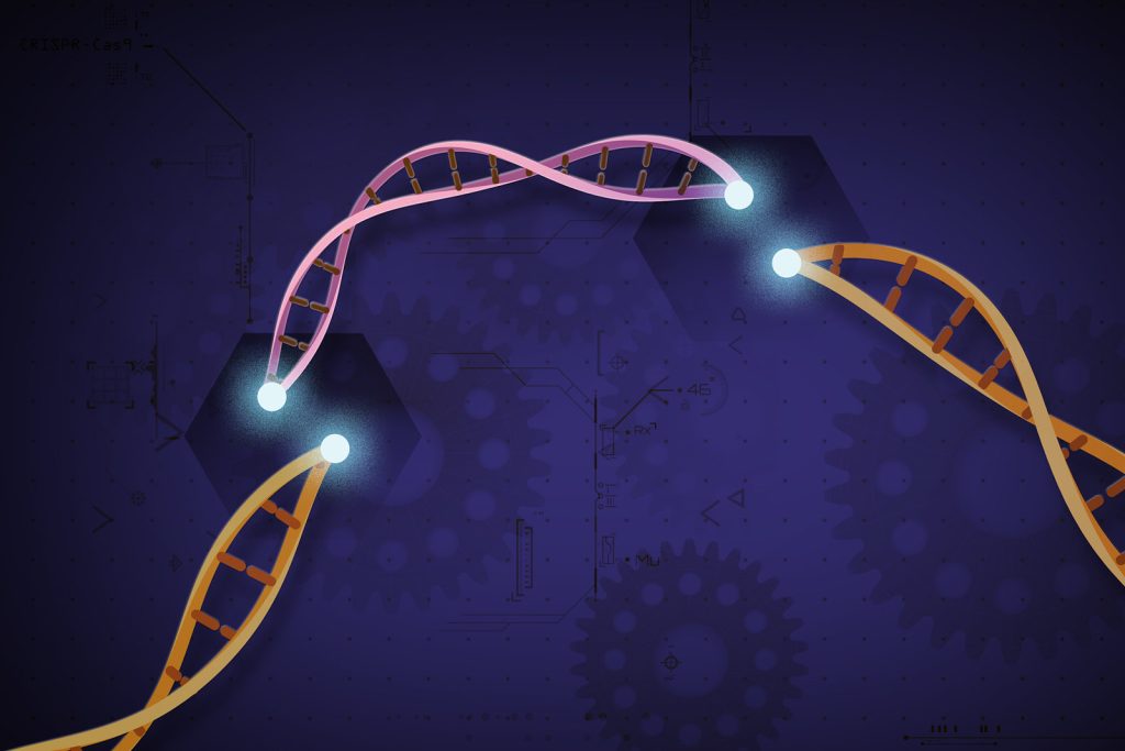
CRISPR-Cas9 is a customisable tool that lets scientists cut and insert small pieces of DNA at precise areas along a DNA strand. This lets scientists study our genes in a specific, targeted way. Credit: Ernesto del Aguila III, National Human Genome Research Institute, NIH

Researchers from the Karolinska Institutet in Sweden have identified potential pitfalls in the use of the gene editing technique CRISPR-Cas9, a gene scissors that is used for cancer treatments. Their findings are published in Life Science Alliance.

The study has identified that a cancer cell line, derived from leukaemia, removes a region that encodes a tumour-suppressing gene and genes that control cell growth.

“We found that this elimination often occurs when cancer cells are exposed to stress, such as when using CRISPR, gene scissors, or other treatments such as antibiotics. The elimination changes gene regulation in a unique way, which in turn affects basic biological processes such as DNA replication, cell cycle regulation, and DNA repair,” says Claudia Kutter, research group leader at the Department of Microbiology, Tumor and Cell Biology (MTC), Karolinska Institutet.

This knowledge is important for researchers, clinicians, and biotechnologists to correctly interpret and apply gene editing results. The study also has clinical relevance, as the observed eliminations are in genes associated with cancer, which has implications for cancer research and treatment.

“Shockingly, this elimination has been unintentionally overlooked by many researchers who modify genes in cancer cells by CRISPR screenings. The elimination also occurred more frequently in patients who have undergone cancer treatment. The treated cancer cells had, due to the elimination, a selective advantage, which is bad for the patient’s long-term survival as these cells remained after the treatment,” says Claudia.

“The study mainly serves as a warning signal, but also opens doors for further research aimed at harnessing the potential of gene editing while minimising unintended consequences,” Claudia concludes.

Source: Karolinska Institutet

European Populations are More Genetically Diverse than Expected

Source: CC0

Researchers have found that previous studies analysing the genomes of people with European ancestry may have reported inaccurate results by not fully accounting for population structure. By considering mixed genetic lineages, researchers at the National Human Genome Research Institute (NHGRI) demonstrated that previously inferred links between a genomic variant for lactase and traits such as a person’s height and low-density lipoprotein cholesterol (LDL-C) level may not be valid.

The study, published in Nature Communications, shows that people with European ancestry, who were previously treated as a genetically homogenous group in large-scale genetic studies, have clear evidence of mixed genetic lineages, known as admixture. As such, the results from previous genome-wide association studies that do not account for admixture in their examinations of people with European ancestry should be re-evaluated.

“By reading population genetics papers, we realised that the pattern of genetic makeup in Europe is too detailed to be viewed on a continental level,” said Daniel Shriner, PhD, staff scientist in the NIH Center for Research on Genomics and Global Health and senior author of the study. “What is clear based on our analysis, is when data from genetic association studies of people of European ancestry are evaluated, researchers should adjust for admixture in the population to uncover true links between genomic variants and traits.”

To look at European genetic ancestry, the researchers collated data in published genetic association studies and generated a reference panel of genomic data that included 19 000 individuals of European ancestry across 79 populations in Europe and European Americans in the US, capturing ancestral diversity not seen in other large catalogues of human genomic variation.

As an example, the researchers investigated the lactase gene, which encodes a protein that helps digest lactose and is highly varied across Europe. Using the new reference panel, they analysed how a genomic variant of the lactase gene is related to traits such as height, body mass index and LDL-C.

When the researchers considered the genetic admixture of the European population in their analysis, they found that the genomic variant of the lactase gene is not linked to height or LDL-C level. In contrast, the same variant does influence body mass index.

“The findings of this study highlight the importance of appreciating that the majority of individuals in populations around the world have mixed ancestral backgrounds and that accounting for these complex ancestral backgrounds is critically important in genetic studies and the practice of genomic medicine,” says Charles Rotimi, PhD, NIH Distinguished Investigator, director of the Center for Research on Genomics and Global Health and senior author of the study.

While the lactase gene is one example of a gene that may be incorrectly linked to some traits based on previous analyses, the researchers say it’s likely that there are other false associations in the literature and that some true associations are yet to be found. Information about how genomic variants are related to different traits helps researchers estimate polygenic risk scores and may give clues about a person’s ability to respond safely to drug treatments.

While the differences in any two people’s genomes are less than 1%, the small percentage of genomic variation can give clues about where a person’s ancestors might have come from and how different families might be related. Information about who a person is biologically descended from, known as genetic ancestry, can give important clues about genetic risks for common diseases.

“Finding true genetic associations will help researchers be more efficient and careful with how further research is conducted,” said first author Mateus Gouveia, PhD, research fellow in the Center for Research on Genomics and Global Health. “We hope that by accounting for mixed ancestries in future genomic analyses, we can improve the predictive value of polygenic risk scores and facilitate genomic medicine.”

The reference panel generated in this study is available to the scientific community for use in other studies, with additional information provided in the paper.

Source: NIH/National Human Genome Research Institute

A Single Gene-editing Infusion may Control Inherited High LDL Cholesterol

CRISPR-Cas9 is a customisable tool that lets scientists cut and insert small pieces of DNA at precise areas along a DNA strand. This lets scientists study our genes in a specific, targeted way. Credit: Ernesto del Aguila III, National Human Genome Research Institute, NIH

A single infusion of a CRISPR-based gene-editing therapy significantly reduced low-density lipoprotein cholesterol (LDL-C, the ‘bad cholesterol’) in people who carry one gene for the inherited condition that results in very high LDL-C levels and a high risk of heart attack at an early age, according to findings presented at the American Heart Association’s Scientific Sessions 2023.

“Instead of daily pills or intermittent injections over decades to lower bad cholesterol, this study reveals the potential for a new treatment option – a single-course therapy that may lead to deep LDL-C lowering for decades,” said senior study author Andrew M. Bellinger, M.D., Ph.D., chief scientific officer at Verve Therapeutics in Boston.

The investigational treatment, VERVE-101, uses DNA-editing technology to permanently turn off the PCSK9 gene in the liver. PCSK9 is a gene that plays a critical role in controlling blood LDL-C through its regulation of the LDL receptor. People with heterozygous familial hypercholesterolaemia (ie, one gene for the disorder inherited from one parent) are treated with oral lipid-lowering medications such as statins as well as PCSK9 inhibitors to bring levels under control, though this only occurs in a small percentage of patients. The study presented is the first human trial of VERVE-101.

Earlier this year, the results of the researchers’ one-year animal study were published in Circulation. In that animal study, VERVE-101 lowered PSCK9 levels 67%-83% and LDL-C 49%-69%, depending on the dose. After a single dose, the reductions have now lasted 2.5 years, supporting the idea that VERVE-101 may potentially be an effective long-term or permanent treatment for high LDL-C.

The ongoing, first-in-human study included 7 men and 2 women in New Zealand or the United Kingdom: average age of 54 years; 8 white adults; and 1 Asian adult. Each participant was diagnosed with heterozygous familial hypercholesterolemia and had extremely high bad cholesterol levels (average measure of 201mg/dL) despite taking the maximum-tolerated LDL cholesterol-lowering medication.

“These numbers are consistent with the fact that, despite available treatments, only about 3% of patients living with heterozygous familial hypercholesterolemia globally have reached target treatment goals,” Bellinger said.

The majority of study participants had pre-existing severe coronary artery disease and had already experienced a heart attack, or undergone coronary bypass surgery or stenting to allow adequate blood flow to heart muscle. None were taking PCSK9 inhibitors while enrolled in the study.

Each participant received a single intravenous infusion of VERVE-101, with the first cohort (n=3) receiving a low dose of 0.1 mg/kg and other cohorts receiving escalating doses, after consultation with an independent safety monitoring board. The highest dose received was 0.6 mg/kg.

The study found that the highest-two VERVE-101 doses:

  • reduced LDL-C by 39% and 48% in the two participants receiving 0.45mg/kg of the drug and 55% in the sole participant receiving 0.6mg/kg;
  • reduced blood PCSK9 protein levels by 47%, 59% and 84% in the three participants receiving the 0.45 mg/kg or 0.6 mg/kg doses; and
  • reduced LDL-C at six months in the sole participant receiving 0.6mg/kg, with follow-up ongoing.

“We were thrilled to see that the previous testing we had done of VERVE-101 in animal models translated faithfully to these findings in humans,” Bellinger said.

Most adverse events encountered were mild and unrelated to treatment. Serious adverse cardiovascular events, specifically a cardiac arrest, a myocardial infarction and an arrhythmia, occurred in two patients who had underlying advanced coronary artery disease. “All safety events were reviewed with the independent data safety monitoring board, who recommended continuation of trial enrolment with no protocol changes required,” Bellinger said.

Studies involving a larger number of patients and with a control group will be required to fully document the efficacy and safety of VERVE-101, noted Bellinger.

The study is still enrolling patients to receive the highest-two doses of VERVE-101. After a year’s follow-up, each participant will go into a long-term follow-up study for an additional 14 years, as required by the FDA for all participants in any human genome editing trials.

Among the study’s limitations is that this is an interim report with a few participants who all received the treatment; therefore, no participants receiving an alternate treatment or no treatment were available for direct comparison. Results in the study were measured by reductions in LDL-C, not changes in the occurrence of heart attacks; however, LDL-C reduction is a well-known, validated endpoint among patients with heterozygous familial hypercholesterolaemia and coronary artery disease.

Source: American Heart Assoication

Scientists Identify Stress-linked Gene in Treatment-resistant Depression

Photo by Andrew Neel on Unsplash

It has long been appreciated that major depressive disorder (MDD) has genetic as well as environmental influences. In a new study in Biological Psychiatry, researchers identify a gene that interacted with stress to mediate aspects of treatment-resistant MDD in an animal model.

Jing Zhang, PhD, at Fujian Medical University and senior author of the study, said, “Emerging evidence suggests that MDD is a consequence of the co-work of genetic risks and environmental factors, so it is crucial to explore how stress exposure and risk genes co-contribute to the pathogenesis of MDD.”

To do that, the authors used a mouse model of stress-induced depression called chronic social defeat stress (CSDS) in which mice are exposed to aggressor mice daily for two weeks. They focused on a gene called LHPP, which interacts with other signalling molecules at neuronal synapses. Increased expression of LHPP in the stressed mice aggravated the depression-like behaviours by decreasing expression of BDNF and PSD95 by dephosphorylating two protein kinases, CaMKIIα and ERK, under stress exposure.

Dr Zhang noted, “Interestingly, LHPP mutations (E56K, S57L) in humans can enhance CaMKIIα/ERK-BDNF/PSD95 signaling, which suggests that carrying LHPP mutations may have an antidepressant effect in the population.”

MDD is an extremely heterogeneous condition. Differences in the types of depression experienced by people influence the way they respond to treatment. A large subgroup of people with depression fail to respond to standard antidepressant medications and have “treatment-resistant” symptoms of depression. These patients often respond to different medications, such as ketamine or esketamine, or to electroconvulsive therapy. Notably, esketamine markedly alleviated LHPP-induced depression-like behaviours, whereas the traditional drug fluoxetine did not, suggesting that the mechanism might underlie some types of treatment-resistant depression.

John Krystal, MD, Editor of Biological Psychiatry, said of the work, “We have limited understanding of the neurobiology of treatment-resistant forms of depression. This study identifies a depression risk mechanism for stress-related behaviours that fail to respond to a standard antidepressant but respond well to ketamine. This may suggest that the risk mechanisms associated with the LHPP gene shed light on the poorly understood biology of treatment-resistant forms of depression.”

Dr Zhang added, “Together, our findings identify LHPP as an essential player driving stress-induced depression, implying targeting LHPP as an effective strategy in MDD therapeutics in the future.”

Source: Elsevier

Study Finds Epigenetic Changes Behind Cancer Progression

Photo by Sangharsh Lohakare on Unsplash

The journey a cell makes from healthy to metastatic cancer is mostly driven by epigenetic changes, according to a new computational study that has been recently published in the journal Nature.

Every cell makes its own proteins by accessing its genetic information, but genetic mutations may ruin the function of the affected proteins. In oncology, this is regarded as the genetics of cancer. The last decades, however, have seen the rise of a new field: the epigenetics of cancer.

Epigenetic modifications do not change the information but temporarily modify the cell’s ability to read some of its own genes and produce the associated proteins instead. This forms a vast epigenetic program that controls general cell functions and, when altered, it may put it at the starting line of malignant transformation. Is there a way to track these changes and understand the epigenetics of cancer transition?

Now an international team of researchers has made headway towards this goal. They analysed 1.7 million cells from 225 samples from primary and metastatic origin, from 205 patients of 11 different cancer types. For each cell, the team obtained the full transcriptome, exome and epigenome. This covers virtually all gene mutations, gene accessibility and its consequences. With the help of enormous computational resources, they were able deduce the whole functional status of each analysed cell and link it to its particular cancer type.

The results of the work demonstrate that many regions in the DNA are differentially activated or inactivated in a cancer-specific manner, creating a signature for each tumour. These differences are relevant for cancer progression and many correspond to already identified hallmarks of cancer, the steps a cell must undergo to become malignant. Dr Eduard Porta, group leader at the Josep Carreras Leukaemia Research Institute (IJC-CERCA), is part of the team and contributed with his experience in the analysis of large amounts of biological data.

Epigenetic changes at the DNA level stand out as an underlying cause of cancer, according to the new publication. Particularly, the accessibility of enhancer regions, a kind of master regulator acting upon many genes at once. Taken together, the results converges into a short list of genes that can be used as markers for good or poor prognosis, valuable information for the clinical management of patients.

The analysis has also identified the cellular pathways of these important genes, making it possible to track their distant interactions. Sometimes, the affected genes are so fundamental that is impossible to drug them directly without side effects but, knowing the full pathway, researchers may develop strategies to target the weakest link in the chain, maximising the therapeutic benefits while minimising undesirable effects.

Source: Josep Carreras Leukaemia Research Institute

Twin Study Reveals Epigenetic Signature for Obesity

Photo by I Yunmai on Unsplash

A susceptibility to gain weight may be written into the epigenetic information of human cells, a Washington State University study indicates.

The proof-of-concept study with a set of 22 twins found an epigenetic signature in buccal cells appearing only for the twins who were obese compared to their thinner siblings. The findings could lead to the development of a simple cheek swab test for an obesity biomarker and enable earlier prevention, the researchers said.

“Obesity appears to be more complex than simple consumption of food. Our work indicates there’s a susceptibility for this disease and molecular markers that are changing for it,” said Michael Skinner, a WSU professor of biology and corresponding author of the study published in the journal Epigenetics.

The study focused on twins to help eliminate the role of genetics and instead focus on epigenetics, molecular processes which are separate from DNA but influence how genes are expressed. The fact that the epigenetic signature was found in cheek cells rather than fat cells also suggests that the obesity signature is likely found throughout the human system.

The signature’s systemic nature also suggests that something may have occurred early in one twin’s life that triggered obesity susceptibility, Skinner added. It’s also possible that it was inherited by one twin and not the other.

For this study, Skinner worked with lead author Glen Duncan, director of the Washington State Twin Registry based at WSU, to identify 22 twin pairs, both identical and fraternal, who were discordant for obesity: one sibling had a body mass index (BMI) of 30 or higher, the standard for obesity defined by the Centers of Disease Control and Prevention, while the other sibling was in the normal range of 25 and below.

The research team analysed cells from cheek swabs provided by the twins. In the cells from the twin siblings who were obese, they found similar epigenetic changes to DNA methylation regions, areas where molecular groups made of methane attach to DNA, regulating gene expression or turning genes on or off.

The study would need to be replicated with larger groups of people to develop a biomarker test for obesity, the authors said.

The goal would be able to identify people earlier in life before they become obese so health care providers might help create interventions such as lifestyle changes, medication or both, said Duncan.

“Ultimately we would like to have some kind of preventative measure instead of our usual approach which is treatment,” he said. “It’s a simple fact that it’s better to prevent a disease, then try to treat it after you have it.”

Source: Washington State University

Genetic Analysis Reveals Secrets of Vlad Dracula the Impaler

Mediaeval tyrant and inspiration for vampires, protein analysis reveals health secrets about Vlad the Impaler

New research analysing ancient protein residues left in letters written by the sadistic 15th century tyrant – and vampire inspiration – Vlad Dracula the Impaler suggests that he suffered from a number of health conditions. One of these conditions seemingly confirms one of the more outlandish tales about him – that he cried tears of blood.

Vlad the Impaler got his nickname because he impaled thousands of people on stakes: enemies (mainly the Ottoman Empire), criminals and anyone suspected of conspiring against his rule. He was eventually defeated in 1460, but the newly invented printing press spread the tale of his gruesome deeds all over Europe. Tales surrounding him may have inspired the iconic character of Bram Stoker’s Count Dracula in 1897. Nevertheless, more modern vampire stories such as Netflix’s ‘Castlevania’ make use of Vlad as inspiration.

This terrifying reputation made him an interesting topic for a bit of genetic archaeology in a paper published in Analytical Chemistry. Using sophisticated proteomic techniques, scientists analysed three letters written in 1457 and 1475 by the voivode of Wallachia, Vlad III, also known as Vlad the Impaler, or Vlad Dracula. This allowed them to tease out information about the man who wrote the letters as well as general information about the environmental conditions of 15th century Wallachia, a place of regional trade and conflict as well as disease transmission.

While centuries-old paper is unlikely to hold entire DNA strands, scientists were still able to piece together genetic information about the writer. The technique depends on the notion that a person’s writing hand will tend to rest on the paper being written upon, rubbing off a surprising amount of organic molecules in the process. They applied ethylene vinyl acetate to the papers, and with mass spectrometry, they discovered over 500 peptides – short chains of amino acids – with about 100 being of human origin, which they looked up in database searches.

Figure 1. (a) First letter (archive catalog number is II 365), dated August 4, 1475, here investigated, also showing the positions of the EVA strips (brownish rectangles) applied to its surface for capturing biological material; (b) mapping of the fluorescence of phenylalanine, tyrosine, and tryptophan under flash UV illumination (see the original article). Anal. Chem. 2023, 95, 34, 12732-12744

The researchers noted that while many mediaeval people may have handled these papers, it is also presumable that the most prominent ancient proteins can be attributed to the one who wrote and signed them – Prince Vlad the Impaler.

First, they discovered proteins pointing to ciliopathy, which affects the cellular cilia or the cilia anchoring structures, the basal bodies or ciliary function. This can manifest in a wide range of disorders, ranging from cerebral malformation to liver disease and intellectual disability.

They also uncovered signs of an undetermined inflammatory disease which likely involved his skin and respiratory tract.

Proteomics data also suggests that, according to some stories, he might also have suffered from a pathological condition called haemolacria – he could shed tears admixed with blood. This appears to confirm what some stories said about Vlad – that he sometimes cried tears of blood. While it is a known medical condition, it would have no doubt been terrifying for superstitious mediaeval people to behold when seen in someone with a reputation like Vlad the Impaler’s.

Non-human peptides also proved to be a window into the conditions of the time, hinting at common foods, pests and diseases. Database searches of the identified, as potential endogenous original components, 3 proteins from bacteria, 24 from viruses, 4 from fungi, 17 from insects (suggesting fruit flies), and 5 from plants (including rice, wheat and thale cress). Of the bacteria, they noted that some peptides related to Enterobacterales are specific to Yersinia pestis, the pathogenic bacterium causing plague, whereas another group is specific to E. coli.