Category: Genetics

Phase 1 Clinical Trial of a Gene Therapy for Alzheimer’s
Image source: Pixabay/CC0

Researchers at University of California San Diego School of Medicine have received a grant to conduct a first-in-human Phase 1 clinical trial of a gene therapy for treating Alzheimer’s disease (AD) or Mild Cognitive Impairment (MCI), a condition often preceding dementia.

Gene therapy is an experimental technique that uses genes or gene products for the treatment or prevention of diseases by altering the DNA of living cells. Viral vectors are commonly used to insert the DNA changes into the target cells’ nuclei, but non-viral vectors also exist though they are generally less efficient.

The clinical trial, developed by principal investigator Mark Tuszynski, MD, PhD, professor of neuroscience and director of the Translational Neuroscience Institute at UC San Diego School of Medicine, delivers the brain-derived neurotrophic factor (BDNF) gene into the brains of qualifying trial participants where it is hoped it will stimulate BDNF production in cells.

BDNF belongs to a family of growth factors (proteins) found in the brain and central nervous system that support existing neurons and promote growth and differentiation of new neurons and synapses. BDNF is particularly important in brain regions susceptible to degeneration in AD.

“We found in earlier studies that delivering BDNF to the part of the brain that is affected earliest in Alzheimer’s disease — the entorhinal cortex and hippocampus — was able to reverse the loss of connections and to protect from ongoing cell degeneration,” said Tuszynski. “These benefits were observed in aged rats, aged monkeys and amyloid mice.”

The three-year-long trial seeks to recruit 12 participants with either diagnosed AD or MCI to receive AAV2-BDNF treatment, with another 12 persons serving as a control group over that period.

This will be the first safety and efficacy assessment of AAV2-BDNF in humans. A previous gene therapy trial from 2001 to 2012 using AAV2 and a different protein called nerve growth factor (NGF) found increased growth, axonal sprouting and activation of functional markers in the brains of participants.

“The BDNF gene therapy trial in AD represents an advance over the earlier NGF trial,” said Tuszynski. “BDNF is a more potent growth factor than NGF for neural circuits that degenerate in AD. In addition, new methods for delivering BDNF will more effectively deliver and distribute it into the entorhinal cortex and hippocampus.”

Source: UC San Diego

SARS-CoV-2 Does Not Alter Human DNA

Image source: Pixabay

Despite controversial claims, the SARS-CoV-2 virus likely does not integrate its genetic material into the genes of humans, according to a study published in the Journal of Virology.

A prior study reported the virus’s genetic material was found to have integrated into human DNA in cells in petri dishes, though the scientists conducting the newer research now say this was resulted from genetic artefacts in the testing.

Study co-lead author Majid Kazemian, a Purdue University assistant professor of biochemistry and computer science, said that this finding has two important implications.

“Relatively little is known about why some individuals persistently test positive for the virus even long after clearing the infection,” he explained. “This is important because it’s not clear whether such individuals have been re-infected or whether they continue to be infectious to others. So-called ‘human genome invasion’ by SARS-CoV-2 has been suggested as an explanation for this observation, but our data do not support this case.

“If the virus was able to integrate its genetic material into the human genome, that could have meant that any other mRNA could do the same. But because we have shown that this is not supported by current data, this should allay any concerns about the safety of mRNA vaccines,” he concluded.

It is indeed possible for virus genetic material to be incorporated into the DNA of humans and other animals, which are referred to as ‘chimeric events’. These have happened over many millions of years; human DNA contains approximately 100 000 pieces of ‘fossil DNA’ from viruses that have been accumulated throughout our evolution, accounting for nearly 10% of the genetic material in our cells. Some viral fragments even play a role in diseases such as cancer.

Recent scientific journal articles have raised controversy by claiming that the SARS-CoV-2 virus can also cause these chimeric events. Even before this study demonstrated it was not the case, the researchers suspected it was unlikely, said co-lead author Dr Ben Afzali, an Earl Stadtman Investigator of the National Institutes of Health’s National Institute of Diabetes and Digestive and Kidney Diseases.

“While an earlier study suggested that, in cells infected with SARS-CoV-2, genetic material from the virus copied and pasted itself into human DNA, our group thought this seemed unlikely,” Dr Afzali said. “SARS-CoV-2, like HIV, has its genetic material in the form of RNA but, unlike HIV, does not have the machinery to convert the RNA into DNA. SARS-CoV-2 is unlikely to paste itself into the genome and coronaviruses, in general, does not go near human DNA. As our study shows, we find it highly improbable that SARS-CoV-2 could integrate into the human genome.”

Christiane Wobus, associate professor of microbiology and immunology at the University of Michigan Medical School, another co-lead author on the study, said that the collective understanding of RNA viruses is that SARS-CoV-2 integrating into the human genome was extremely unlikely, it was still worth asking the question.

“Unexpected findings in science—when confirmed independently—lead to paradigm shifts and propel fields forward. Therefore, it is good to be open-minded and examine unexpected results carefully, which I believe we did in our study,” she said. “However, we did not find conclusive evidence for SARS-CoV-2 integration, but instead showed that during the RNA sequencing methodology, chimeras are produced at a very low level as an artifact of the laboratory technique.”

To examine any possible integration event, the researchers came up with a novel technique involving extraction the genetic material from infected cells and then boosting the amount of the genetic material 30-fold. With any chimeric events in the host cell DNA, these bits of genetic material from SARS-CoV-2 would have also increased those by 30. The data did not show this.

“We found the frequency of host-virus chimeric events was, in fact, not greater than background noise,” Kazemian stated. “When we enriched the SARS-CoV-2 sequences from the bulk RNA of infected cells, we found that the chimeric events are, in all likelihood, artifacts. Our work does not support the claim that SARS-CoV-2 fuses or integrates into human genomes.”

Source: Medical Xpress

Journal information: Bingyu Yan et al, Host-virus chimeric events in SARS-CoV2 infected cells are infrequent and artifactual, Journal of Virology (2021). DOI: 10.1128/JVI.00294-21

Researchers Close in on Genetic Cure for Congenital Deafness

Researchers are a step closer in the quest to use gene therapy to enable people born deaf to hear, having uncovered a new role for a key protein.

The study, published in Molecular Biology of the Cell, focused on a large gene responsible for an inner-ear protein called otoferlin. Otoferlin mutations are linked to severe congenital hearing loss, a common type of deafness in which patients can hear almost nothing.

“For a long time otoferlin seemed to be a one-trick pony of a protein,” explained Colin Johnson, associate professor of biochemistry and biophysics in the Oregon State UniversityCollege of Science. “A lot of genes will find various things to do, but the otoferlin gene had appeared only to have one purpose and that was to encode sound in the sensory hair cells in the inner ear. Small mutations in otoferlin render people profoundly deaf.”

Because the otoferlin gene is too big as it normally is to package into a delivery vehicle for molecular therapy, Prof Johnson’s team explored the use of a shortened version.

Research led by graduate student Aayushi Manchanda showed the shortened version needed to have part of the gene known as the transmembrane domain, for a surprising reason: without it, the sensory cells matured slowly.

“That was surprising since otoferlin was known to help encode hearing information but had not been thought to be involved in sensory cell development,” Johnson said.

For years, scientists in Prof Johnson’s lab have been working with the otoferlin molecule and in 2017 they identified a shortened form of the gene that can function in the encoding of sound.

To find out if the transmembrane domain of otoferlin needed to be part of the shortened version of the gene, Manchanda shortened the transmembrane domain in zebrafish.

Zebrafish are a small freshwater species that is very popular as a research organism. They grow rapidly, from a cell to a swimming fish in about five days, and share a remarkable similarity to humans at the molecular, genetic and cellular levels due to the conservation of mammalian genes early in their evolution. Embryonic zebrafish are transparent and easily maintained, and are amenable to genetic manipulation.

“The transmembrane domain tethers otoferlin to the cell membrane and intracellular vesicles but it was not clear if this was essential and had to be included in a shortened form of otoferlin,” Manchanda said. “We found that the loss of the transmembrane domain results in the sensory hair cells producing less otoferlin as well as deficits in hair cell activity. The mutation also caused a delay in the maturation of the sensory cells, which was a surprise. Overall the results argue that the transmembrane domain must be included in any gene therapy construct.”

At the molecular level, Manchanda found that a lack of transmembrane domain led to otoferlin not properly linking the neurotransmitter-filled synaptic vesicles to the cell membrane, resulting in less neurotransmitter being released.

“Our study suggests otoferlin’s ability to tether the vesicles to the cell membrane is a key mechanistic step for neurotransmitter release during the encoding of sound,” Manchanda said.

Source: EurekaAlert!

New Study Finds Genetic Switch Role in Melanoma

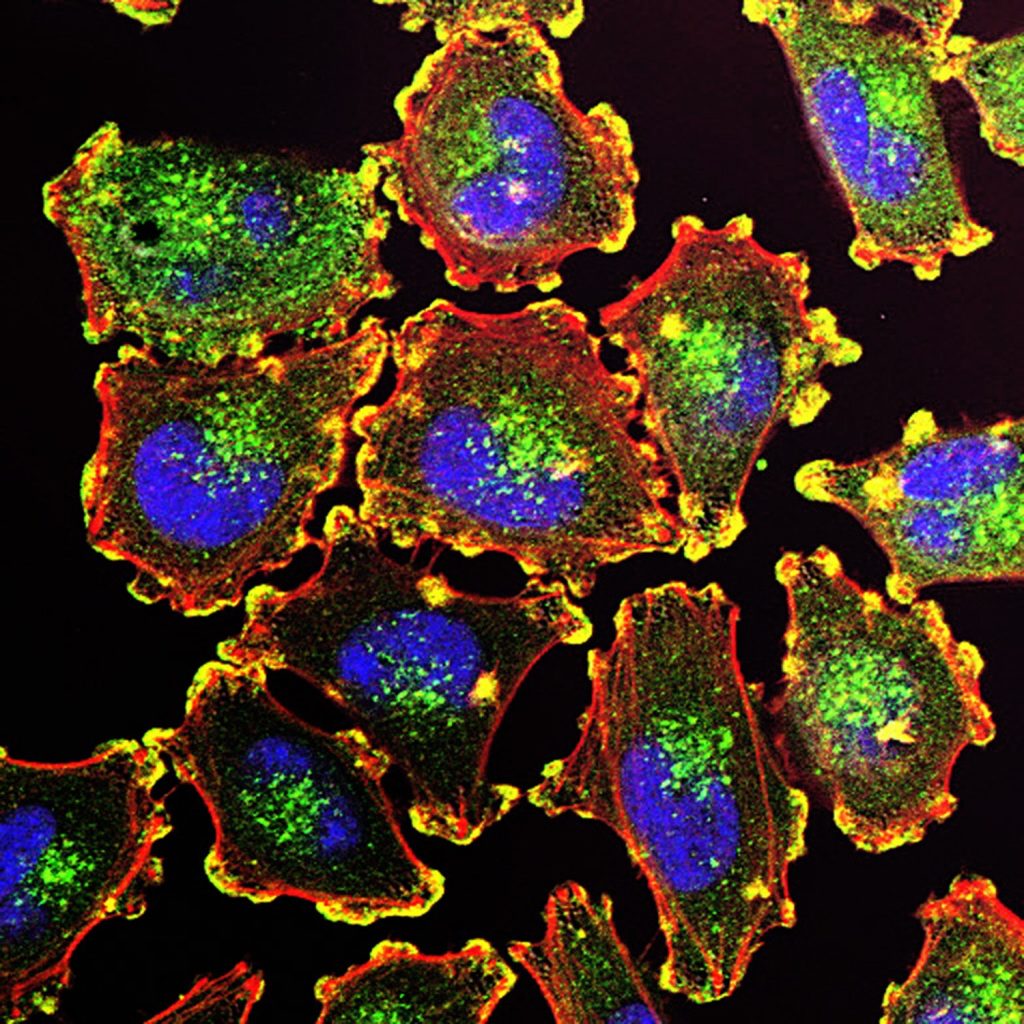
The ability of cancer cells to move and spread depends on actin-rich core structures such as the podosomes (yellow) shown here in melanoma cells. Cell nuclei (blue), actin (red), and an actin regulator (green) are also shown. Source: National Cancer Institute
Metastatic melanoma cells

A study published in the journal Cell Reports reveals that a genetic switch that could potentially be targeted to develop new treatments for melanoma by keeping the switch turned off.

Melanoma causes the majority of skin cancer-related deaths, despite only making up roughly 1 percent of skin cancers. The incidence of malignant melanoma is rapidly increasing around the world, and this increase is occurring at a faster rate than that of any other cancer except lung cancer in women. Treatments exist for this serious disease, but the effectiveness of these drugs can vary depending on the individual.

“We’ve been able to correlate the activity of this genetic switch to melanin production and cancer,” said Salk study corresponding author Marc Montminy, a professor in the Clayton Foundation Laboratories for Peptide Biology.

Melanoma develops when melanocytes, the pigment-producing skins in the cell, mutate and begin to multiply out of control. These mutations can cause proteins such as CRTC3 to prompt the cell to produce an abnormal amount of pigment, or to migrate and be more invasive.

While it was known that the CRTC family of proteins (CRTC1, CRTC2, and CRTC3) is involved in pigmentation and melanoma, obtaining precise details about the individual proteins has proven difficult. “This is a really interesting situation where different behaviours of these proteins, or genetic switches, can actually give us specificity when we start thinking about therapies down the road,” said first author Jelena Ostojic, a former Salk staff scientist and now a principal scientist at DermTech.

When the researchers deleted the CRTC3 gene in mice caused a color change in the animal’s coat color, demonstrating that the protein is needed for melanin production. They also found that when melanoma cells lacked the protein, they migrated and invaded less, meaning they were less aggressive, suggesting that inhibiting the protein could help treat the disease.

The team also described the connection between two cellular signalling systems that work on the CRTC3 protein in melanocytes. These two systems tell the cell to either proliferate or make the pigment melanin. Montminy likens this process to a relay race: essentially, a baton (chemical message) is passed from one protein to another until it reaches the CRTC3 switch, either turning it on or off.

“The fact that CRTC3 was an integration site for two signaling pathways—the relay race—was most surprising,” says Montminy, who holds the J.W. Kieckhefer Foundation Chair. “CRTC3 makes a point of contact between them that increases specificity of the signal.”

Next, the team plans to further investigate the mechanism of how CTRC3 impacts the balance of melanocyte differentiation to develop a better understanding of its role in cancer.

Source: Salk Institute

Muscle Atrophy Gene Identified from Mice Sent into Space

Dragon cargo capsule arriving at the International Space Station. Image by SpaceX-Imagery from Pixabay

Researchers from the University of Tsukuba have found a new gene involved in muscle atrophy when they sent mice into space to explore effects of weightlessness on skeletal muscles.

Extended periods of skeletal muscle inactivity or mechanical unloading (bed rest, immobilisation, spaceflight and reduced step) can result in a significant loss of muscle mass and strength which ultimately lead to muscle atrophy. Spaceflight is one of the leading models of understanding muscle atrophy from disuse.

As the molecular and cellular mechanisms involved in disuse skeletal muscle atrophy have been studied, several different signaling pathways have been studied to understand their regulatory role in this process. However, large gaps exist in the understanding of the regulatory mechanisms involved, as well as their functional significance.

Prior studies examining the effects of reduced gravity on muscle mass and function have used a ground control group which cannot be directly compared to the space experimental group. Researchers from the University of Tsukuba set out to explore the effects of gravity in mice subjected to the same housing conditions, such as the stresses of launch, landing and cosmic radiation. “In humans, spaceflight causes muscle atrophy and can lead to serious medical problems after return to Earth” says senior author Professor Satoru Takahashi. “This study was designed based on the critical need to understand the molecular mechanisms through which muscle atrophy occurs in conditions of microgravity and artificial gravity.”

Two groups of six mice each were housed onboard the International Space Station for 35 days. One group was subjected to artificial gravity (1g) and the other was left in microgravity. All mice were returned to Earth aboard a Dragon capsule and the team compared the effects of the different onboard environments on skeletal muscles. “To understand what was happening inside the muscles and cells, at the molecular level, we examined the muscle fibers. Our results show that artificial gravity prevents the changes observed in mice subjected to microgravity, including muscle atrophy and changes in gene expression,” explained Prof Takahashi. 

Transcriptional analysis of gene expression showed that the artificial gravity environment prevented altered expression of atrophy-related genes, and also identified other genes possibly associated with atrophy. Specifically, a gene called Cacng1 was identified as possibly having a functional role in myotube atrophy, which previously had no known function, and was shown to have increased activity when muscle atrophy was present.

When muscle fibres were cultured in vitro, ones which had Cacng1 expression upregulated were decreased in diameter by 27.5%. A similar effect was seen in newborn mice with upregulated Cacng1.

This work validated the use of 1g artificial gravity environments in spaceflight for examining the effects of microgravity in muscles. These studies add to the body of knowledge surrounding the mechanisms of muscle atrophy, possibly improving the treatment of related diseases.

Source: Tsukuba University

Journal information: Okada, R., et al. (2021) Transcriptome analysis of gravitational effects on mouse skeletal muscles under microgravity and artificial 1 g onboard environment. Scientific Reports. doi.org/10.1038/s41598-021-88392-4.

Oesophageal Cancer Unleashes ‘Fossil Viruses’ Hidden in Human DNA

Colorectal cancer cells. Photo by National Cancer Institute on Unsplash.

Scientists have discovered that many oesophageal cancers turn on ancient viral DNA that was embedded in our genome hundreds of millions of years ago.

“It was surprising,” said principal investigator Adam Bass, MD, the Herbert and Florence Irving Professor of Medicine at Columbia University Vagelos College of Physicians and Surgeons and Herbert Irving Comprehensive Cancer Center. 

“We weren’t specifically searching for the viral elements, but the finding opens up a huge new array of potential cancer targets that I think will be extremely exciting as ways to enhance immunotherapy.”

The idea that bits of ancient retroviruses within the human genome—known as endogenous retroviral elements, or ERVs—play a role in cancer is not new. About 8% of our genome is made up of these remnants of viral infections that have accumulated over the last 100 million years. While ERV sequences have degraded over the aeons and cannot produce viral particles, the viral fossils sometimes wind up inside other genes, disrupting their normal activities, or act as switches that turn on cancer-causing genes.

Recent research however shows that ERVs may also have a cancer fighting role if they are transcribed into strands of RNA.

“When cells activate lots of ERVs, a lot of double-stranded RNA is made and gets into the cell cytoplasm,” Dr Bass explained. “That creates a state that’s like a viral infection and can cause an inflammatory response. In that way, ERVs may make the cancer more susceptible to immunotherapy, and many researchers are working on ways to trick cancer cells into activating ERVs.”

In the new study, Bass and colleagues created organoids of oesophageal mouse tissue to follow the development of cancer from normal cells to malignancy.

With these organoids, Bass discovered that a specific cancer-promoting gene in oesophageal cancers called SOX2 triggers the expression of many ERVs.  

As ERV expression and the accumulation of double-stranded RNAs that can result can be toxic to cells, the researchers found that there is a specific enzyme called ADAR1 that rapidly degrades these double-stranded RNAs.

ADAR1 has been implicated in oesophageal cancer by other researchers, although its role had been unclear. Levels of ADAR1 are known to correlate with poor survival. “The cancers are dependent on ADAR1 to prevent an immune reaction that can be very toxic to the cells,” Bass says.

Some patients with oesophageal cancer are currently treated with immunotherapy, which has been shown to increase survival by several months. “We have a lot of enthusiasm that blocking ADAR1 may both have direct efficacy for oesophageal cancers and that ADAR1 inhibition may have even great effects by enhancing the efficacy of cancer immunotherapy in patients with oesophageal cancer,” Bass said.

Beyond the results regarding ADAR1 and ERVs, the process of modelling the development of oesophageal cancer via genomic engineering of organoids also uncovered a variety of other processes in oesophageal cancer that could help develop new therapies.

“The way we used organoids to build cancers up from the normal cell is a powerful system for uncovering cancer-causing activities and testing therapeutic targets,” said Bass. “By making individual genome alterations in these models one at a time, we can see which combinations of genetic alterations lead to cancer and then determine specific mechanisms of tumor formation.”

In this study, the organoids started with overexpression of the SOX2 gene, a commonly amplified factor that promotes the development of squamous cancers.

In the study, Dr Bass’ team built a panel of organoids emulating conditions ranging from normal oesophagus to fully transformed cancer.

By comparing normal and cancerous organoids, the team could dissect how the activity of SOX2 differs in normal and cancerous tissues. “It’s important to understand the difference, since potential treatments need to target the cancer functions but have lesser impact upon normal tissue,” he says. “It’s relatively easy to kill cancer cells. The problem is, how do you kill cancer cells but spare other cells?”

Results from the organoids showed that when SOX2 is overactive—and two tumour suppressors are inactivated—SOX2 interacts with other factors, activating an array of cancer-causing genes in addition to their effects upon induction of ERVs.  

“These findings reveal new vulnerabilities in SOX2 oesophageal cancers,” Bass said, “that will now allow us to begin developing therapies that can precisely target the cancer cell and improve the treatment of patients.”

Source: Columbia University Irving Medical Center

Journal information: Zhong Wu, et al. Reprogramming of the esophageal squamous carcinoma epigenome by SOX2 promotes ADAR1 dependence. Nature Genetics, 2021; DOI: 10.1038/s41588-021-00859-2

Liver Genes May Dictate Heart Disease Risk

Neon outline of a human heart. Photo by Olivier Collett on Unsplash

A new study by has discovered that the liver could influence people’s susceptibility to obesity and cardiovascular disease.

Drawing on data from the UK Biobank with over 700 000 individuals, scientists from Brunel University London and Imperial College London found that heart disease is far more common among people with genes that previously been linked to the control of functions such as the metabolism of fat and glucose.

Published in Nature Communications, it’s hoped the research could help clinicians identify those with the greatest risk of future heart disease, possibly allowing for interventions at an earlier stage.

“We were looking at liver enzymes, which are a reflection of our liver function, to identify which genes in the human genome control liver function and what else might be associated with those genes,” explained project co-lead Dr Raha Pazoki, a lecturer in biomedical sciences at Brunel.

“One of the things we found, for instance, was that these genes are linked to obesity and the distribution of fat in the body and the percentage of fat in the liver – they are implicated in our metabolism and how our bodies process fats and glucose.”

The team used a method known as Mendelian randomisation, which makes use of large-scale genetic datasets to replicate a controlled, randomised trial. Mendelian randomisation is a research method which provides evidence on putative causal relations between modifiable risk factors and disease, using genetic variants as natural experiments. The scientists examined individuals’ genomes based on the number of hazardous liver genes they carry, then investigated how an abundance of these hazardous genes impacts an individual’s susceptibility to heart disease.

“We found that when we looked at coronary heart disease, for example, there is an abundance of disease in those who carry hazardous liver genes compared to those who don’t. We can therefore say that there is a causal link between liver function and cardiovascular disease.”

Source: Medical Xpress

Journal information: Genetic analysis in European ancestry individuals identifies 517 loci associated with liver enzymes, Nature Communications (2021). DOI: 10.1038/s41467-021-22338-2

Severe COVID and Male Balding Gene Linked

Photo by Brett Sayles from Pexels

While COVID has been long known to be more dangerous in men than women, research which is still in its early stages shows that some of this increased risk could be from having a gene for male balding. 

A team of researchers in the US first suspected the link when they noticed that men with a common form of hormone-sensitive hair loss, known as androgenetic alopecia, were also more likely to be hospitalised with COVID.  They presented their findings May 6 at the virtual spring meeting of the European Academy of Dermatology and Venereology (EADV).

“Among hospitalized men with COVID-19, 79% presented with androgenetic alopecia compared to 31%-53% that would be expected in a similar aged match population,” said researchers led by Dr Andy Goren, chief medical officer at Applied Biology Inc in California. 

The researchers noted that androgenetic alopecia is due to the activity of the androgen receptor (AR) gene, which can lead to balding in some men. An enzyme called TMPRSS2, key to COVID infection, is also androgen-sensitive, and might be affected by the AR gene as well, explained Dr Goren’s group.

One key segment on the AR gene seems to affect both COVID severity and male balding.

In the new study, the Irvine group enrolled 65 men hospitalised with COVID, and conducted a genetic analysis on them. The results showed that participants with certain structural differences in the AR gene were at greater risk of developing severe COVID. Speaking in a meeting press release, Goren said the AR gene anomaly “could be used as a biomarker to help identify male COVID-19 patients most at risk for ICU admissions.”

He added that he believes that “the identification of a biomarker connected with the androgen receptor is another piece of evidence highlighting the important role of androgens [male hormones] in COVID-19 disease severity.”

Dr Teresa Murray Amato  has seen many severe cases of COVID. She is chair of emergency medicine at Long Island Jewish Forest Hills in New York City. Though not connected to the new research, but said it “did show a significant correlation between a higher number of androgen receptors and a higher incidence of ICU admissions for patients infected with COVID-19.”

Dr Amato added that, “While the study is small and the exact association is not completely understood, it may show at least one answer to why men were more likely to be admitted to ICU and have overall higher morality with COVID-19 infections.”

According to Amato, further investigations are necessary to determine whether “medications that block androgen receptors will be useful in treating a subset of [COVID-19] patients.”

Since the findings were presented at a medical meeting, they should be considered preliminary until published in a peer-reviewed journal.

Source: Medical Xpress

‘Chemical Nose’ Sniffs Out Cancer-causing DNA Folds

Artistic depiction of DNA double helix. Image by lisichik from Pixabay

Small folds in DNA structure have been implicated in breast cancer and other diseases have been elusive until a team of researchers developed a “chemical nose” to seek them out.

In the journal Nature Chemistry, chemists at  UC Riverside describe the development and testing of a “chemical nose” enabling them to “smell” unusual folds in DNA.

“If a DNA sequence is folded, it could prevent the transcription of a gene linked to that particular piece of DNA,” explained study author and UCR chemistry professor Wenwan Zhong. “In other words, this could have a positive effect by silencing a gene with the potential to cause cancer or promote tumors.”

DNA folding could also have a negative effect, however.

“DNA folds could potentially keep viral proteins from being produced to minimize immune response,” Prof Zhong said.

DNA folds have also been examined as potential targets for chemotherapy.   

To date, scientists have been unable to easily determine the effects of DNA folding on living organisms, as they lacked the proper tools to study them. In order to create one that can study the tiny structures, TUCR organic chemistry professor Richard Hooley and colleagues modified an existing concept that has previously been used to detect other things, such as chemical components in different vintages of wine.

The chemical system could be configured to seek out any kind of molecule, but it could not detect DNA in the way it was currently used. The addition of nonstandard components Prof Hooley’s group enabled the nose to sniff out its DNA target.

“Humans detect smells by inhaling air containing odor molecules that bind to multiple receptors inside the nose,” explained Prof Hooley. “Our system is comparable because we have multiple receptors able to interact with the DNA folds we’re looking for.”

The chemical nose consists of three parts: host molecules, fluorescent guest molecules, and DNA, which is the target. The guest molecules glow when the sought-after folds are present in a sample.

DNA is made of four nucleic acids: guanine, adenine, cytosine and thymine which are combined into the familiar double helix of DMA. Sections that are rich in guanine sometimes fold differently and create what’s called a G-quadruplex.

The parts of the genome that make these quadruplex structures are extremely complex, though the researchers have found that their folds are known to regulate gene expression, and aid in maintaining cell health.

The researchers sought to demonstrate that they could detect a single type of quadruplex composed of four guanines. Prof Zhong said that having accomplished this, the research team will try to build on their success.

“Now we think we can do more,” she said. “There are other three-dimensional structures in DNA, and we want to understand those as well.”

For their next step, the researchers will examine how effects that damage DNA influence the ways they fold. In addition, they also plan to study RNA folding since RNA is also important for cellular functioning.

“RNA has even more complex structures than DNA, and is more difficult to analyze, but understanding its structure has great potential for disease research,” Prof Zhong said. 

Source: UC Riverside News

Foetal Genetics Drive Birth Defects and Pregnancy Loss

Pregnant belly with ultrasound image superimposed. Image by Rudy and Peter Skitterians from Pixabay

A study by Yale researchers has shown that the genetics of the foetus and placenta drive developmental abnormalities, including those that lead to pregnancy loss and autism.

In the US, a birth defect is diagnosed in about 3% of children born every year, according to the Centers for Disease Control and Prevention. Birth defects also occur in most cases of pregnancy loss, and also result in many deaths in the first year of life. The causes are thought to be a complex interplay of environmental and biological factors.

“Mothers often feel that they are responsible for these defects,” explained senior author Dr Harvey Kliman,  Research Scientist at the Department of Obstetrics, Gynecology & Reproductive Services, Yale School of Medicine. “But it’s not their fault. This new research points to the genetics of these children as being the most important cause.”

Dr Kliman and his team examined placental data for nearly 50 sets of identical and non-identical twins. They discovered identical twins had similar numbers of abnormal cell growths called trophoblast inclusions (TIs), which are markers for a number of developmental abnormalities, while non-identical twins showed a markedly different TI count.

While identical twins share the same DNA, non-identical twins only share half of their DNA. Identical twins were found to often have the same number of TIs or were within one of having the same TI count. Non-identical twins had TI counts that differed by four or five on average.

“This work suggests that developmental abnormalities are much more likely to be due to the genetics of the child, and not the mother’s fault,” Dr Kliman explained.

The findings were reported in the journal Placenta.  

First author Julia Katz, a former Yale undergraduate and now medical student at Hofstra University, provided the inspiration for the study.

Katz and her brother, Jesse, who was born underweight and with several congenital abnormalities, are non-identical twins. “I had a lot of guilt, growing up, about why my twin had certain conditions that I didn’t,” Katz explained. “I think mothers also tend to blame themselves.”

After a lecture,  Katz approached Dr Kliman and asked him what causes babies to be born undersized, a conversation which led to a discussion about developmental abnormalities and Katz’s desire to find out more about her and her twin’s genetics — including looking at her own placental slides from birth.

It also led Dr Kliman, Katz, and co-author Parker Holzer, a graduate student in the Yale Department of Statistics and Data Science, to conduct the new study.

“Julia’s need to resolve this burden is what propelled our study,” Dr Kliman said. “Hopefully, this finding will help many other people, as well.”

“This experience has shown me that if you have a question, ask it,” Katz added. “And if you don’t get an answer, try to answer it yourself.”

Source: News-Medical.Net

Journal information: Katz, J., et al. (2021) Genetics, not the uterine environment, drive the formation of trophoblast inclusions: Insights from a twin study. Placenta. doi.org/10.1016/j.placenta.2021.04.010.