Category: Genetics

Research Reveals Many More Epigenetic Influences on Offspring

Pregnant with ultrasound image
Source: Pixabay

New research suggests that epigenetic information, which turns DNA sections on or off, and is normally reset between generations, is more frequently carried from mother to offspring than previously thought. The findings were published in Nature Communications.

Despite not directly altering the DNA sequence, epigenetic mechanisms can regulate gene expression through chemical modifications of DNA bases and changes to the chromosomal superstructure in which DNA is packaged.

These epigenetic changes can be induced through various such as diet and stress. While epigenetic modifications are reversible, it was thought that they rarely remain through generations in humans despite persisting through multiple cycles of cell replication.

Epigenetic changes can be influenced by environmental variations such as our diet, but these changes do not alter DNA and are normally not passed from parent to offspring.

The new research reveals that the supply of a specific protein in the mother’s egg can affect the genes that drive skeletal patterning of offspring. 

Chief investigator Professor Marnie Blewitt said the findings initially left the team surprised.

“It took us a while to process because our discovery was unexpected,” Professor Blewitt said.

“Knowing that epigenetic information from the mother can have effects with life-long consequences for body patterning is exciting, as it suggests this is happening far more than we ever thought.

“It could open a Pandora’s box as to what other epigenetic information is being inherited.”

The research examined the protein SMCHD1, an epigenetic regulator discovered by Prof Blewitt in 2008, and Hox genes, which control the identity of each vertebra during embryonic development in mammals. The epigenetic regulator prevents these genes from being activated too soon.

In this study, the researchers discovered that the amount of SMCHD1 in the mother’s egg affects the activity of Hox genes and influences the patterning of the embryo. Without maternal SMCHD1 in the egg, offspring were born with altered skeletal structures.  

First author and PhD researcher Natalia Benetti said this was clear evidence that epigenetic information had been inherited from the mother, rather than just DNA.

“While we have more than 20 000 genes in our genome, only that rare subset of about 150 imprinted genes and very few others have been shown to carry epigenetic information from one generation to another,” Benetti said.

“Knowing this is also happening to a set of essential genes that have been evolutionarily conserved from flies through to humans is fascinating.”

The research showed that SMCHD1 in the egg, which only persists for two days after conception, has a life-long impact.

SMCHD1 variants are linked to developmental disorder Bosma arhinia microphthalmia syndrome (BAMS) and facioscapulohumeral muscular dystrophy (FSHD), a form of muscular dystrophy. The researchers say their findings could have implications for women with SMCHD1 variants and their children in the future.

Research is underway on using on SMCHD1 to design novel therapies to treat developmental disorders, such as Prader Willi Syndrome and the degenerative disorder FSHD.

Source: The Walter and Eliza Hall Institute of Medical Research

Quantum Leap for Genetic Disease Therapy with Baculovirus DNA Repair Kit

DNA repair
Source: Pixabay/CC0

Genetic mutations behind a genetic kidney disease affecting children and young adults have been fixed in patient-derived kidney cells with a high-capacity DNA ‘repair kit’. The advance, developed by University of Bristol scientists, is published in Nucleic Acids Research.

In this new study, the international team describe how they created a DNA repair vehicle to genetically fix faulty podocin, a common genetic cause of inheritable Steroid Resistant Nephrotic Syndrome (SRNS).

Podocin is a protein normally located on the surface of specialised kidney cells and is essential for kidney function. Faulty podocin, however, remains stuck inside the cell and never makes it to the surface, terminally damaging the podocytes. Since the disease cannot be cured with medications, gene therapy which repairs the genetic mutations causing the faulty podocin offers hope for patients.

Typically, human viruses have been utilised in gene therapy applications to carry out genetic repairs. These are used as a ‘Trojan Horse’ to enter cells carrying the errors. Currently dominating systems include lentivirus (LV), adenovirus (AV) and adeno-associated virus (AAV), which are all relatively harmless viruses that readily infect humans. Their viral shells however restrict the amount of cargo they can carry and deliver, namely the DNA kit necessary for efficient genetic repair. This limits the scope of their application in gene therapy.

By applying synthetic biology techniques, the team led by Dr Francesco Aulicino and Professor Imre Berger, re-engineered baculovirus, a insect virus which has a nearly unlimited cargo capacity.

“What sets apart baculovirus from LV, AV, and AAV is the lack of a rigid shell encapsulating the cargo space.” said Dr Francesco Aulicino, who led the study. The shell of baculovirus resembles a hollow stick, simply lengthening when the cargo increases. This allows a much more sophisticated tool-kit can be delivered by the baculovirus.

First, baculovirus had to be equipped to penetrate human cells which it normally would not do. “We decorated the baculovirus with proteins that enabled it to enter human cells very efficiently.” explained Dr Aulicino. The scientists then used their engineered baculovirus to deliver much larger DNA pieces than was previously possible, and build these into the genomes of a whole range of human cells.

The DNA in the human genome comprises 3 billion base-pairs making up ~25,000 genes, which encode for the proteins that are essential for cellular functions. If faulty base-pairs occur in our genes, faulty proteins are made which can make us ill, resulting in hereditary disease. Gene therapy promises repair of hereditary disease at its very root, by rectifying such errors in our genomes. Gene editing approaches, in particular CRISPR/Cas-based methods, have greatly advanced the field by enabling genetic repair with base-pair precision.

The team used patient-derived podocytes carrying the disease-causing error in the genome to demonstrate the aptitude of their technology. By creating a DNA repair kit, comprising protein-based scissors and the nucleic acid molecules that guide them – and the DNA sequences to replace the faulty gene, the team delivered with a single engineered baculovirus a healthy copy of the podocin gene concomitant with the CRISPR/Cas machinery to insert it with base-pair precision into the genome. This was able to reverse the disease-causing phenotype and restore podocin to the cell surface.

Professor Imre Berger explained: “We had previously used baculovirus to infect cultured insect cells to produce recombinant proteins for studying their structure and function.” This method, called MultiBac, has been highly successful to make very large multiprotein complexes with many subunits, in laboratories world-wide. “MultiBac already exploited the flexibility of the baculovirus shell to deliver large pieces of DNA into the cultured insect cells, instructing them to assemble the proteins we were interested in.” When the scientists realised that the same property could potentially transform gene therapy in human cells, they created this new DNA repair kit.

Dr Aulicino added: “There are many avenues to utilise our system. In addition to podocin repair, we could show that we can simultaneously correct many errors in very different places in the genome efficiently, by using our single baculovirus delivery system and the most recent editing techniques available.”

Source: University of Bristol

CRISPR Editing can Destabilise the Genome, Study Finds

DNA repair
Source: Pixabay/CC0

A new study published in Nature Biotechnology identifies risks in the use of CRISPR gene editing, which is employed in a number of therapies. Looking at its use in T cells, the researchers detected a loss of genetic material in a significant percentage – up to 10% of the treated cells. They explain that such loss can lead to destabilisation of the genome, which might cause cancer.

The study was led by Drs Adi Barzel, Dr Asaf Madi and Dr Uri Ben-David at Tel Aviv University.

Developed about a decade ago, CRISPR cleaves DNA sequences at certain locations in order to delete unwanted segments, or alternately repair or insert beneficial segments. It has already proved impressively effective in treating a range of diseases – cancer, liver diseases, genetic syndromes, and more. In 2020 at the University of Pennsylvania, the first approved clinical trial ever to use CRISPR took T cells from a donor, and expressed an engineered receptor targeting cancer cells, while using CRISPR to destroy genes coding for the original receptor – which otherwise might have caused the T cells to attack cells in the recipient’s body. 

In the present study, the researchers sought to examine whether the potential benefits of CRISPR therapeutics might be offset by risks resulting from the cleavage itself, assuming that broken DNA is not always able to recover.

Dr Ben-David and his research associate Eli Reuveni explained: “The genome in our cells often breaks due to natural causes, but usually it is able to repair itself, with no harm done. Still, sometimes a certain chromosome is unable to bounce back, and large sections, or even the entire chromosome, are lost. Such chromosomal disruptions can destabilise the genome, and we often see this in cancer cells. Thus, CRISPR therapeutics, in which DNA is cleaved intentionally as a means for treating cancer, might, in extreme scenarios, actually promote malignancies.”

To examine the extent of potential damage, the researchers repeated the 2020 Pennsylvania experiment, cleaving the T cells’ genome in exactly the same locations – chromosomes 2, 7, and 14. Using single-cell RNA sequencing, they analysed each cell separately and measured the expression levels of each chromosome in every cell.

They detected a significant loss of genetic material in some of the cells. For example, when chromosome 14 had been cleaved, about 5% of the cells showed little or no expression of this chromosome. When all chromosomes were cleaved simultaneously, the damage increased, with 9%, 10%, and 3% of the cells unable to repair the break in chromosomes 14, 7, and 2 respectively. The three chromosomes did differ, however, in the extent of the damage they sustained. 

Dr Madi and his student Ella Goldschmidt explained: “Single-cell RNA sequencing and computational analyses enabled us to obtain very precise results. We found that the cause for the difference in damage was the exact place of the cleaving on each of the three chromosomes. Altogether, our findings indicate that over 9% of the T-cells genetically edited with the CRISPR technique had lost a significant amount of genetic material. Such loss can lead to destabilisation of the genome, which might promote cancer.”

Based on their findings, the researchers caution that extra care should be taken when using CRISPR therapeutics. They also propose alternative, less risky, methods, for specific medical procedures, and recommend further research into two kinds of potential solutions: reducing the production of damaged cells or identifying damaged cells and removing them before the material is administered to the patient.

Dr Barzel and his PhD student Alessio Nahmad conclude: “Our intention in this study was to shed light on potential risks in the use of CRISPR therapeutics,” adding that as scientists, they “examine all aspects of an issue, both positive and negative, and look for answers.”

Source: EurekAlert!

Scientists Untangle the Secrets of DNA Compression in Sperm

Genetics
Image source: Pixabay

During sperm production, an enormous amount of DNA has to be packed into a very small space without breaking anything. Protamines are the key to this compression, wrapping the DNA thread tightly, but humans have a second type of protamine which had an unknown purpose. Insights into this key mechanism are described in PLoS Genetics.

During the production of human sperm cells, DNA has to be packed into a tiny space, not unlike trying to cram too many clothes into a tiny suitcase to go on holiday. DNA is normally in a comparatively loose tangle. In sperm cells, however, it is enormously compressed. The 23 DNA threads have a total length of one metre and have to be packed into a head just three thousandths of a millimetre in diameter. All of this must happen without the delicate DNA threads tearing or becoming inextricably tangled up.

We often sit on packed suitcases to close them, and the body uses a similar trick during spermatogenesis. “If DNA were to take up as much space as a watermelon under normal circumstances, sperm cells would then only be as big as a tennis ball,” explained Professor Hubert Schorle from the University Hospital Bonn.

This process is termed hypercondensation. In their loose state, DNA threads are wrapped around numerous spherical protein molecules, the histones. In this state, they resemble 23 tiny strings of pearls. During hypercondensation, the histones are first exchanged for transition proteins, which are in turn are replaced by so-called protamines. Due to their chemical composition, protamines exert a very strong attraction on DNA, causing it to wrap itself in very firm and tightly loops around the protamine

“Most mammals seem to produce only one type of protamine, PRM1,” explained Dr Lena Arévalo, a postdoctoral researcher in Schorle’s group. “In humans, but also rodents like mice, it’s different — they have a second type, PRM2.” Until now, it was unclear what this second protamine is needed for. It was however known that some parts of it are successively cut off during sperm development.

These cut-off parts that appear to be immensely important, according to the new study. When mice produce only a truncated PRM2 molecule that lacks the cut-off snippets, they are infertile. “The removal of transition proteins during hypercondensation is impaired,” Dr Arévalo said. “In addition, the condensation seems to proceed too quickly, causing the DNA strands to break.”

It is possible that a defective protamine 2 can also lead to infertility in human males. The team now plans to investigate this hypothesis further, thanks to their lab being the only one so far that has successfully generated and bred PRM and PRM2 deficient mouse lines.

Source: University of Bonn

Once-off Gene Therapy Reduces Haemophilia B Bleeding in Patients

https://www.pexels.com/photo/a-close-up-shot-of-bags-of-blood-4531306/
Photo by Charlie-Helen Robinson on Pexels

A single gene therapy injection could dramatically reduce the bleeding risk faced by people with haemophilia B, according to the results of a Phase I/II clinical trial published in the New England Journal of Medicine.

Low levels of the factor IX (FIX) protein, needed for clot formation, are behind haemophilia B. The FIX protein gene is on the X chromosome, so the severe form of haemophilia B is much more common in men, though it can occur in women due to X chromosome inactivation.

To prevent excessive bleeding, patients with haemophilia B typically need regular replacement therapy consisting of weekly injections of recombinant FIX. Despite advances in treatment, patients may continue to see debilitating joint damage.

The new treatment, from University College London, Royal Free Hospital and biotechnology company Freeline Therapeutics, is a type of adeno-associated virus (AAV) gene therapy candidate, called FLT180a, is being developed to treat severe and moderately severe cases of haemophilia B.

The Phase I/II multi-centre clinical trial, called B-AMAZE, and the related long-term follow up study found that a single treatment with FLT180a led to sustained production of FIX protein from the liver in 9 of 10 patients, across four different dose levels, removing the need for regular replacement therapy.

Out of 17 male patients aged 18 or over who underwent screening, 10 with severe or moderately severe haemophilia B took part in the 26-week trial of FLT180a, will be followed-up to assess safety and durability of FIX expression for 15 years.

Lead author Professor Pratima Chowdary of the Royal Free Hospital said: “Removing the need for haemophilia patients to regularly inject themselves with the missing protein is an important step in improving their quality of life. The long term follow up study will monitor the patients for durability of expression and surveillance for late effects.”

One patient, Elliott Mason, told the BBC: “I’ve not had any treatment since I had my therapy, it’s all a miracle really, well it’s science, but it feels quite miraculous to me.

“My life is completely normal, there’s nothing that I have to stop and think ‘how might my haemophilia affect this?’.”

AAV gene therapy works uses a packaging from the proteins found in the outer coat of the virus to deliver a functional copy of a gene directly to patient tissues. Newly synthesised proteins are released into the blood and a one-time infusion can have long-term effects.

Over several weeks to several months, patients took immunosuppressive drugs to prevent their immune systems from rejecting the therapy, and all reported known side effects.

Though the therapy was well tolerated, patients all experienced adverse events, with an abnormal blood clot in one who received the highest FLT180a dose and had the highest levels of FIX protein.

Professor Amit Nathwani, who co-authored the study, said: “Gene therapy is still a young field that pushes the boundaries of science for people with severe genetic diseases.

“The B-AMAZE long-term data add to the growing body of evidence that gene therapy has the potential to free patients from the challenges of having to adhere to lifelong therapy or could provide treatment where none exists today.”

In nine out of the ten patients, the treatment led to a sustained increase in FIX protein production, which led to a decrease in excessive bleeding. They also no longer required weekly injections of FIX protein.

After 26 weeks, five patients had normal levels of FIX protein, three had low but increased levels, and one patient treated at the highest dose had an abnormally high level.

Pamela Foulds, MD, Chief Medical Officer of Freeline, said: “The B-AMAZE long-term data continue to support our confidence that a single dose of FLT180a could protect people with haemophilia B from bleeding and the need for lifelong FIX replacement through durable expression of FIX at protective levels.”

Source: EurekAlert!

‘Silent’ Mutations in Genes Found to be Mostly Harmful

Genetics
Image source: Pixabay

New research with yeast strains carrying ‘silent’ gene mutations, where misspellings occur but do not affect the protein they code for, shows that they are not harmless as previously assumed – rather, they are mostly harmful. The findings, which appear in the journal Nature,  could overturn decades of thinking if they are applicable to organisms such as humans.

In the early 1960s, University of Michigan alumnus Marshall Nirenberg and a few other scientists deciphered the genetic code, determining the rules by which information in DNA molecules is translated into proteins.

They identified three-letter units in DNA sequences, known as codons, that specify each of the 20 amino acids that make up proteins, work for which Nirenberg later shared a Nobel Prize with two others.

Occasionally, single-letter misspellings occur, which are known as point mutations. Point mutations that alter the resulting protein sequences are called nonsynonymous mutations, while those that do not are called silent or synonymous mutations.

Around one-quarter to one-third of point mutations in protein-coding DNA sequences are synonymous. Ever since the genetic code was cracked, those mutations have generally been assumed to be neutral, or nearly so.

But a new study involving the genetic manipulation of yeast cells in the laboratory demonstrates that most synonymous mutations are strongly harmful.

The strong nonneutrality of most synonymous mutations – if found to be true for other genes and in other organisms – would have major implications for the study of human disease mechanisms, population and conservation biology, and evolutionary biology, according to the study authors.

“Since the genetic code was solved in the 1960s, synonymous mutations have been generally thought to be benign. We now show that this belief is false,” said the study’s senior author, Professor Jianzhi “George” Zhang at the University of Michigan.

“Because many biological conclusions rely on the presumption that synonymous mutations are neutral, its invalidation has broad implications. For example, synonymous mutations are generally ignored in the study of disease-causing mutations, but they might be an underappreciated and common mechanism.”

In the past decade, anecdotal evidence has suggested that some synonymous mutations are nonneutral, which Prof Zhang and his colleagues wanted to investigate.

They chose to address this question in budding yeast (Saccharomyces cerevisiae) because the organism’s short generation time (about 80 minutes) and small size allowed them to measure the effects of a large number of synonymous mutations relatively quickly, precisely and conveniently.

With gene editing, they constructed more than 8000 mutant yeast strains, each carrying a synonymous, nonsynonymous or nonsense mutation in one of 21 selected genes.

Then they quantified the “fitness” of each mutant strain by measuring how quickly it reproduced relative to the nonmutant strain. Darwinian fitness, simply put, refers to the number of offspring an individual has. In this case, measuring the reproductive rates of the yeast strains showed whether the mutations were beneficial, harmful or neutral.

To their surprise, the researchers found that 75.9% of synonymous mutations were significantly deleterious, while 1.3% were significantly beneficial.

“The previous anecdotes of nonneutral synonymous mutations turned out to be the tip of the iceberg,” said study lead author Xukang Shen, a graduate student research assistant in Prof Zhang’s lab.

“We also studied the mechanisms through which synonymous mutations affect fitness and found that at least one reason is that both synonymous and nonsynonymous mutations alter the gene-expression level, and the extent of this expression effect predicts the fitness effect.”

Prof Zhang said the researchers knew beforehand, based on the anecdotal reports, that some synonymous mutations were likely nonneutral.

“But we were shocked by the large number of such mutations,” he said. “Our results imply that synonymous mutations are nearly as important as nonsynonymous mutations in causing disease and call for strengthened effort in predicting and identifying pathogenic synonymous mutations.”

The U-M-led team said that while there is no particular reason why their results would be restricted to yeast, confirmations in diverse organisms are required to verify the generality of their findings.

Source: University of Michigan

One in 500 Men Carry an Extra Sex Chromosome, Increasing Disease Risk

Photo by Cottonbro on Pexels

Around one in 500 men could be carrying an extra sex chromosome (X or Y), putting them at increased risk of diseases such as type 2 diabetes, atherosclerosis and thrombosis, according to a study published in Genetics in Medicine.

Researchers from the universities of Cambridge and Exeter analysed genetic data on 200 000 men aged 40 to 70 from UK Biobank. They found 356 men who carried either an extra X chromosome or an extra Y chromosome.

Some men have an extra X or Y chromosome – XXY or XYY, which is usually not obvious without a genetic test. Men with extra X chromosomes, a condition known as Klinefelter syndrome, are sometimes identified during investigations of delayed puberty and infertility; however, most are unaware that they have this condition. Men with an extra Y chromosome tend to be taller as boys and adults, but otherwise they have no distinctive physical features.

In today’s study, the researchers identified 213 men with an extra X chromosome and 143 men with an extra Y chromosome. As the participants in UK Biobank tend to be ‘healthier’ than the general population, this suggests that around one in 500 men may carry an extra X or Y chromosome.

Only a small minority of these men had a diagnosis of sex chromosome abnormality on their medical records or by self-report: fewer than one in four (23%) men with XXY and only one of the 143 XYY men (0.7%) had a known diagnosis.

By linking genetic data to routine health records, the team found that men with XXY have much higher chances of reproductive problems, including a three-fold higher risk of delayed puberty and a four-fold higher risk of being childless. These men also had significantly lower blood concentrations of testosterone. Men with XYY appeared to have a normal reproductive function.

Men with either XXY or XYY had higher risks of several other health conditions: a three-fold higher risk of developing type 2 diabetes, six-fold risk of venous thrombosis, three-fold risk of pulmonary embolism, and four-fold risk of chronic obstructive pulmonary disease (COPD).

It is unclear why an extra chromosome should increase the risk, said the researchers, or why the risks were so similar regardless of which sex chromosome was duplicated.

Yajie Zhao, a PhD student at the Medical Research Council (MRC) Epidemiology Unit at the University of Cambridge, the study’s first author, said: “Even though a significant number of men carry an extra sex chromosome, very few of them are likely to be aware of this. This extra chromosome means that they have substantially higher risks of a number of common metabolic, vascular, and respiratory diseases — diseases that may be preventable.”

Professor Ken Ong, also from the MRC Epidemiology Unit at Cambridge and joint senior author, added: “Genetic testing can detect chromosomal abnormalities fairly easily, so it might be helpful if XXY and XYY were more widely tested for in men who present to their doctor with a relevant health concern.

“We’d need more research to assess whether there is additional value in wider screening for unusual chromosomes in the general population, but this could potentially lead to early interventions to help them avoid the related diseases.”

Professor Anna Murray, at the University of Exeter, said: “Our study is important because it starts from the genetics and tells us about the potential health impacts of having an extra sex chromosome in an older population, without being biased by only testing men with certain features as has often been done in the past.”

Previous studies have found that around one in 1,000 females have an additional X chromosome, which can result in delayed language development and accelerated growth until puberty, as well as lower IQ levels compared to their peers.

Source: University of Cambridge

A Blindness Gene That Also Increases Intelligence

DNA repair
Source: Pixabay/CC0

A new study published in Brain shows that a genetic mutation which causes blindness in humans also increases intelligence, possibly through an increase in synaptic activity between the very same neurons damaged by the mutation.

The present study came about when Professors Tobias Langenhan and Manfred Heckmann, came across a paper on a mutation that damages a synaptic protein. The mutation caused patients to go blind, but then doctors noticed that the patients were also of above-average intelligence, something which piqued the two neurobiologists’ interest. “It’s very rare for a mutation to lead to improvement rather than loss of function,” said Prof Langenhan.

The two neurobiologists have been using fruit flies to analyse synaptic functions for many years. “Our research project was designed to insert the patients’ mutation into the corresponding gene in the fly and use techniques such as electrophysiology to test what then happens to the synapses. It was our assumption that the mutation makes patients so clever because it improves communication between the neurons which involve the injured protein,” explained Prof Langenhan. “Of course, you can’t conduct these measurements on the synapses in the brains of human patients. You have to use animal models for that.”

“75 per cent of genes that cause diseases in humans also exist in fruit flies”

Professor Tobias Langenhan

First, in collaboration with Oxford researchers, the scientists showed that the fly protein called RIM looks molecularly identical to that of humans. This was essential in order to be able to study the changes in the human brain in the fly. In the next step, the neurobiologists inserted the genetic mutation into flies. They then took electrophysiological measurements of synaptic activity. “We actually observed that the animals with the mutation showed a much increased transmission of information at the synapses. This amazing effect on the fly synapses is probably found in the same or a similar way in human patients, and could explain their increased cognitive performance, but also their blindness,” concludes Professor Langenhan.

The scientists also found out how the increased transmission at the synapses occurs: the molecular components in the transmitting nerve cell that trigger the synaptic impulses move closer together as a result of the mutation effect and lead to increased release of neurotransmitters. A novel method, super-resolution microscopy, was one of the techniques used in the study. “This gives us a tool to look at and even count individual molecules and confirms that the molecules in the firing cell are closer together than they normally are,” said Prof Langenhan.

“The project beautifully demonstrates how an extraordinary model animal like the fruit fly can be used to gain a very deep understanding of human brain disease. The animals are genetically highly similar to humans. It is estimated that 75% of the genes involving disease in humans are also found in the fruit fly,” explained Professor Langenhan, pointing to further research on the topic: “We have started several joint projects with human geneticists, pathologists and the team of the Integrated Research and Treatment Center (IFB) Adiposity Diseases; based at Leipzig University Hospital, they are studying developmental brain disorders, the development of malignant tumours and obesity. Here, too, we will insert disease-causing mutations into the fruit fly to replicate and better understand human disease.”

Source: Universität Leipzig

People with Blue Eyes Share a Single Ancestor

Eye
Source: Daniil Kuzelev on Unsplash

New research published in Human Genetics shows that people with blue eyes trace their ancestry back to a single individual. Researchers tracked down a genetic mutation which took place 6–10 000 years ago and is the cause of the eye colour of all blue-eyed humans without albinism alive on the planet today.

While blue eyes evolved only once, blonde hair has evolved at least twice: in Melanesian populations, blonde hair evolved independently to European populations, involving a mutation in a different gene.

“Originally, we all had brown eyes,” said Professor Hans Eiberg from the University of Copenhagen. “But a genetic mutation affecting the OCA2 gene in our chromosomes resulted in the creation of a ‘switch’, which literally ‘turned off’ the ability to produce brown eyes.” The OCA2 gene codes for the P protein, which is involved melanin production. This ‘switch’, located in the gene next to OCA2, does not completely shut off production but instead is limited to reducing the production of melanin in the iris, effectively ‘diluting’ brown eyes to blue. The switch’s effect on OCA2 is very specific therefore. If the OCA2 gene is completely destroyed or turned off, albinism would be the result.

Eye colours from brown to green depend on the amount of melanin in the iris, but blue-eyed individuals only have a small degree of variation in the amount of melanin in their eyes. “From this we can conclude that all blue-eyed individuals are linked to the same ancestor,” said Professor Eiberg. “They have all inherited the same switch at exactly the same spot in their DNA.” Brown-eyed individuals, by contrast, have considerable individual variation in the area of their DNA that controls melanin production.

Professor Eiberg and his team studied mitochondrial DNA and compared the eye colour of blue-eyed individuals in countries as diverse as Jordan, Denmark and Turkey. His research stretches back to 1996, when he first implicated the OCA2 gene as being responsible for eye colour.

The mutation of brown to blue eyes does not confer any evolutionary advantage, as with others such as hair colour.

As Professor Eiberg explained, “it simply shows that nature is constantly shuffling the human genome, creating a genetic cocktail of human chromosomes and trying out different changes as it does so.”

Source: University of Copenhagen

Rare COVID Vaccine Blood Clots May Result from Genetics

Photo by Spencer Davis on Unsplash

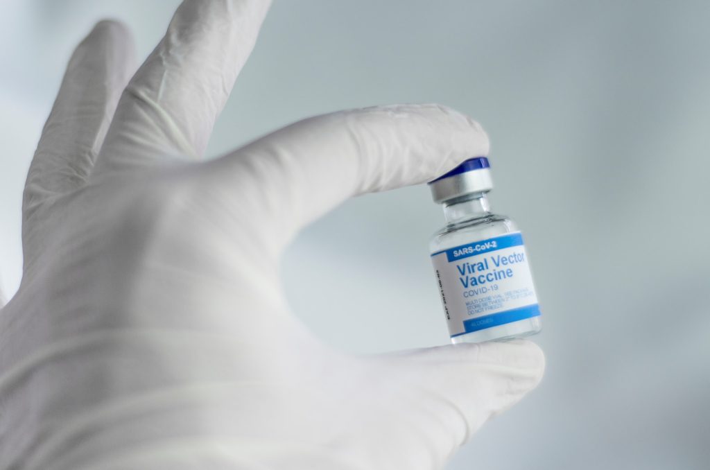
Scientists have discovered that the rare blood clot side-effect associated with some COVID vaccines could be the result of a specific gene variant, which could make a genetic screening test possible.

Vaccine-induced thrombotic thrombocytopenia (VITT), a rare disorder causing thrombosis and thrombocytopenia (low blood platelet counts), was linked to AstraZeneca’s COVID vaccine in early 2021, leading some countries to pause or restrict its use. It is also associated with the Johnson & Johnson vaccine, which also uses a viral vector.

Now, a new study may help to explain what’s causing the rare side effect. The study by Flinders University and SA Pathology is now available on the medRxiv preprint server and is awaiting peer review.

Examining five unrelated individuals who all had the clotting complication after vaccination, the researchers found that all of the patients had unusually structured antibodies against a protein called platelet factor 4 (PF4), which is involved in blood clotting.

In addition, all five shared a specific version of a gene responsible for producing these antibodies.

“We knew previously that PF4 was directly involved in the clotting disorder, and we knew that aberrant antibodies against PF4 are responsible, but what we don’t know is how and why some people develop them,” explained lead author Dr Jing Jing Wang.

The antibodies were all found to be derived from the same amino acid sequence. The researchers then found that all of the patients carried a specific variant of one gene, called IGLV3-21*02, most commonly occurring in people of European descent.

“The other specific amino acid sequences of these antibodies from each patient were derived from separate basic sequences but had all evolved to carry very similar properties, making them very potent attackers of the PF4 protein,” explained research team leader Professor Tom Gordon.

“Together, this suggests that it is the combination of a variant in a gene and the evolution of this antibody towards targeting the PF4 protein in a destructive manner, which is leading to this harmful side-effect.”

Though why the antibody is found in such a tiny number of vaccine recipients remains unknown, the identification of the gene could enable a genetic screening tool to identify patients who are at risk of this severe complication.

“It also provides a unique opportunity for targeted, specific therapy development aimed at neutralising this highly damaging but very specific antibody,” said Dr Wang.

Source: Flinders University