Category: Gastrointestinal

Deprescribing Efforts Shows that PPI Risks are Less than Expected

Photo by Towfiqu Barbhuiya on Unsplash

Whether it’s costs, safety risks or “pill fatigue” they’re trying to reduce, many health systems and clinics have started working on ways to encourage deprescribing of medications that patients may not need. Now, a new study published in the BMJ shows the potential promise, and pitfalls, of a massive effort to reduce overuse of proton pump inhibitors (PPIs), widely prescribed for heartburn.

The findings also reveal that some of the feared risks from PPIs may be overblown.

The U.S. study tracks the impact of an intervention that imposed limits on PPI prescription size and refills for patients without a documented reason to be on the medication, discontinued old prescriptions, and provided education to patients and clinicians on alternatives.

The effort was carried out in one region of the Veterans Health Administration system, called VISN 17, and involved a quarter of a million patients, making it one of the largest ever studies on deprescribing.

Key findings

In all, the intervention led to a massive reduction in PPI use: a nearly 30% reduction in prescriptions of PPIs compared to other VA regions.

But the drive to reduce potentially unnecessary PPI use had one unintended consequence: a drop in prescribing to veterans who actually have an ongoing need to take PPIs because their other medicines carry a high risk of gastrointestinal bleeding. Strong evidence shows that PPIs are effective for preventing gastrointestinal bleeding and they are recommended in clinical guidelines.

Reassuringly, no matter the reason for taking PPIs, the deprescribing effort didn’t lead to increases in health care visits with gastrointestinal diagnoses. Nor did it lead to increases in gastrointestinal bleeding in patients at high risk, which suggests that the deprescribing initiative itself was safe.

Interestingly, the rate of purported negative PPI effects, such as kidney disease, stroke, heart attack or pneumonia, didn’t go down in VISN17 relative to the other regions. Hip fractures, another risk linked with PPIs in past studies, only went down by a small percentage.

This supports evidence from other high-quality studies that suggest PPIs may be a marker of patients at risk for certain adverse outcomes, but that the drugs are unlikely to be the cause.

For this reason, the main benefits to deprescribing PPIs have more to do with cost and hassle of taking more pills than clinical risk reduction.

More about the study

The new VA-funded study uses data from multiple years before and after VISN 17 implemented its PPI deprescribing program for most veterans living in Texas, and parts of New Mexico and Oklahoma.

It was led by a multi-institutional team that includes investigators from University of Michigan and the VA Center for Clinical Management Research (CCMR) in Ann Arbor; the University of Pennsylvania and the VA Center for Health Equity Research and Promotion (CHERP) in Philadelphia; and the Yale School of Medicine and VA Center for Pain Research, Informatics, Multi-morbidities, and Education (PRIME).

“This intervention worked so well because it was involuntary to some degree – refills could no longer be on autopilot for patients without a clear indication for the medication,” says Jacob Kurlander, MD, MS, first author of the study and a gastroenterologist at Michigan Medicine, U-M’s academic medical center, and the Lieutenant Colonel Charles S. Kettles VA Ann Arbor Medical Center. “At the same time, what we saw is that is that patients who benefit from PPIs for bleeding prevention – which is sometimes overlooked by doctors – got swept up in this effort, too.”

This signals that deprescribing efforts need to take even more care to ensure providers don’t allow a patient who has a need for the drug to inadvertently go off it, Kurlander said.

“Our findings also suggest that PPIs may not be as harmful as some have feared,” he adds.

Before the VISN 17 program started, about 26% of veterans across the country who got their primary care from a VA provider were prescribed a PPI in a six-month period.

By the end of the study period in 2019, only about 15% of veterans in VISN 17 had a PPI prescription, compared with about 22% of those in the other regions.

This means PPI prescribing dropped by 30% within VISN 17, and that there was more than a 7% absolute reduction in PPI use between VISN 17 and other regions by the end of the study period.

The researchers even connected veterans’ VA records with their Medicare data in case they received care outside the VA, and also used information from death certificates to look for causes of cardiovascular-related death. There were no differences between VISN 17 and the other regions.

Bacteria Subtype Linked to Growth in up to 50% of Human Colorectal Cancers

Human colon cancer cells. Credit: National Cancer Institute

Researchers at Fred Hutchinson Cancer Center have found that a specific subtype of a microbe commonly found in the mouth is able to travel to the gut and grow within colorectal cancer tumours. This microbe is also a culprit for driving cancer progression and leads to poorer patient outcomes after cancer treatment.

The findings, published in Nature, could help improve therapeutic approaches and early screening methods for colorectal cancer, which is the second most common cause of cancer deaths in adults in the U.S. according to the American Cancer Society.

Examining colorectal cancer tumours removed from 200 patients, the Fred Hutch team measured levels of Fusobacterium nucleatum, a bacterium known to infect tumours. In about 50% of the cases, they found that only a specific subtype of the bacterium was elevated in the tumour tissue compared to healthy tissue.

The researchers also found this microbe in higher numbers within stool samples of colorectal cancer patients compared with stool samples from healthy people.

“We’ve consistently seen that patients with colorectal tumours containing Fusobacterium nucleatum have poor survival and poorer prognosis compared with patients without the microbe,” explained Susan Bullman, PhD, Fred Hutch cancer microbiome researcher and co-corresponding study author. “Now we’re finding that a specific subtype of this microbe is responsible for tumour growth. It suggests therapeutics and screening that target this subgroup within the microbiota would help people who are at a higher risk for more aggressive colorectal cancer.”

In the study, Bullman and co-corresponding author Christopher D. Johnston, PhD, Fred Hutch molecular microbiologist, along with the study’s first author Martha Zepeda-Rivera, PhD, a Washington Research Foundation Fellow and Staff Scientist in the Johnston Lab, wanted to discover how the microbe moves from its typical environment of the mouth to a distant site in the lower gut and how it contributes to cancer growth.

First they found a surprise that could be important for future treatments. The predominant group of Fusobacterium nucleatum in colorectal cancer tumours, thought to be a single subspecies, is actually composed of two distinct lineages known as “clades.”

“This discovery was similar to stumbling upon the Rosetta Stone in terms of genetics,” Johnston explained. “We have bacterial strains that are so phylogenetically close that we thought of them as the same thing, but now we see an enormous difference between their relative abundance in tumours versus the oral cavity.”

By separating out the genetic differences between these clades, the researchers found that the tumour-infiltrating Fna C2 type had acquired distinct genetic traits suggesting it could travel from the mouth through the stomach, withstand stomach acid and then grow in the lower gastrointestinal tract. The analysis revealed 195 genetic differences between the clades.

Then, comparing tumour tissue with healthy tissue from patients with colorectal cancer, the researchers found that only the subtype Fna C2 is significantly enriched in colorectal tumour tissue and is responsible for colorectal cancer growth.

Further molecular analyses of two patient cohorts, including over 200 colorectal tumours, revealed the presence of this Fna C2 lineage in approximately 50% of cases.

The researchers also found in hundreds of stool samples from people with and without colorectal cancer that Fna C2 levels were consistently higher in colorectal cancer.

“We have pinpointed the exact bacterial lineage that is associated with colorectal cancer, and that knowledge is critical for developing effective preventive and treatment methods,” Johnston said.

Source: Fred Hutchinson Cancer Center

Certain Gut Bacteria Linked to Reduced Cardiovascular Disease Risk

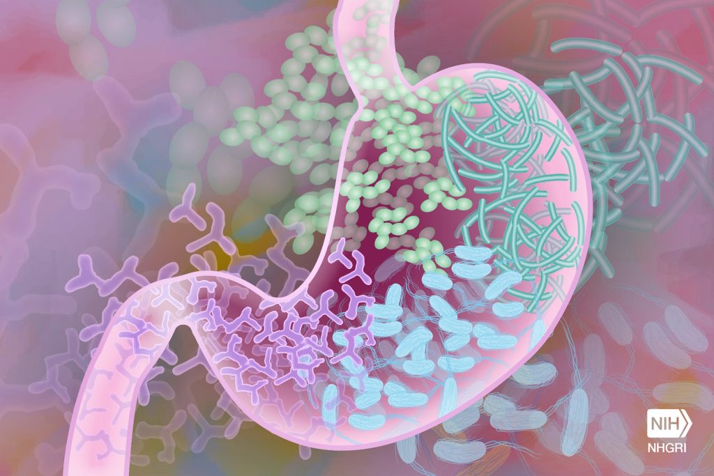
Gut Microbiome. Credit Darryl Leja National Human Genome Research Institute National Institutes Of Health

Changes in the gut microbiome have been implicated in a range of diseases including type 2 diabetes, obesity, and inflammatory bowel disease. Now, a team of researchers has found that microbes in the gut may affect cardiovascular disease as well. In a study published in Cell, the team has identified specific species of bacteria that consume cholesterol in the gut and may help lower cholesterol and heart disease risk in people.

Researchers at the Broad Institute of MIT and Harvard along with Massachusetts General Hospital analysed metabolites and microbial genomes from more than 1400 participants in the Framingham Heart Study, a decades-long project focused on risk factors for cardiovascular disease.

The team discovered that bacteria called Oscillibacter take up and metabolise cholesterol from their surroundings, and that people carrying higher levels of the microbe in their gut had lower levels of cholesterol. They also identified the mechanism the bacteria likely use to break down cholesterol. The results suggest that interventions that manipulate the microbiome in specific ways could one day help decrease cholesterol in people. The findings also lay the groundwork for more targeted investigations of how changes to the microbiome affect health and disease.

“Our research integrates findings from human subjects with experimental validation to ensure we achieve actionable mechanistic insight that will serve as starting points to improve cardiovascular health,” said Xavier, who is a core institute member and a professor at Harvard Medical School and Massachusetts General Hospital.

Postdoctoral researcher Chenhao Li and research scientist Martin Stražar, both in Xavier’s lab, were co-first authors on the study.

Cholesterol cues

In the past decade, other researchers have uncovered links between composition of the gut microbiome and elements of cardiovascular disease, such as a person’s triglycerides and blood sugar levels after a meal. But scientists haven’t been able to target those connections with therapies in part because they lack a complete understanding of metabolic pathways in the gut.

In the new study, the Broad team gained a more complete and detailed picture of the impact of gut microbes on metabolism. They combined shotgun metagenomic sequencing, which profiles all of the microbial DNA in a sample, with metabolomics, which measures the levels of hundreds of known and thousands of unknown metabolites. They used these tools to study stool samples from the Framingham Heart Study.

“The project outcomes underline the importance of high-quality, curated patient data,” Stražar said. “That allowed us to note effects that are really subtle and hard to measure and directly follow up on them.”

More than 16 000 associations between microbes and metabolic traits were found, one of them particularly strong: People with several species of bacteria from the Oscillibacter genus had lower cholesterol levels than those who lacked the bacteria. The researchers found that species in the Oscillibacter genus were surprisingly abundant in the gut, representing on average 1 in every 100 bacteria.

The researchers then wanted to figure out the biochemical pathway the microbes use to break down cholesterol. To do this, they first needed to grow the organism in the lab. Fortunately, the lab has spent years collecting bacteria from stool samples to create a unique library that also included Oscillibacter.

After successfully growing the bacteria, the team used mass spectrometry to identify the most likely byproducts of cholesterol metabolism in the bacteria. This allowed them to determine the pathways the bacteria uses to lower cholesterol levels. They found that the bacteria converted cholesterol into intermediate products that can then be broken down by other bacteria and excreted from the body. Next, the team used machine-learning models to identify the candidate enzymes responsible for this biochemical conversion, and then detected those enzymes and cholesterol breakdown products specifically in certain Oscillibacter in the lab.

The team found another gut bacterial species, Eubacterium coprostanoligenes, that also contributes to decreased cholesterol levels. This species carries a gene that the scientists had previously shown is involved in cholesterol metabolism. In the new work, the team discovered that Eubacterium might have a synergistic effect with Oscillibacter on cholesterol levels, which suggests that new experiments that study combinations of bacterial species could help shed light on how different microbial communities interact to affect human health.

Microbial messages

The human gut microbiome remains mostly unmapped, but the team believes they have paved the way for the discovery of other similar metabolic pathways impacted by gut microbes, which could be targeted therapeutically.

“There are many clinical studies trying to do faecal microbiome transfer studies without much understanding of how the microbes interact with each other and the gut,” Li said. “Hopefully stepping back by focusing on one particular bug or gene first, we’ll get a systematic understanding of gut ecology and come up with better therapeutic strategies like targeting one or a few bugs.”

“Because of the large number of genes of unknown function in the gut microbiome, there are gaps in our ability to predict metabolic functions,” Li added. “Our work highlights the possibility that additional sterol metabolism pathways may be modified by gut microbes. There are potentially a lot of new discoveries to be made that will bring us closer to a mechanistic understanding of how microbes interact with the host.”

Source: Broad Institute of MIT and Harvard

Common Respiratory Viruses Trigger Most Cases of Intussusception in Children

Photo by Andrea Piacquadio on Unsplash

Viral infections trigger more cases of intussusception in young children than previously thought, according to a new study. The research, led by Murdoch Children’s Research Institute (MCRI) and published in Clinical Infectious Diseases, found that during the COVID lockdowns, hospital admissions for intussusception, a medical emergency involving obstruction of the intestine, among young children significantly decreased.

For the study, 12 years of data was analysed across Victoria, NSW and Queensland. A total of 5589 intussusception cases were recorded between January, 2010 and April, 2022. Of those, 3179 were children under the age of two.

During the lockdown periods, Victoria and NSW experienced a decline in hospital admissions for intussusception among children under two by 62.7% and 40.1%, respectively. The rate of intussusception cases has now returned to normal levels.

MCRI and Monash University researcher Dr Ben Townley said the magnitude of the decline supported that common respiratory diseases such as colds, the flu and respiratory syncytial virus (RSV), were behind a significant proportion of intussusception cases.

“Reductions in intussusception hospital admissions were seen in all age groups, however most occurred in children less than two years of age,” he said.

“Intussusception is the leading cause of acute bowel obstruction in infants and young children and without prompt diagnosis and management, can be fatal.

“Countries with prolonged COVID lockdowns and suppression strategies saw reductions in common respiratory viruses, which influenced the drop in intussusception admissions.”

Victoria experienced the greatest lockdown duration, with Melbourne having six lockdown periods, for a total of 263 days. Greater Sydney had 159 days and Brisbane had 18 days in lockdown.

MCRI Professor Jim Buttery said the decrease in intussusception cases was greater than expected given previous research into the causes of the condition.

“Our analysis found commons viruses play a larger role than previously recognised in triggering intussusception,” he said.

Professor Buttery said the findings raised the possibility that emerging vaccines like the new RSV vaccines may help prevent intussusception.

“When a new vaccine against common childhood respiratory viruses is introduced, we may find there are some unexpected benefits, like protecting more children from intussusception,” he said.

Researchers from Sydney Children’s Hospital Network, University of Melbourne and Queensland Health also contributed to the findings.

Source: Murdoch Childrens Research Institute

Probing the Gut’s Ability to Change Size According to Nutrient Intake

Source: CC0

The gut has considerable plasticity among animals, shrinking as much 50% in cases of fasting such as hibernating and able to rapidly return to normal size on refeeding. Now, scientists from the University of Copenhagen used fruit flies to investigate the signalling mechanisms and cellular changes that regulate this rapidly renewable tissue, which could reveal insights into diseases such as colorectal cancer. Their results are published in Nature Communications.

“Taking advantage of the broad genetic toolbox available in the fruit fly, we have investigated the mechanisms underpinning nutrient-dependent gut resizing,” says Dr Ditte S. Andersen.

The results show that nutrient deprivation results in an accumulation of progenitor cells that fail to differentiate into the mature cells causing the gut to shrink.

Upon refeeding these stalled progenitor cells readily differentiate into mature cells to promote regrowth of the gut.

Ditte S. Andersen continues: “We have identified activins as critical regulators of this process. In nutrient restrictive conditions, activin signalling is strongly repressed, while it is reactivated and required for progenitor maturation and gut resizing in response to refeeding. Activin-dependent resizing of the gut is physiologically important as inhibition of activin signalling reduces survival of flies to intermittent fasting.”

Regulators of organ plasticity are essential for host adaptation to an ever-changing environment, however, the same signals are often deregulated in cancers. Indeed, mutations affecting activin signalling are frequent in cancer cells in a variety of tissues. This study provides a starting point for investigating the link between aberrant activin signalling and the development of colorectal cancers and sets the stage for exploring the efficiency of anti-activin therapeutic strategies in treating colorectal cancers.

Source: University of Copenhangen

Metabolic Diseases may be Driven by Gut Microbiome, Loss of Ovarian Hormones

Photo by Ravi Patel on Unsplash

The gut microbiome interacts with the loss of female sex hormones to exacerbate metabolic disease, including weight gain, fat in the liver and the expression of genes linked with inflammation, researchers report in the journal Gut Microbes.

The findings, using rodent models, may shed light on why women are at significantly greater risk of metabolic diseases such as obesity and Type 2 diabetes after menopause, when ovarian production of female sex hormones diminishes.

“Collectively, the findings demonstrate that removal of the ovaries and female hormones led to increased permeability and inflammation of the gut and metabolic organs, and the high-fat diet exacerbated these conditions,” said Kelly S. Swanson, the director of the Division of Nutritional Sciences and a professor in nutrition at the University of Illinois Urbana-Champaign who is a corresponding author of the paper. “The results indicated that the gut microbiome responds to changes in female hormones and worsens metabolic dysfunction.”

“This is the first time it has been shown that the response of microbiome to the loss of ovarian hormone production can increase metabolic dysfunction,” said first author Tzu-Wen L. Cross, a professor of nutrition science and the director of the Gnotobiotic Animal Facility at Purdue University. Cross was a doctoral student at the U. of I. when she began the research.

“The gut microbiome is sensitive to sex hormone changes and can further impact the risk of disease development.”

Cross said early microbiome research, beginning around 2005, looked at how the microbiome contributes to obesity development, but most of those studies focused on males.

“Metabolic dysfunction that is driven by the loss of ovarian-function in menopausal women – and how much the gut microbiome contributes to that – has not been studied. The aetiology is clearly very complex, but those gut-microbiome related factors are certainly components that we speculated play a role,” she said.

The scientists created diet-induced obesity in female mice and simulated the loss of female sex hormones by removing the ovaries in half of the population to examine any metabolic and inflammatory changes, including those to enzymes in the gut. The diets for both groups of mice were identical except for the proportion of fat, which constituted 60% or 10% of calories for those in the high-fat and low-fat groups, respectively.

In the second leg of the study, faecal samples were harvested from mice with or without ovaries and implanted in germ-free mice to study the impact on weight gain and metabolic and inflammatory activity in the gut, liver and fat tissue.

“The mice that were recipients of the gut microbiome of ovariectomized mice gained more weight and fat mass, and they had greater expression of genes in the liver associated with inflammation, obesity, Type 2 diabetes, fatty liver disease and atherosclerosis compared with those in the control group,” Swanson said.

Assessing the severity of fatty tissue and triglyceride concentrations in the liver, the scientists found that the triglyceride levels were significantly higher and fatty deposits in the liver and groin were greater in the mice that consumed the high-fat diet compared with all other treatment groups.

Those on the high-fat diet and those without ovaries had significantly larger fat cells, which are associated with cell death and the infiltration of macrophages. Along with elevated expression of the genes associated with inflammation and macrophage markers, these mice had lower expression of genes that are involved with glucose and lipid metabolism.

In the donor mice without ovaries that consumed the low-fat diet, the scientists found increased levels of beta-glucuronidase, an enzyme produced by the colon and some intestinal bacteria that breaks down and recycles steroidal metabolites such as oestrogen and various toxins, including carcinogens.

The scientists also examined the expression of genes coding for tight-junction proteins, which affect cell membranes’ permeability. They found that the mice without ovaries and those fed the high-fat diet had lower levels of these proteins in the liver and colon, which suggested their gut barriers were more permeable, compromised by either their diet or the absence of female hormones.

In the livers of the recipient mice that received transplants from donors without ovaries, the scientists found elevated expression levels of the gene for arginase-1, which plays a critical role in the elimination of nitrogenous waste. High levels of this protein have been associated with cardiovascular problems such as hypertension and atherosclerosis.

Source: University of Illinois at Urbana-Champaign, News Bureau

No Difference in Short-term Complications for Obesity Surgeries

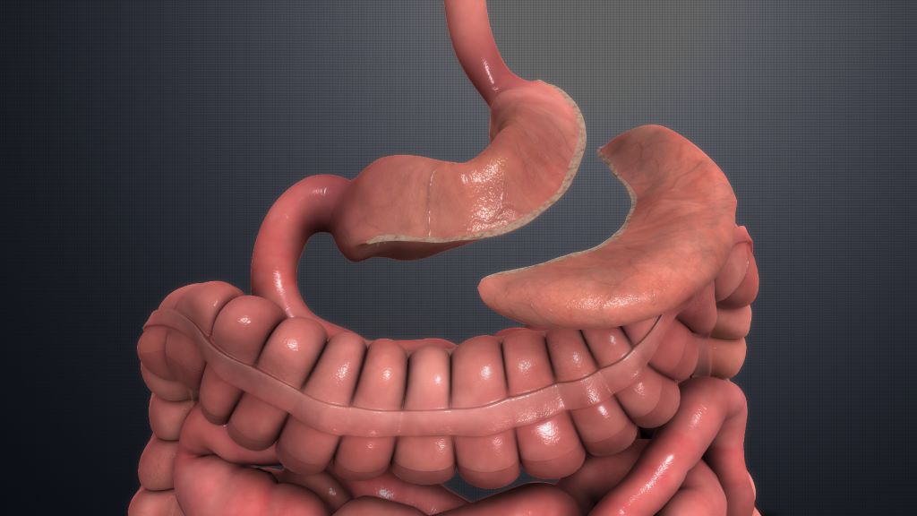
Sleeve gastrectomy. Credit: Scientific Animations CC4.0

The two most common obesity surgeries, gastric bypass and gastric sleeve, have few short-term complications and show no significant differences, according to study findings published in the journal JAMA Network Open. These are the first results of a multicentre randomised controlled trial comparing obesity surgeries conducted by the University of Gothenburg.

Patients undergoing obesity surgery will normally have a BMI of at least 40, or 35 if they also have other serious medical conditions related to obesity.

The most common procedures are gastric bypass, where a large part of the stomach and part of the small intestine are bypassed, and gastric sleeve, where a large part of the stomach is surgically removed.

The aim of the current study was to compare the short-term risks of the different procedures.

The study is the largest of its kind. 1735 adult patients planned for surgery in Norway and Sweden between 2015 and 2022 agreed to participate, and they were randomly assigned to either gastric bypass or gastric sleeve.

Relatively few complications

Surgical time was longer for gastric bypass, averaging 68 minutes compared to 47 minutes for gastric sleeve, but hospitalisation after surgery was one day regardless of method.

The follow-ups also gave equivalent results for the two methods.

At 30 days after surgery, both groups had relatively few complications such as haemorrhage, leakage, blood clots and infections.

No deaths occurred during the follow-up period of a total of 90 days.

“For both surgical procedures, the risk of complications is very low, especially from an international perspective, and there is no statistically significant or clinically relevant difference between the groups,” says Suzanne Hedberg, first author of the study.

Many stakeholders and many opinions

“Many people have had surgery, or are on waiting lists for surgery, and there are lots of discussions and opinions about the different methods. What the study shows is that patients and doctors can now choose their surgical method without considering short-term surgical risks,” she says.

Suzanne Hedberg defended her thesis in surgery at Sahlgrenska Academy, University of Gothenburg in April 2023, and is a consultant at Sahlgrenska University Hospital.

The study, included in her thesis, is the first publication with results from BEST (Bypass Equipoise Sleeve Trial), a Scandinavian registry-based randomised controlled multicentre study comparing the two methods of obesity surgery.

The main outcome of the trial which analyses the risk of complications and weight progression over 5 years, is expected to be completed in 2028.

“For the ongoing studies, we are off to a good start with equivalent groups, laying a good foundation for further comparisons of more long-term results,” concludes Suzanne Hedberg.

Source: University of Gothenburg

Gut Microbiome Composition Affects Sensitivity to Respiratory Viruses

Gut Microbiome. Credit Darryl Leja National Human Genome Research Institute National Institutes Of Health

The composition of microbiota found in the gut influences how susceptible mice are to respiratory virus infections and the severity of these infections, according to Georgia State University researchers. The findings, published in the journal Cell Host & Microbe, report that segmented filamentous bacteria, a bacterial species found in the intestines, protected mice against influenza virus infection when these bacteria were either naturally acquired or administered.

This protection against infection also applied to respiratory syncytial virus (RSV) and severe acute SARS-CoV-2. To maintain this protection, the study noted that segmented filamentous bacteria required immune cells in the lungs called basally resident alveolar macrophages.

In this study, the researchers investigated how differences in specific microbial species can impact outcomes of respiratory virus infections and how they might do so, which hasn’t been well defined previously.

They studied mice with discrete microbiome differences and mice differing in only the presence or absence of segmented filamentous bacteria.

Viral titers in the lung were measured several days after infection and varied significantly depending on the nature of the microbiome of the different animal groups.

“These findings uncover complex interactions that mechanistically link the intestinal microbiota with the functionality of basally resident alveolar macrophages and severity of respiratory virus infection,” said Dr. Andrew Gewirtz, co-senior author of the study and Regents’ Professor in the Institute for Biomedical Sciences at Georgia State.

The study found that in segmented filamentous bacteria-negative mice, basally resident alveolar macrophages were quickly depleted as respiratory virus infection progressed.

However, in segmented filamentous bacteria-colonised mice, basally resident alveolar macrophages were altered to resist influenza virus infection depletion and inflammatory signaling.

The basally resident alveolar macrophages disabled influenza virus, in large part by activating a component of the immune system referred to as the complement system.

“We find it remarkable that the presence of a single common commensal bacterial species, amidst the thousands of different microbial species that inhabit the mouse gut, had such strong impacts in respiratory virus infection models and that such impacts were largely attributable to reprogramming of basally resident alveolar macrophages,” said D. Richard Plemper, co-senior author of the study, Regents’ Professor and director of the Center for Translational Antiviral Research at Georgia State.

“If applicable to human infections, these findings will have major implications for the future risk assessment of a patient to advance to severe disease.”

“We find it highly unlikely that segmented filamentous bacteria is the only gut microbe capable of impacting the phenotype of alveolar macrophages, and consequently, proneness to respiratory virus infection,” Gewirtz said.

“Rather, we hypothesize that gut microbiota composition broadly influences proneness to respiratory virus infection. Microbiota mediated programming of basally resident alveolar macrophages may not only influence the severity of acute respiratory virus infection, but may also be a long-term post-respiratory virus infection health determinant.”

Source: Georgia State University

Researchers Figure out Why Cancer Immunotherapy can Cause Colitis

Gastrointestinal inflammation. Credit: Scientific Animations CC4.0

Researchers have identified a mechanism behind immunotherapy sometimes causing colitis. They also found a way to deliver immunotherapy’s cancer-killing impact without the unwelcome side effect. The researchers, from the University of Michigan Health Rogel Cancer Center, published their findings in Science.

“This is a good example of how understanding a mechanism helps you to develop an alternative therapy that’s more beneficial. Once we identified the mechanism causing the colitis, we could then develop ways to overcome this problem and prevent colitis while preserving the anti-tumour effect,” said senior study author Gabriel Nunez, MD, professor of pathology at Michigan Medicine.

Immunotherapy is a promising treatment for several types of cancer. But immune checkpoint inhibitors can also cause severe side effects, including colitis. Colitis can cause severe gastrointestinal discomfort, causing some patients to discontinue their cancer treatment because of it.

The problem facing researchers was that while patients were developing colitis, the laboratory mice were not, preventing them from studying the cause of this side effect.

To get past this, the Rogel team, led by first author Bernard C. Lo, PhD, created a new mouse model, injecting microbiota from wild-caught mice into the traditional mouse model.

In this model, the mice did develop colitis after administration of antibodies used for tumour immunotherapy. Now, researchers could trace back the mechanism to see what was causing this reaction.

In fact, colitis developed because of the composition of the gut microbiota, which caused immune T cells to be hyper-activated while regulatory T cells that put the brakes on T cell activation were deleted in the gut. This was happening within a specific domain of the immune checkpoint antibodies.

Researchers then removed that domain, which they found still resulted in a strong anti-tumour response but without inducing colitis.

“Previously, there were some data that suggested the presence of certain bacteria correlated with response to therapy. But it was not proven that microbiota were critical to develop colitis. This work for the first time shows that microbiota are essential to develop colitis from immune checkpoint inhibition,” Nunez said.

To follow up what they saw in mice, researchers reanalysed previously reported data from studies of human cells from patients treated with immune checkpoint antibodies, which reinforced the role of regulatory T cells in inducing colitis.

The Rogel team plans additional studies to further understand the mechanisms causing colitis and seeks clinical partners to move this knowledge to a clinical trial.

Source: Michigan Medicine – University of Michigan

The Inability to Burp – A Rarely Discussed Condition that Affects Quality of Life

Photo by Kyle Glenn on Unsplash

The inability to burp, called retrograde cricopharyngeus dysfunction (R-CPD), is caused by failure of the throat’s cricopharyngeal muscle to relax to allow the outward passage of gas. An interview-based study in Neurogastroenterology & Motility reveals the impact of R-CPD on quality of life.

The study included 199 adults affected by the condition, most of whom reported abdominal bloating, socially awkward gurgling noises, excessive flatulence, and difficulty vomiting. Only half discussed their symptoms with their primary care clinician, and 90% felt they did not receive adequate help. Participants also reported embarrassment, anxiety/depression, negative impacts on relationships, and work disruption due to R-CPD.

“R-CPD encompasses more than just the physical challenge of being unable to burp; it also significantly impacts people’s daily lives, relationships, and mental well-being,” said corresponding author Jason N. Chen, a medical student at Texas Tech University Health Sciences Center. Future efforts should concentrate on raising awareness about R-CPD, which can help increase identification and treatment rates.”

Source: Wiley