Category: Gastrointestinal

Intestinal Nutrient Sensors Create ‘Gut Instincts’ for Digestion

Source: CC0

Rare hormone producing cells in the gut secrete hormones in response to incoming food and play key roles in managing digestion and appetite. Researchers have now developed new tools to identify potential ‘nutrient sensors’ on these hormone producing cells and study their function. This could result in new strategies to interfere with the release of these hormones and provide avenues for the treatment of a variety of metabolic or gut motility disorders.

The work, led by led by the Hubrecht Institute and Roche’s Institute of Human Biology, is reported in Science.

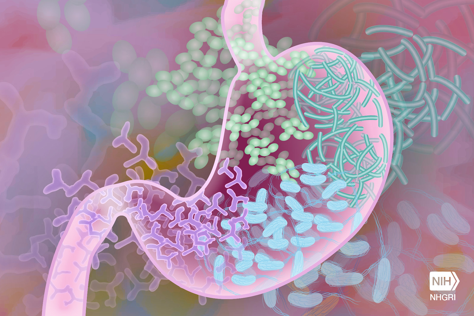
The intestine acts as a vital barrier. It protects the body from harmful bacteria and highly dynamic pH levels, while allowing nutrients and vitamins to enter the bloodstream. The gut is also home to endocrine cells, which secrete many hormones that regulate bodily functions. These enteroendocrine cells (endocrine cells of the gut) are very rare cells that release hormones in response to various triggers, such as stretching of the stomach, energy levels and nutrients from food. These hormones in turn regulate key aspects of physiology in response to the incoming food, such as digestion and appetite. Thus, enteroendocrine cells are the body’s first responders to incoming food, and instruct and prepare the rest of the body for what is coming.

Understanding hormone release

Medications that mimic gut hormones, most famously GLP-1, are promising for the treatment of multiple metabolic diseases. The ability to directly manipulate endocrine cells to adjust hormone secretion could open up new therapeutic options. However, it has been challenging to understand how gut hormone release can be influenced effectively. Researchers have had trouble identifying the sensors on cells.

Enteroendocrine cells represent less than 1% of cells in the intestinal epithelium. In addition, the sensors on these cells are expressed in low amounts. Current studies mainly rely on mouse models, but the signals to which mouse cells respond are likely different from those to which human cells respond. Therefore, new models and approaches were required to study these signals.

Enteroendocrine cells in organoids

The Hubrecht team has previously developed methods to derive large quantities of enteroendocrine cells in human organoids. Organoids contain the same cell types of the organ they are derived from. Therefore, they are useful to explore the development and function of cells. Using a special protein, Neurogenin-3, the researchers could generate high numbers of endocrine cells in organoids of the intestine.

Enteroendocrine cells have different sensors and hormone profiles in different regions of the gut. In order to study these rare cells, the researchers needed to make organoids of all these different regions.

Stomach organoids

In the current study, the team managed to enrich enteroendocrine cells in organoids of other parts of the digestive system, including the stomach. Like the real stomach, stomach organoids respond to known inducers of hormone release and secrete large amounts of the hormone Ghrelin. Ghrelin is also called the ‘hunger hormone’ because it plays a key role in signaling hunger to the brain. The Ghrelin production of the stomach organoids confirms that these organoids can be used to study hormone secretion in enteroendocrine cells.

Enteroendocrine cell sensors

Since enteroendocrine cells are rare, researchers have struggled to profile many of these cells. In the current study, the team identified a so-called surface marker, called CD200, on human cells. The researchers used this surface marker to isolate a large number of human enteroendocrine cells from organoids and study their sensors. This revealed numerous receptor proteins that had not yet been identified in enteroendocrine cells.

The team stimulated the organoids with molecules that would activate these receptors and identified multiple new sensory receptors that control hormone release. When the researchers inactivated these receptors using CRISPR-based gene editing, hormone secretion was often blocked.

Therapeutic applications

With these data, the researchers can now predict how human enteroendocrine cells respond when certain sensory receptors are activated. Their findings thus pave the way for additional studies to explore the effects of these receptor activations. The enteroendocrine cell-enriched organoids will allow the team to perform larger, unbiased studies to identify new regulators of hormone secretion. These studies may eventually lead to therapies for metabolic diseases and gut motility disorders.

Source: Hubrecht Institute

Antigens in Foods Suppress Gut Tumours by Activating Immune Cells

Photo by Pixabay on Pexels

Researchers led by Hiroshi Ohno at the RIKEN Center for Integrative medical sciences (IMS) in Japan have discovered that food antigens like milk proteins help keep tumours from growing in our guts, specifically the small intestines. Experiments revealed how these proteins trigger the intestinal immune system, allowing it to effectively stop the birth of new tumours. The study was published in the scientific journal Frontiers in Immunology.

Food antigens get a lot of negative press because they are the source of allergic reactions to foods such as peanuts, shellfish, bread, eggs, and milk. Even if not allergenic, these antigens, along with the many others found in plants and beans, are still considered foreign objects that need to be checked out by the immune system. Ohno and his team have previously reported that food antigens activate immune cells in the small intestines, but not the large intestines. At the same time, some immune cells activated by gut bacteria are known to suppress tumours in the gut. In the new study, the RIKEN IMS researchers bring these two lines of thought together and tested whether food antigens suppress tumours in the small intestines.

The team began with a mouse model with a mutated tumour-suppression gene. Like people with familial adenomatous polyposis, when this gene malfunctions, the mice develop tumours throughout the small and large intestines. The first experiment was fairly simple. They fed these mice normal food or antigen-free food and found that the ones that got normal food had fewer tumours in the small intestines, but the same amount in the large intestines.

Next, they added a common representative antigen called albumin – which can be found in meat and was not in the normal food – to the antigen-free diet, making sure that the total amount of the protein equalled the amount of protein in the normal diet. When the mice were given this diet, tumours in the small intestine were suppressed just as they has been with normal food. This means that tumour suppression was directly related to the presence of antigen, not the nutritional value of the food or any specific antigen.

Mice that got the plain antigen-free diet had many fewer T cells than those that got the normal food or the antigen-free food with milk protein. Further experiments revealed the biological process that makes this possible.

These findings have clinical implications. Similar to antigen-free diets, clinical elemental diets include simple amino acids, but not proteins. This reduces digestive work and can help people with severe gastrointestinal conditions, such as Crohn’s disease or irritable bowel syndrome. According to Ohno, “small intestinal tumours are much rarer than those in the colon, but the risk is higher in cases of familial adenomatous polyposis, and therefore the clinical use of elemental diets to treat inflammatory bowel disease or other gastrointestinal conditions in these patients should be considered very carefully.”

Elemental diets are sometimes adopted by people without severe gastrointestinal conditions or allergies as a healthy way to lose weight or reduce bloating and inflammation. The new findings suggest that this could be risky and emphasises that these kinds of diets should not be used without a doctor’s recommendation.

Source: RIKEN

The Gut Microbiome can Affect Symptoms of Hypopituitarism

Gut Microbiome. Credit Darryl Leja National Human Genome Research Institute National Institutes Of Health

In research published in PLOS Genetics, scientists have shown that the balance of bacteria in the gut can influence symptoms of hypopituitarism in mice. They also showed that aspirin was able to improve hormone deficiency symptoms in mice with this condition.

People with mutations in a gene called Sox3 develop hypopituitarism, where the pituitary gland doesn’t make enough hormones. It can result in growth problems, infertility and poor responses of the body to stress.

The scientists at the at the Francis Crick Institute removed Sox3 from mice, causing them to develop hypopituitarism around the time of weaning (starting to eat solid food).

They found that mutations in Sox3 largely affect the hypothalamus in the brain, which instructs the pituitary gland to release hormones. However, the gene is normally active in several brain cell types, so the first task was to ask which specific cells were most affected by its absence.

The scientists observed a reduced number of cells called NG2 glia, suggesting that these play a critical role in inducing the pituitary gland cells to mature around weaning, which was not known previously. This could explain the associated impact on hormone production.

The team then treated the mice with a low dose of aspirin for 21 days. This caused the number of NG2 glia in the hypothalamus to increase and reversed the symptoms of hypopituitarism in the mice.

Although it’s not yet clear how aspirin had this effect, the findings suggest that it could be explored as a potential treatment for people with Sox3 mutations or other situations where the NG2 glia are compromised.

An incidental discovery revealed the role of gut bacteria in hormone production

When the National Institute for Medical Research (NIMR) merged with the Crick in 2015, mouse embryos were transferred from the former building to the latter, and this included the mice with Sox3 mutations.

When these mice reached the weaning stage at the Crick, the researchers were surprised to find that they no longer had the expected hormonal deficiencies.

After exploring a number of possible causes, lead author Christophe Galichet compared the microbiome – bacteria, fungi and viruses that live in the gut – in the mice from the Crick and mice from the NIMR, observing several differences in its makeup and diversity. This could have been due to the change in diet, water environment, or other factors that accompanied the relocation.

He also examined the number of NG2 glia in the Crick mice, finding that these were also at normal levels, suggesting that the Crick-fed microbiome was somehow protective against hypopituitarism.

To confirm this theory, Christophe transplanted faecal matter retained from NIMR mice into Crick mice, observing that the Crick mice once again showed symptoms of hypopituitarism and had lower numbers of NG2 glia. 

Although the exact mechanism is unknown, the scientists conclude that the make-up of the gut microbiome is an example of an important environmental factor having a significant influence on the consequences of a genetic mutation, in this case influencing the function of the hypothalamus and pituitary gland.

Source: Francis Crick Institute

Breastfeeding Shapes the Gut Microbiome and Protects against Asthma

Photo by Wendy Wei

Human breast milk regulates a baby’s mix of microbes, known as the microbiome, during the infant’s first year of life, in turn lowers the child’s risk of developing asthma, according to a new study published in Cell.

Led by researchers at NYU Langone Health and the University of Manitoba, the study results showed that breastfeeding beyond three months supports the gradual maturation of the microbiome in the infant’s digestive system and nasal cavity, the upper part of the respiratory tract. Conversely, stopping breastfeeding earlier than three months disrupts the paced development of the microbiome and was linked to a higher risk of preschool asthma.

Some components in breast milk, such as complex sugars called human milk oligosaccharides, can only be broken down with the help of certain microbes. This provides a competitive advantage to microbes capable of digesting these sugars. By contrast, infants who are weaned earlier than three months from breast milk and who then rely solely on formula feeding, become home to a different set of microbes –ones that will help the infant to digest the components in formula. While many of these microbes that thrive on formula do eventually end up in all babies, the researchers showed that their early arrival is linked to an increased risk of asthma.

“Just as a pacemaker regulates the rhythm of the heart, breastfeeding and human milk set the pace and sequence for microbial colonisation in the infant’s gut and nasal cavity, ensuring that this process occurs in an orderly and timely manner,” said study co-senior investigator and computational biologist Liat Shenhav, PhD. “Healthy microbiome development is not only about having the right microbes. They also need to arrive in the right order at the right time,” said Dr Shenhav, an assistant professor at NYU Grossman School of Medicine, its Institute for Systems Genetics, and the school’s Department of Microbiology.

For the study, Dr Shenhav, who is also an assistant professor at NYU’s Courant Institute of Mathematical Sciences, worked in collaboration with study co-senior investigator Meghan Azad, PhD, director of the Manitoba Interdisciplinary Lactation Center, and a professor of paediatrics and child health, at the University of Manitoba.

Another key study finding was that the bacterium Ruminococcus gnavus appeared much sooner in the guts of children who were weaned early from breast milk than in those of children who were exclusively breastfed. The bacterium is known to be involved in the production of molecules called short-chain fatty acids, and the formation and breakdown of the amino acid tryptophan. Both tryptophan and its metabolites have been linked to immune system regulation and disruption in previous research, including an increased risk of asthma. The study authors noted that beyond aiding in digestion, an infant’s microbiome plays a crucial role in the immune system’s development.

The study tracked the ebb and flow of microbes in the guts and noses of infants during the first year of life, as well as details on breastfeeding and the composition of their mothers’ milk. All the children and their mothers were participating in the CHILD Cohort Study, a long-term research project that has been studying the same 3500 Canadian children at different stages of life from the womb well into adolescence.

The data provided by the CHILD Cohort Study enabled researchers to detangle the impact of breastfeeding on an infant’s microbiome from a range of other environmental factors, including prenatal smoke exposure, antibiotics, and the mother’s asthma history.

Even when these factors were accounted for, they found that breastfeeding duration remained a powerful determinant for the child’s microbial makeup over time. They also used these microbial dynamics and data on milk components to train a machine learning model that accurately predicted asthma years in advance. Finally, they created a statistical model to learn causal relationships, which showed that the primary way breastfeeding reduces asthma risk is through shaping the infant’s microbiome.

“The algorithms we developed provide valuable insights into microbial dynamics during an infant’s first year of life and how these microbes interacted with the infant,” said Dr Shenhav. “These insights allowed us to move beyond identifying associations, enhancing our ability to make predictions and explore causal relationships.

“Our research highlights the profound impact of breastfeeding on the infant microbiome and breastfeeding’s essential role in supporting respiratory health. By uncovering the mechanisms behind the protective effects of breast milk, as demonstrated in this study, we aim to inform national guidelines on breastfeeding and weaning from breast milk in a data-driven manner.

“With further research, our findings could also contribute to developing strategies to prevent asthma in children who cannot be breastfed for at least three months,” she added.

Source: NYU Langone Health / NYU Grossman School of Medicine

Antibiotic Usage can Damage the Intestine’s Protective Mucus Layer

Source: CC0

Researchers at Umeå University and Tartu University have found that a history of repeated antibiotic use causes defects in the normally protective mucus barrier of the gut, due to antibiotic-driven alterations in the microbiota. In a further study in a different collaboration, the researchers found a bacteria-independent mechanism through which antibiotics can damage the mucus barrier directly.

The results have been published in the scientific journals Gut Microbes and Science Advances.

“Together, these two studies suggest that antibiotics can damage the mucus layer through at least two independent mechanisms, and that they may have long-lasting effects through an altered gut bacteria. This further supports the notion that antibiotics should be administered in a responsible manner,” says Björn Schröder, Docent in Infection Biology in the Department of Molecular Biology at Umeå University.

Previous research has shown the consequences of short-term antibiotic treatments on the intestinal environment, but it is less clear how repeated antibiotic use in past years can affect our guts.

To address this question, Björn Schröder and his group at Umeå University teamed up with a research group at Tartu University in Estonia, who have built a deeply characterised cohort of individuals that provided stool samples and health records.

The researchers selected individuals who had taken at least five courses of antibiotics in the past, but not within six months before the stool collection, and compared their microbiota composition to individuals who had not taken any antibiotics within the last 10 years.

“The analysis revealed changes to the gut bacteria composition, even though the antibiotics were taken a long time ago. These results indicate that repeated antibiotic use has a lasting effect on gut bacteria composition that can persist at least months after the last treatment,” says Kertu-Liis Krigul, PhD student at Tartu University.

After transplantation of the human microbiota into mice and using specialised methods to analyse the mucus function in the gut, the researchers found that the function of the mucus layer was disrupted in mice transplanted with bacteria from humans with a history of repeated antibiotic use. Expansion of the mucus was reduced, and the mucus layer became penetrable, allowing bacteria to move closer to the intestinal lining.

“Looking at the bacteria present in the gut in more detail, we could see that bacteria known to feed on the mucus layer were present at higher levels in these mice. This further supports a role for the gut bacteria in determining how well the mucus barrier can function,” says Rachel Feeney, PhD student at the Department of Molecular Biology at Umeå University.

A separate study carried out in another international collaboration, further showed that antibiotics can also directly disrupt the mucus barrier in a gut bacteria-independent manner.

By giving the antibiotic vancomycin to normal and ‘bacteria-free’ mice, the researchers were able to show that this antibiotic can act directly on the mucus barrier, independent of the gut bacteria. Complementary experiments on intestinal tissue were carried out at Umeå University and showed that the antibiotic could disrupt the mucus expansion within a few minutes of application.

Source: Umeå University

Inflammation Leaves a Long-lasting Impression on Intestinal Stem Cells

Irritable bowel syndrome. Credit: Scientific Animations CC4.0

Researchers at Baylor College of Medicine, the University of Michigan and collaborating institutions have discovered that inflammation in the gut leaves long-term marks on intestinal stem cells (ISCs) that reduce their ability to heal the intestine, even after inflammation has receded. This is important because it affects ISCs’ response to future challenges. The study appeared in Cell Stem Cell.

“We study graft-vs-host disease (GVHD), a major cause of mortality after bone marrow transplantation, a potentially curative therapy for many blood diseases. One of our goals is to better understand GVHD and identify strategies to control it,” said corresponding author Dr Pavan Reddy, professor and director of the Dan L Duncan Comprehensive Cancer Center at Baylor and previously at the University of Michigan.

“GVHD is an inflammatory reaction in which immune T cells from the bone marrow transplant donor attack the host gut cells, mainly ISCs,” said first author Dr Dongchang Zhao, in Reddy’s lab.

Although many ISCs perish during GVHD, survivors remain. However, it’s not known whether they are fully functional or can return to their full functionality after the resolution of GVHD, which has fundamental implications for host resilience and repair.

“In the current study, we investigated the consequences of inflammation on ISCs in well-defined clinically relevant models of GVHD,” Reddy said.

“Using cellular and animal models, we found that exposure to inflammation drove ISCs to change their metabolism in ways that resulted in the accumulation of succinate, a product of cellular processes, which in turn reprogramed the epigenome,” Zhao said.

The epigenome is a system of chemical marks on the DNA that regulates the genes expressed by the cell. Inflammation-led epigenome reprogramming changed the expression of genes involved in cell reproduction. Overall, reprogrammed ISCs were less able of regenerating, a first step toward healing the intestine.

“We then investigated whether ISCs would be able to recuperate their regenerative ability after inflammation had resolved,” Reddy said. “We found that ISCs had not overcome their initial exposure to inflammation. Despite mitigating GVHD inflammation for 28 days, ISCs retained a reduced regenerative capacity that led to poor recovery and increased mortality from challenges, such as non-lethal radiation exposure, in animal models. More research is on the way to design strategies to help ISCs ‘forget’ their encounter with inflammation and enhance their resilience against immune attacks.”

Source: Baylor College of Medicine

Recurrent Chlamydia Results from Bacteria Settling in the Intestine

Immunofluorescence staining of human gastric cells grown in a microplate and infected with Chlamydia trachomatis. Blue: cell nuclei, green: C. trachomatis, grey: actin. (Image: Pargev Hovhannisyan / Universität Würzburg)

A phenomenon is known from everyday clinical practice that can occur after successful antibiotic treatment: when people who have already been treated come to the doctor with a new chlamydia infection, they are often infected with exactly the same strains of bacteria as the previous infection.

“It is therefore reasonable to assume that the bacteria find a niche in the body where they are not yet vulnerable, that they form a permanent reservoir there and can become active again later,” says Professor Thomas Rudel, chlamydia expert and Head of the Chair of Microbiology at the Biocentre of Julius-Maximilians-Universität (JMU) Würzburg in Germany. This phenomenon is known as persistence. It is problematic because the chlamydia that persist in the body become increasingly resistant to antibiotics over time.

Intestinal Organoids Experimentally Infected with Chlamydia

Experiments on mouse models have shown that chlamydia can persist in the intestines of animals. In humans the bacteria also seem to make themselves at home there. This is reported by the research groups of Thomas Rudel and Sina Bartfeld in the journal PLOS Pathogens. Professor Bartfeld worked at JMU until 2021; she now heads the Department of Medical Biotechnology at Technische Universität Berlin.

The researchers identified the intestine as a niche with the help of artificial organs in miniature format, so-called organoids. These are structures produced in the laboratory from human intestinal cells that are very similar in structure and function to the model organ.

The teams from Würzburg and Berlin tried to infect the intestinal organoids with chlamydia. They discovered that the inner cell layer of the organoids is very resistant to the bacteria: the pathogens could only penetrate there if the cell epithelium was damaged. From the blood side, however, the chlamydia were able to infect very efficiently. “In this case, we repeatedly found the persistent forms of the bacteria, which can be clearly identified with their typical shape under the electron microscope,” says JMU researcher Pargev Hovhannisyan, first author of the publication.

Clinical Studies and Further Experiments Must Follow

Transferred to the human organism, this would mean that chlamydia infection with subsequent persistence can only occur with difficulty via the inner side of the intestine, but very easily via the blood. However, whether this actually happens in the human body has yet to be confirmed in clinical studies, says Thomas Rudel.

The next step for Thomas Rudel and Sina Bartfeld is to to find out whether the chlamydia select certain cell types for their persistence – no easy task, as the intestine consists of hundreds of different cell types. But perhaps it is also factors from the surrounding tissue that trigger persistence. These and other details are now to be investigated.

Source: University of Würzburg

Study Shows that Probiotics in Pregnancy Benefit Mothers and Offspring

Photo by SHVETS production: https://www.pexels.com/photo/focused-pregnant-black-woman-taking-vitamins-on-couch-6991899/

Giving probiotics to pregnant mice can enhance both the immune system and behaviour of the mothers and their offspring, according to a new study led by The Ohio State University Wexner Medical Center and College of Medicine.

“These results suggest that certain probiotics given to mothers during pregnancy can improve their offsprings’ behaviour and may affect the metabolism of common amino acids in our diets. Probiotics may also help counteract the negative effects of prenatal stress,” said study senior author Tamar Gur, MD, PhD, at OSU. 

Study findings are published online in the journal Brain, Behavior, and Immunity

Many studies have attested to the benefits of probiotics, which are considered safe to take during pregnancy. Researchers led by first author Jeffrey Galley, PhD found that a specific probiotic, Bifidobacterium dentium, may change how the body processes certain amino acids, such as tryptophan. During pregnancy, tryptophan helps control inflammation and brain development. 

“We have strong evidence this specific probiotic helped reduce stress-related problems in both mothers and their offspring, including helping the babies gain weight and improving their social behaviour,” said Gur, who also is an associate professor of psychiatry, neuroscience and obstetrics and gynaecology at Ohio State. 

Gur’s research team has studied how prenatal stress can lead to abnormal brain development and behavioural changes in offspring. So far, they’ve found that stress is linked to changes in brain inflammation and amino acid metabolism, as well as long-term reductions in social behaviour and abnormal microbiomes in offspring.

This study enhances their understanding of how gut microbes and probiotics can influence amino acid metabolism and help with behaviour and immune issues related to prenatal stress. The study also highlights the many benefits of this specific probiotic, even without the presence of stress.

“Now, we aim to understand the mechanisms behind these changes and explore ways to prevent or treat these effects,” Gur said. “Since prenatal stress is common in many pregnancies, we want to develop methods to reduce its negative effects.”

Source: Ohio State University Wexner Medical Center

Ketogenic Diet Reduces Friendly Gut Bacteria and Raises Cholesterol Levels

Photo by Jose Ignacio Pompe on Unsplash

A study from the University of Bath reveals that ketogenic low-carbohydrate diets can increase cholesterol levels and reduce beneficial gut bacteria, specifically Bifidobacterium.

Published in Cell Reports Medicine, the research from the Centre for Nutrition, Exercise, and Metabolism involved 53 healthy adults for up to 12 weeks. Participants followed either a moderate sugar diet (control), a low-sugar diet (less than 5% of calories from sugar), or a ketogenic (keto) low-carbohydrate diet (less than 8% of calories from carbohydrates).

Key findings include:

•Increased Cholesterol: The keto diet raised cholesterol levels, particularly in small and medium sized LDL particles. The diet increased apolipoprotein B (apoB), which causes plaque buildup in arteries. In contrast, the low-sugar diet significantly reduced cholesterol in LDL particles.

•Reduced Favourable Gut Bacteria: The keto diet altered gut microbiome composition, notably decreasing Bifidobacteria, beneficial bacteria often found in probiotics. This bacteria has wide ranging benefits: producing b vitamins, inhibiting pathogens and harmful bacteria and lowering cholesterol. Sugar restriction did not significantly impact the gut microbiome composition.

•Glucose Tolerance: The keto diet reduced glucose tolerance, meaning the adults’ bodies became less efficient at handling carbohydrates.

•Both Diets Resulted In Fat Loss: Keto Diet resulted in an average of 2.9kg fat mass loss per person, whilst the sugar restricted diet followed with an average 2.1kg fat mass loss per person at 12 weeks.

•Metabolism: Researchers also noticed that the keto diet caused significant changes in lipid metabolism and muscle energy use, shifting the body’s fuel preference from glucose to fats.

•Physical Activity Levels: Both sugar restriction and keto diets achieved fat loss without changing physical activity levels. Previous studies from the Centre for Nutrition, Exercise and Metabolism have shown that skipping breakfast or intermittent fasting cause reductions in physical activity.

Lead researcher Dr. Aaron Hengist highlighted the concerning cholesterol findings:

“Despite reducing fat mass, the ketogenic diet increased the levels of unfavourable fats in the blood of our participants, which, if sustained over years, could have long-term health implications such as increased risk of heart disease and stroke.”

Dr. Russell Davies, who led the microbiome research, explained the impact on gut health:

“Dietary fibre is essential for the survival of beneficial gut bacteria like Bifidobacteria. The ketogenic diet reduced fibre intake to around 15 grams per day, half the NHS recommended intake. This reduction in Bifidobacteria might contribute to significant long-term health consequences such as an increased risk of digestive disorders like irritable bowel disease, increased risk of intestinal infection and a weakened immune function.”

Professor Javier Gonzalez, who oversaw the research, commented on the glucose findings:

“The ketogenic diet reduced fasting glucose levels but also reduced the body’s ability to handle carbs from a meal. By measuring proteins in muscle samples taken from participants’ legs, we think this is probably an adaptive response to eating less carbohydrates day-to-day and reflects insulin resistance to storing carbs in muscle. This insulin resistance is not necessarily a bad thing if people are following a ketogenic diet, but if these changes persist when people switch back to a higher carbohydrate diet it could increase the risk of developing type 2 diabetes in the long-term”

In light of this new research, the academics conclude that if you’re considering a diet, a low sugar one will be better for most people. More work is needed to understand how individuals may benefit from each type of diet. The government recommends that free sugars (those added to food or drink or found naturally in honey, syrups, fruit juices and smoothies) should be restricted to less than 5% of total energy intake. Professor Dylan Thompson, who also oversaw the work, said:

“The ketogenic diet is effective for fat loss, but it comes with varied metabolic and microbiome effects that may not suit everyone. In contrast, sugar restriction supports government guidelines for reducing free sugar intake, promoting fat loss without apparent negative health impacts.”

Source: University of Bath

Specific Type of Dietary Fibre Could Stimulate GLP-1 Release

Gut Microbiome. Credit Darryl Leja National Human Genome Research Institute National Institutes Of Health

New research led by Frank Duca, associate professor at the University of Arizona, suggests that consuming foods rich in beta-glucan, a type of fibre found in oats and barley, can reduce body weight and obesity by stimulating the release of glucagon-like peptide-1 (GLP-1). The study, published in The Journal of Nutrition, analysed the impact of different fibres on gut microbiota.

“We know that fibre is important and beneficial; the problem is that there are so many different types of fibre,” Duca said. “We wanted to know what kind of fibre would be most beneficial for weight loss and improvements in glucose homeostasis so that we can inform the community, the consumer and then also inform the agricultural industry.”

Not all fibre is created equal

The researchers looked at the effect of five different plant-based fibres in rodent diets: pectin, beta-glucan, wheat dextrin, starch and cellulose. Only beta-glucan resulted in reduction of body weight and fat, as well as improvements in glucose homeostasis. Beta-glucan is a unique fibre that is found in many foods, including oats, barley, mushrooms and yeasts, and future studies will examine how different sources of beta-glucan could differ in their effectiveness.

Changes in metabolites – the molecules produced when gut bacteria interact with fibre – seemed to be responsible for the weight-loss effects,  particularly a specific metabolite called butyrate. Butyrate is a key fuel source for colon cells, promoting a healthy gut barrier to reduce systemic inflammation. Butyrate also induces the release of gut peptides, or messengers that regulate the functions of the gut, such GLP-1.

Drugs like semaglutide are synthetic versions of GLP-1, which stimulate insulin and can also help people feel full. One key difference of naturally occurring GLP-1 is its rapid degradation near the intestine, whereas semaglutide is made to last longer and target the brain.

“Part of the benefits of consuming dietary fibre is through the release of GLP-1 and other gut peptides that regulate appetite and body weight,” Duca said. “However, we don’t think that’s all of the effect. We think that there are other beneficial things that butyrate could be doing that are not gut peptide related, such as improving gut barrier health and targeting peripheral organs like the liver.”

Duca is researching other types of fibre that can be beneficial for weight reduction. In a previous study, the Duca Lab discovered that barley flour was the most effective in promoting weight loss compared to several other commercially available flours. Other studies involving oligofructose have also demonstrated beneficial effects. In the future, Duca hopes to collaborate with other researchers to develop enhanced fibres that can optimise the release of butyrate.

Source: University of Arizona