Category: Gastrointestinal

Scientists Find a Molecule that Promotes Gut Healing and Stifles Tumour Growth

Irritable bowel syndrome. Credit: Scientific Animations CC4.0

Researchers at Karolinska Institutet have found a molecule that can both help the intestines to heal after damage and suppress tumour growth in colorectal cancer. The discovery could lead to new treatments for inflammatory bowel disease (IBD) and cancer. The results are published in the journal Nature.

Many patients with inflammatory bowel disease (IBD) such as Crohn’s disease or ulcerative colitis do not respond to available treatments, highlighting the need to identify novel therapeutic strategies. In this study, researchers propose that promoting mucosal healing through tissue regeneration could be a valid alternative to immunosuppressive drugs.  

“However, it’s virtually impossible to promote tissue regeneration without the risk of inducing tumour growth, as cancer cells can hijack the body’s natural healing processes and start to grow uncontrollably,” says lead author Srustidhar Das, research specialist in Eduardo Villablanca’s research group at Karolinska Institutet. “We’ve now identified a molecule that can help the intestines to heal after damage while suppressing tumour growth in colorectal cancer.” 

New drug candidates 

In their search for new ways to treat IBD, the researchers have identified a handful of molecules with drug-candidate potential. They found that activation of a protein called the Liver X receptor (LXR) can promote regeneration and suppress tumour growth in colorectal cancer. 

“The discovery of both these functions was astonishing,” says last author Eduardo J. Villablanca, docent at Karolinska Institutet. “We now need to study how LXR controls tumour formation more closely.” 

The researchers used a collection of advanced technologies to conduct their study, which included mapping the transcriptome of intestinal cells. The researchers also cultivated what are known as 3D organoids: small, three-dimensional cell structures that mimic the function and structure of the body’s own organs, albeit in miniature format. 

They then used spatial transcriptomics to map the gene expression in the different tissues, a technique that has been developed at SciLifeLab by scientists from the Royal Institute of Technology (KTH) and Karolinska Institutet in Sweden. 

Third most common cancer 

Patients, the third most common type in Sweden, are often treated with chemotherapy and radiotherapy, but this can cause irritation and swelling of the bowel mucosa with subsequent chronic intestinal inflammation. 

“Thus, this new therapeutic molecule has the potential to treat not only IBD patients but also cancer patients to prevent chronic bowel disorders after radiotherapy and/or chemotherapy,” says Eduardo J. Villablanca. 

Source: Karolinska Institutet

Parkinson’s Drug Found to Promote Pathogenic Gut Bacteria

Fig. 1: Chemical imaging of active gut microbes. After brief incubation with heavy water, culture medium and a drug, various chemical bonds (here C-D and C-H) in the stool sample are shown in yellow and green, their ratio in yellow-purple (left). Selected microbes are detected in the same image section with fluorescence-labelled oligonucleotide probes in cyan. The activity of the detected microbes can be determined based on the amount of C-D bonds. C: Xiaowei Ge (Boston University)

An international team of scientists have revealed that the widely prescribed Parkinson’s disease drug entacapone significantly disrupts the human gut microbiome by inducing iron deficiency. This international study, provides new insights into the often-overlooked impact of human-targeted drugs on the microbial communities that play a critical role in human health. The findings, published in Nature Microbiology, suggest however that iron supplementation can help counteract these impacts.

While it is well established that antibiotics can significantly disrupt the human gut microbiome, emerging research shows that a wide range of human-targeted drugs – particularly those used to treat neurological conditions – can also profoundly affect the microbial communities living in our bodies. Despite their intended therapeutic effects on different organs, these drugs can inadvertently disrupt the balance of gut microbes, leading to potential health consequences. Until now, most studies investigating these interactions relied either on patient cohort analyses affected by many confounding factors or on experiments using isolated gut bacteria, which do not fully capture the complexity of the human microbiome.

Investigating drug–bug interactions

The team, which included some from the University of Vienna, used a novel experimental approach. The researchers studied the effects of two drugs – entacapone and loxapine, a medication for schizophrenia – on faecal samples from healthy human donors. They incubated the samples with therapeutic concentrations of these drugs, then analysed the impact on the microbial communities using advanced molecular and imaging techniques, including heavy water labelling combined with Stimulated Raman Spectroscopy (SRS). The team discovered that loxapine and even more so entacapone severely inhibited many microbiome members, while E. coli dramatically expanded in the presence of entacapone.

“The results were even more striking when we examined microbial activity, rather than just their abundance,” explained Fatima Pereira, lead author of the study and former Postdoctoral researcher at the University of Vienna. “The heavy water-SRS method allowed us to observe the subtle yet significant changes in the gut microbiome, which are often missed in traditional abundance-based measurements.”

Entacapone induces iron starvation, favours pathogenic microbes

The researchers hypothesised that entacapone might interfere with iron availability in the gut, a crucial resource for many microbes. Their experiments confirmed that adding iron to faecal samples containing entacapone counteracted the drug’s microbiome-altering effects. Further investigation revealed that E. coli, which thrived under these conditions, carried a highly efficient iron-uptake system (enterobactin siderophore). This system allowed the bacteria to overcome iron starvation and proliferate, even in the presence of the drug.

“By showing that entacapone induces iron deficiency, we have uncovered a new mechanism of drug-induced gut dysbiosis, in which the drug selects for E. coli and other potentially pathogenic microbes well adapted to iron limiting conditions,” said Michael Wagner, scientific director of the Excellence Cluster and vice-head of the Centre for Microbiology and Environmental Systems Science (CeMESS) at the University of Vienna.

Wider implications for drug–microbiome interactions

This discovery has broader implications for understanding how other human-targeted drugs might affect the gut microbiome. Several drugs, including entacapone, contain metal-binding catechol groups, suggesting that this mechanism could be a more common pathway for drug-induced microbiome alterations.

The findings also present an opportunity to mitigate the side effects of drugs like entacapone. By ensuring sufficient iron availability to the large intestine, it may be possible to reduce dysbiosis and the gastrointestinal issues that often accompany Parkinson’s disease treatment.

“The next step is to explore how we can modify drug treatments to better support the gut microbiome,” said Wagner. “We are looking at strategies to selectively deliver iron to the large intestine, where it can benefit the microbiome without interfering with drug absorption in the small intestine.”

Source: University of Vienna

Genetically Tailored Diets for IBS may Soon be Possible

Irritable bowel syndrome. Credit: Scientific Animations CC4.0

An international study has found that genetic variations in human carbohydrate-active enzymes may affect how people with irritable bowel syndrome (IBS) respond to a carbohydrate-reduced diet.

The research, which is published in Clinical Gastroenterology & Hepatologyshows that IBS patients with genetic defects in carbohydrate digestion had a better response to certain dietary interventions. This could lead to tailored treatments for IBS, using genetic markers to predict which patients benefit from specific diets.

Irritable bowel syndrome (IBS) is a digestive disorder affecting up to 10% of the global population. It is characterised by abdominal pain, bloating, diarrhoea, or constipation. Despite its prevalence, treating IBS remains a challenge as symptoms and responses to dietary or pharmacological interventions vary significantly.

Patients often connect their symptoms to eating certain foods, especially carbohydrates, and dietary elimination or reduction has emerged as an effective treatment option, though not all patients experience the same benefits.

Nutrigenetics (the science investigating the combined action of our genes and nutrition on human health) has highlighted how changes in the DNA can affect the way we process food. A well-known example is lactose intolerance, where the loss of function in the lactase enzyme hinders the digestion of dairy products.

Now, this pioneering new study suggests that genetic variations in human carbohydrate-active enzymes (hCAZymes) may similarly affect how IBS patients respond to a carbohydrate-reduced (low-FODMAP) diet.

The team have now revealed that individuals with hypomorphic (defective) variants in hCAZyme genes are more likely to benefit from a carbohydrate-reduced diet.

The study, involving 250 IBS patients, compared two treatments: a diet low in fermentable carbohydrates (FODMAPs) and the antispasmodic medication otilonium bromide. Strikingly, of the 196 patients on the diet, those carrying defective hCAZyme genes showed marked improvement compared to non-carriers, and the effect was particularly pronounced in patients with diarrhoea-predominant IBS (IBS-D), who were six times more likely to respond to the diet. In contrast, this difference was not observed in patients receiving medication, underscoring the specificity of genetic predisposition in dietary treatment efficacy.

These findings suggest that genetic variations in hCAZyme enzymes, which play a key role in digesting carbohydrates, could become critical markers for designing personalised dietary treatments for IBS. The ability to predict which patients respond best to a carbohydrate-reduced diet has the potential to strongly impact IBS management, leading to better adherence and improved outcomes.

Study leader Dr D’Amato, Gastrointestinal Genetics Research group at CIC bioGUNE and the Department of Medicine and Surgery at LUM University in in Italy.

In the future, incorporating knowledge of hCAZyme genotype into clinical practice could enable clinicians to identify in advance which patients are most likely to benefit from specific dietary interventions. This would not only avoid unnecessary restrictive diets for those unlikely to benefit but also open the door to personalised medicine in IBS.

Source: University of Nottingham

Gut Health Signals could Transform Arthritis Treatment

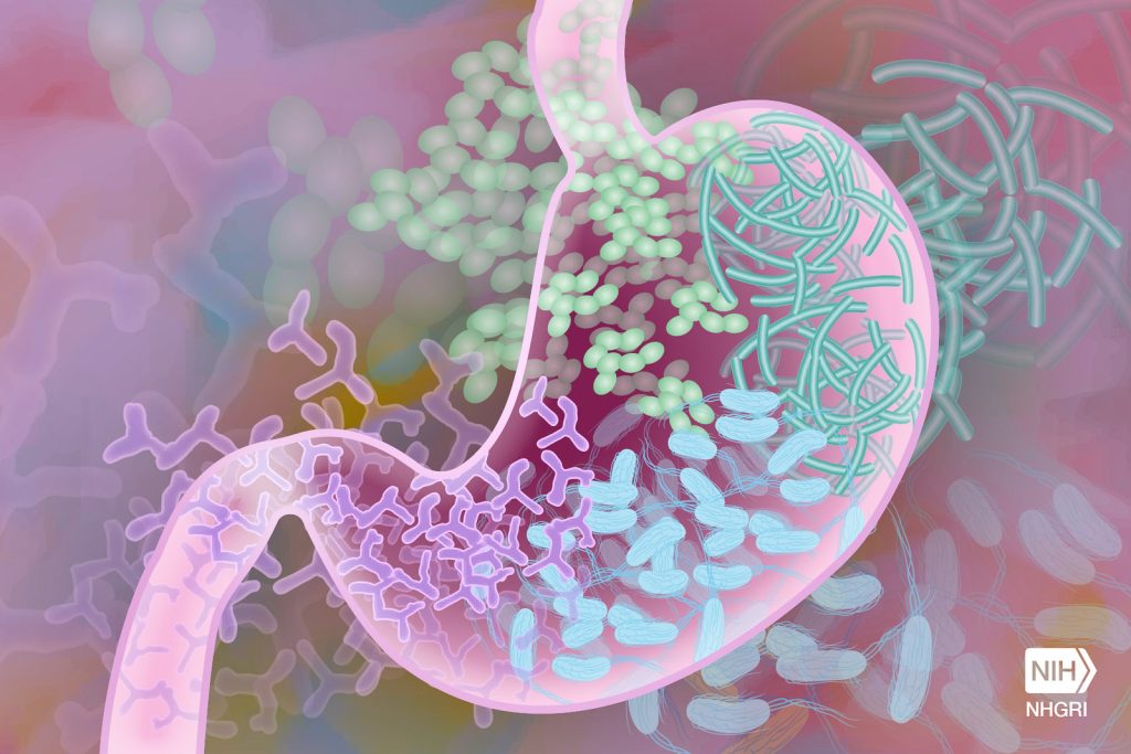
Gut Microbiome. Credit Darryl Leja National Human Genome Research Institute National Institutes Of Health

Changes in the gut microbiome before rheumatoid arthritis is developed could provide a window of opportunity for preventative treatments, new research suggests.

Bacteria associated with inflammation is found in the gut in higher amounts roughly 10 months before patients develop clinical rheumatoid arthritis, according to a longitudinal study by researchers at the University of Leeds. 

This new research might give us a major opportunity to act sooner to prevent rheumatoid arthritis.

Dr Christopher Rooney, Leeds Institute of Medical Research

Previous research has linked rheumatoid arthritis to the gut microbiome, which is the ecosystem of microbes in your intestines. But this new study, published in the Annals of the Rheumatic Diseases, reveals a potential intervention point. 

Lead researcher Dr Christopher Rooney, NIHR Academic Clinical Lecturer at the University of Leeds and Leeds Teaching Hospitals NHS Trust, said: “Patients at risk of rheumatoid arthritis are already experiencing symptoms such as fatigue and joint pain, and they may know someone in their family who has developed the disease. As there is no known cure, at-risk patients often feel a sense of hopelessness, or even avoid getting tested.  

“This new research might give us a major opportunity to act sooner to prevent rheumatoid arthritis.” 

Major opportunity for treatment

Funded by Versus Arthritis, the longitudinal study was conducted on 19 patients at risk of rheumatoid arthritis, with samples taken five times during a 15-month period.  

Five of these patients progressed to clinical arthritis, and the research showed they had gut instability with higher amounts of bacteria including Prevotella, which is associated with rheumatoid arthritis, about ten months before progression. The remaining 14, whose disease didn’t progress, had largely stable amounts of bacteria in their gut. 

Potential treatments that the researchers want to test at the 10-month window include changes to diet like eating more fibre, taking prebiotics or probiotics, and improving dental hygiene to keep harmful bacteria from periodontal disease away from the gut. 

The exact relationship between gut inflammation and rheumatoid arthritis development remains unclear. In a small number of patients within the study, the gut changes occurred before there were any changes to the joints observed by a rheumatologist, but more research is needed to determine whether these influence each other. 

Although bacteria is associated with rheumatoid arthritis, the researchers want to make it clear that there is no evidence this is contagious. 

Lucy Donaldson, director for research and health intelligence at Versus Arthritis, said: “At Versus Arthritis, we welcome the findings of this study which could give the clinicians of the future a crucial window of opportunity to delay – or even prevent – the onset of rheumatoid arthritis. This success is testament to the dedication of UK researchers who are working to personalise treatment and prevent chronic conditions that have significant impacts on a person’s ability to work, raise families and live independently.” 

The study initially took data from 124 individuals who had high levels of CCP+, an antibody that attacks healthy cells in the blood, which indicates risk of developing rheumatoid arthritis. The researchers compared their samples to 22 healthy individuals and seven people who had a new rheumatoid arthritis diagnosis.  

The findings from this larger group showed that the gut microbiome was less diverse in the at-risk group, compared to the healthy control group. 

The longitudinal study, which took samples from 19 patients over 15 months, revealed the changes in bacteria at ten months before progression to rheumatoid arthritis. 

The Leeds research team will now carry out an analysis of treatments that have already been trialled, to inform future testing of treatments at this potential 10-month intervention point. 

Source: University of Leeds

Study Suggests that High-intensity Exercise Suppresses Appetite – Especially in Women

Photo by Ketut Subiyanto on Unsplash

A vigorous workout does more to suppress hunger levels in healthy adults than does moderate exercise, and females may be especially susceptible to this response, according to a small study published in the Journal of the Endocrine Society.

The study examines the effects of exercise intensity on ghrelin levels and appetite between men and women. Ghrelin is known as the “hunger hormone” and is associated with perceptions of hunger.

“We found that high intensity exercise suppressed ghrelin levels more than moderate intensity exercise,” said lead author Kara Anderson, PhD, of the University of Virginia. “In addition, we found that individuals felt ‘less hungry’ after high intensity exercise compared to moderate intensity exercise.”

Ghrelin circulates in acylated (AG) and deacylated (DAG) forms, which are known to affect appetite. Data on the impact of exercise intensity on AG and DAG levels, and their effects on appetite, is sparse and primarily limited to males, the study noted.

To address this shortfall, the study examined eight males and six females. Participants fasted overnight and then completed exercises of varying intensity levels, determined by measurements of blood lactate, followed by self-reported measurements of appetite.

Females had higher levels of total ghrelin at baseline compared with males, the study noted. But only females demonstrated “significantly reduced AG” following the intense exercise, according to the findings.

“We found that moderate intensity either did not change ghrelin levels or led to a net increase,” the study noted. These findings suggest that exercise above the lactate threshold “may be necessary to elicit a suppression in ghrelin.”

Researchers also acknowledged that more work is needed to determine the extent to which the effects of exercise differ by sex. Ghrelin has been shown to have wide-ranging biological effects in areas including energy balance, appetite, glucose homeostasis, immune function, sleep, and memory.

“Exercise should be thought of as a ‘drug,’ where the ‘dose’ should be customised based on an individual’s personal goals,” Anderson said. “Our research suggests that high-intensity exercise may be important for appetite suppression, which can be particularly useful as part of a weight loss program.”

Source: The Endocrine Society

Intestinal Nutrient Sensors Create ‘Gut Instincts’ for Digestion

Source: CC0

Rare hormone producing cells in the gut secrete hormones in response to incoming food and play key roles in managing digestion and appetite. Researchers have now developed new tools to identify potential ‘nutrient sensors’ on these hormone producing cells and study their function. This could result in new strategies to interfere with the release of these hormones and provide avenues for the treatment of a variety of metabolic or gut motility disorders.

The work, led by led by the Hubrecht Institute and Roche’s Institute of Human Biology, is reported in Science.

The intestine acts as a vital barrier. It protects the body from harmful bacteria and highly dynamic pH levels, while allowing nutrients and vitamins to enter the bloodstream. The gut is also home to endocrine cells, which secrete many hormones that regulate bodily functions. These enteroendocrine cells (endocrine cells of the gut) are very rare cells that release hormones in response to various triggers, such as stretching of the stomach, energy levels and nutrients from food. These hormones in turn regulate key aspects of physiology in response to the incoming food, such as digestion and appetite. Thus, enteroendocrine cells are the body’s first responders to incoming food, and instruct and prepare the rest of the body for what is coming.

Understanding hormone release

Medications that mimic gut hormones, most famously GLP-1, are promising for the treatment of multiple metabolic diseases. The ability to directly manipulate endocrine cells to adjust hormone secretion could open up new therapeutic options. However, it has been challenging to understand how gut hormone release can be influenced effectively. Researchers have had trouble identifying the sensors on cells.

Enteroendocrine cells represent less than 1% of cells in the intestinal epithelium. In addition, the sensors on these cells are expressed in low amounts. Current studies mainly rely on mouse models, but the signals to which mouse cells respond are likely different from those to which human cells respond. Therefore, new models and approaches were required to study these signals.

Enteroendocrine cells in organoids

The Hubrecht team has previously developed methods to derive large quantities of enteroendocrine cells in human organoids. Organoids contain the same cell types of the organ they are derived from. Therefore, they are useful to explore the development and function of cells. Using a special protein, Neurogenin-3, the researchers could generate high numbers of endocrine cells in organoids of the intestine.

Enteroendocrine cells have different sensors and hormone profiles in different regions of the gut. In order to study these rare cells, the researchers needed to make organoids of all these different regions.

Stomach organoids

In the current study, the team managed to enrich enteroendocrine cells in organoids of other parts of the digestive system, including the stomach. Like the real stomach, stomach organoids respond to known inducers of hormone release and secrete large amounts of the hormone Ghrelin. Ghrelin is also called the ‘hunger hormone’ because it plays a key role in signaling hunger to the brain. The Ghrelin production of the stomach organoids confirms that these organoids can be used to study hormone secretion in enteroendocrine cells.

Enteroendocrine cell sensors

Since enteroendocrine cells are rare, researchers have struggled to profile many of these cells. In the current study, the team identified a so-called surface marker, called CD200, on human cells. The researchers used this surface marker to isolate a large number of human enteroendocrine cells from organoids and study their sensors. This revealed numerous receptor proteins that had not yet been identified in enteroendocrine cells.

The team stimulated the organoids with molecules that would activate these receptors and identified multiple new sensory receptors that control hormone release. When the researchers inactivated these receptors using CRISPR-based gene editing, hormone secretion was often blocked.

Therapeutic applications

With these data, the researchers can now predict how human enteroendocrine cells respond when certain sensory receptors are activated. Their findings thus pave the way for additional studies to explore the effects of these receptor activations. The enteroendocrine cell-enriched organoids will allow the team to perform larger, unbiased studies to identify new regulators of hormone secretion. These studies may eventually lead to therapies for metabolic diseases and gut motility disorders.

Source: Hubrecht Institute

Antigens in Foods Suppress Gut Tumours by Activating Immune Cells

Photo by Pixabay on Pexels

Researchers led by Hiroshi Ohno at the RIKEN Center for Integrative medical sciences (IMS) in Japan have discovered that food antigens like milk proteins help keep tumours from growing in our guts, specifically the small intestines. Experiments revealed how these proteins trigger the intestinal immune system, allowing it to effectively stop the birth of new tumours. The study was published in the scientific journal Frontiers in Immunology.

Food antigens get a lot of negative press because they are the source of allergic reactions to foods such as peanuts, shellfish, bread, eggs, and milk. Even if not allergenic, these antigens, along with the many others found in plants and beans, are still considered foreign objects that need to be checked out by the immune system. Ohno and his team have previously reported that food antigens activate immune cells in the small intestines, but not the large intestines. At the same time, some immune cells activated by gut bacteria are known to suppress tumours in the gut. In the new study, the RIKEN IMS researchers bring these two lines of thought together and tested whether food antigens suppress tumours in the small intestines.

The team began with a mouse model with a mutated tumour-suppression gene. Like people with familial adenomatous polyposis, when this gene malfunctions, the mice develop tumours throughout the small and large intestines. The first experiment was fairly simple. They fed these mice normal food or antigen-free food and found that the ones that got normal food had fewer tumours in the small intestines, but the same amount in the large intestines.

Next, they added a common representative antigen called albumin – which can be found in meat and was not in the normal food – to the antigen-free diet, making sure that the total amount of the protein equalled the amount of protein in the normal diet. When the mice were given this diet, tumours in the small intestine were suppressed just as they has been with normal food. This means that tumour suppression was directly related to the presence of antigen, not the nutritional value of the food or any specific antigen.

Mice that got the plain antigen-free diet had many fewer T cells than those that got the normal food or the antigen-free food with milk protein. Further experiments revealed the biological process that makes this possible.

These findings have clinical implications. Similar to antigen-free diets, clinical elemental diets include simple amino acids, but not proteins. This reduces digestive work and can help people with severe gastrointestinal conditions, such as Crohn’s disease or irritable bowel syndrome. According to Ohno, “small intestinal tumours are much rarer than those in the colon, but the risk is higher in cases of familial adenomatous polyposis, and therefore the clinical use of elemental diets to treat inflammatory bowel disease or other gastrointestinal conditions in these patients should be considered very carefully.”

Elemental diets are sometimes adopted by people without severe gastrointestinal conditions or allergies as a healthy way to lose weight or reduce bloating and inflammation. The new findings suggest that this could be risky and emphasises that these kinds of diets should not be used without a doctor’s recommendation.

Source: RIKEN

The Gut Microbiome can Affect Symptoms of Hypopituitarism

Gut Microbiome. Credit Darryl Leja National Human Genome Research Institute National Institutes Of Health

In research published in PLOS Genetics, scientists have shown that the balance of bacteria in the gut can influence symptoms of hypopituitarism in mice. They also showed that aspirin was able to improve hormone deficiency symptoms in mice with this condition.

People with mutations in a gene called Sox3 develop hypopituitarism, where the pituitary gland doesn’t make enough hormones. It can result in growth problems, infertility and poor responses of the body to stress.

The scientists at the at the Francis Crick Institute removed Sox3 from mice, causing them to develop hypopituitarism around the time of weaning (starting to eat solid food).

They found that mutations in Sox3 largely affect the hypothalamus in the brain, which instructs the pituitary gland to release hormones. However, the gene is normally active in several brain cell types, so the first task was to ask which specific cells were most affected by its absence.

The scientists observed a reduced number of cells called NG2 glia, suggesting that these play a critical role in inducing the pituitary gland cells to mature around weaning, which was not known previously. This could explain the associated impact on hormone production.

The team then treated the mice with a low dose of aspirin for 21 days. This caused the number of NG2 glia in the hypothalamus to increase and reversed the symptoms of hypopituitarism in the mice.

Although it’s not yet clear how aspirin had this effect, the findings suggest that it could be explored as a potential treatment for people with Sox3 mutations or other situations where the NG2 glia are compromised.

An incidental discovery revealed the role of gut bacteria in hormone production

When the National Institute for Medical Research (NIMR) merged with the Crick in 2015, mouse embryos were transferred from the former building to the latter, and this included the mice with Sox3 mutations.

When these mice reached the weaning stage at the Crick, the researchers were surprised to find that they no longer had the expected hormonal deficiencies.

After exploring a number of possible causes, lead author Christophe Galichet compared the microbiome – bacteria, fungi and viruses that live in the gut – in the mice from the Crick and mice from the NIMR, observing several differences in its makeup and diversity. This could have been due to the change in diet, water environment, or other factors that accompanied the relocation.

He also examined the number of NG2 glia in the Crick mice, finding that these were also at normal levels, suggesting that the Crick-fed microbiome was somehow protective against hypopituitarism.

To confirm this theory, Christophe transplanted faecal matter retained from NIMR mice into Crick mice, observing that the Crick mice once again showed symptoms of hypopituitarism and had lower numbers of NG2 glia. 

Although the exact mechanism is unknown, the scientists conclude that the make-up of the gut microbiome is an example of an important environmental factor having a significant influence on the consequences of a genetic mutation, in this case influencing the function of the hypothalamus and pituitary gland.

Source: Francis Crick Institute

Breastfeeding Shapes the Gut Microbiome and Protects against Asthma

Photo by Wendy Wei

Human breast milk regulates a baby’s mix of microbes, known as the microbiome, during the infant’s first year of life, in turn lowers the child’s risk of developing asthma, according to a new study published in Cell.

Led by researchers at NYU Langone Health and the University of Manitoba, the study results showed that breastfeeding beyond three months supports the gradual maturation of the microbiome in the infant’s digestive system and nasal cavity, the upper part of the respiratory tract. Conversely, stopping breastfeeding earlier than three months disrupts the paced development of the microbiome and was linked to a higher risk of preschool asthma.

Some components in breast milk, such as complex sugars called human milk oligosaccharides, can only be broken down with the help of certain microbes. This provides a competitive advantage to microbes capable of digesting these sugars. By contrast, infants who are weaned earlier than three months from breast milk and who then rely solely on formula feeding, become home to a different set of microbes –ones that will help the infant to digest the components in formula. While many of these microbes that thrive on formula do eventually end up in all babies, the researchers showed that their early arrival is linked to an increased risk of asthma.

“Just as a pacemaker regulates the rhythm of the heart, breastfeeding and human milk set the pace and sequence for microbial colonisation in the infant’s gut and nasal cavity, ensuring that this process occurs in an orderly and timely manner,” said study co-senior investigator and computational biologist Liat Shenhav, PhD. “Healthy microbiome development is not only about having the right microbes. They also need to arrive in the right order at the right time,” said Dr Shenhav, an assistant professor at NYU Grossman School of Medicine, its Institute for Systems Genetics, and the school’s Department of Microbiology.

For the study, Dr Shenhav, who is also an assistant professor at NYU’s Courant Institute of Mathematical Sciences, worked in collaboration with study co-senior investigator Meghan Azad, PhD, director of the Manitoba Interdisciplinary Lactation Center, and a professor of paediatrics and child health, at the University of Manitoba.

Another key study finding was that the bacterium Ruminococcus gnavus appeared much sooner in the guts of children who were weaned early from breast milk than in those of children who were exclusively breastfed. The bacterium is known to be involved in the production of molecules called short-chain fatty acids, and the formation and breakdown of the amino acid tryptophan. Both tryptophan and its metabolites have been linked to immune system regulation and disruption in previous research, including an increased risk of asthma. The study authors noted that beyond aiding in digestion, an infant’s microbiome plays a crucial role in the immune system’s development.

The study tracked the ebb and flow of microbes in the guts and noses of infants during the first year of life, as well as details on breastfeeding and the composition of their mothers’ milk. All the children and their mothers were participating in the CHILD Cohort Study, a long-term research project that has been studying the same 3500 Canadian children at different stages of life from the womb well into adolescence.

The data provided by the CHILD Cohort Study enabled researchers to detangle the impact of breastfeeding on an infant’s microbiome from a range of other environmental factors, including prenatal smoke exposure, antibiotics, and the mother’s asthma history.

Even when these factors were accounted for, they found that breastfeeding duration remained a powerful determinant for the child’s microbial makeup over time. They also used these microbial dynamics and data on milk components to train a machine learning model that accurately predicted asthma years in advance. Finally, they created a statistical model to learn causal relationships, which showed that the primary way breastfeeding reduces asthma risk is through shaping the infant’s microbiome.

“The algorithms we developed provide valuable insights into microbial dynamics during an infant’s first year of life and how these microbes interacted with the infant,” said Dr Shenhav. “These insights allowed us to move beyond identifying associations, enhancing our ability to make predictions and explore causal relationships.

“Our research highlights the profound impact of breastfeeding on the infant microbiome and breastfeeding’s essential role in supporting respiratory health. By uncovering the mechanisms behind the protective effects of breast milk, as demonstrated in this study, we aim to inform national guidelines on breastfeeding and weaning from breast milk in a data-driven manner.

“With further research, our findings could also contribute to developing strategies to prevent asthma in children who cannot be breastfed for at least three months,” she added.

Source: NYU Langone Health / NYU Grossman School of Medicine

Antibiotic Usage can Damage the Intestine’s Protective Mucus Layer

Source: CC0

Researchers at Umeå University and Tartu University have found that a history of repeated antibiotic use causes defects in the normally protective mucus barrier of the gut, due to antibiotic-driven alterations in the microbiota. In a further study in a different collaboration, the researchers found a bacteria-independent mechanism through which antibiotics can damage the mucus barrier directly.

The results have been published in the scientific journals Gut Microbes and Science Advances.

“Together, these two studies suggest that antibiotics can damage the mucus layer through at least two independent mechanisms, and that they may have long-lasting effects through an altered gut bacteria. This further supports the notion that antibiotics should be administered in a responsible manner,” says Björn Schröder, Docent in Infection Biology in the Department of Molecular Biology at Umeå University.

Previous research has shown the consequences of short-term antibiotic treatments on the intestinal environment, but it is less clear how repeated antibiotic use in past years can affect our guts.

To address this question, Björn Schröder and his group at Umeå University teamed up with a research group at Tartu University in Estonia, who have built a deeply characterised cohort of individuals that provided stool samples and health records.

The researchers selected individuals who had taken at least five courses of antibiotics in the past, but not within six months before the stool collection, and compared their microbiota composition to individuals who had not taken any antibiotics within the last 10 years.

“The analysis revealed changes to the gut bacteria composition, even though the antibiotics were taken a long time ago. These results indicate that repeated antibiotic use has a lasting effect on gut bacteria composition that can persist at least months after the last treatment,” says Kertu-Liis Krigul, PhD student at Tartu University.

After transplantation of the human microbiota into mice and using specialised methods to analyse the mucus function in the gut, the researchers found that the function of the mucus layer was disrupted in mice transplanted with bacteria from humans with a history of repeated antibiotic use. Expansion of the mucus was reduced, and the mucus layer became penetrable, allowing bacteria to move closer to the intestinal lining.

“Looking at the bacteria present in the gut in more detail, we could see that bacteria known to feed on the mucus layer were present at higher levels in these mice. This further supports a role for the gut bacteria in determining how well the mucus barrier can function,” says Rachel Feeney, PhD student at the Department of Molecular Biology at Umeå University.

A separate study carried out in another international collaboration, further showed that antibiotics can also directly disrupt the mucus barrier in a gut bacteria-independent manner.

By giving the antibiotic vancomycin to normal and ‘bacteria-free’ mice, the researchers were able to show that this antibiotic can act directly on the mucus barrier, independent of the gut bacteria. Complementary experiments on intestinal tissue were carried out at Umeå University and showed that the antibiotic could disrupt the mucus expansion within a few minutes of application.

Source: Umeå University