Category: Endocrinology

Sleep and Growth Hormones Tightly Regulate One Another

Photo by Zhenzhong Liu on Unsplash

As every bodybuilder knows, a deep, restful sleep boosts levels of growth hormone to build strong muscle and bone and burn fat. And as every teenager should know, they won’t reach their full height potential without adequate growth hormone from a full night’s sleep.

But why lack of sleep – in particular the early, deep phase called non-REM sleep — lowers levels of growth hormone has been a mystery.

In a study published in the current issue of the journal Cell, researchers from University of California, Berkeley, dissect the brain circuits in mice that control growth hormone release during sleep and report a novel feedback mechanism in the brain that keeps growth hormone levels finely balanced.

The findings provide a map for understanding how sleep and hormone regulation interact. The new feedback mechanism could open avenues for treating people with sleep disorders tied to metabolic conditions like diabetes, as well as degenerative diseases like Parkinson’s and Alzheimer’s.

“People know that growth hormone release is tightly related to sleep, but only through drawing blood and checking growth hormone levels during sleep,” said study first author Xinlu Ding, a postdoctoral fellow in UC Berkeley’s Department of Neuroscience and the Helen Wills Neuroscience Institute. “We’re actually directly recording neural activity in mice to see what’s going on. We are providing a basic circuit to work on in the future to develop different treatments.”

Because growth hormone regulates glucose and fat metabolism, insufficient sleep can also worsen risks for obesity, diabetes and cardiovascular disease.

The sleep-wake cycle

The neurons that orchestrate growth hormone release during the sleep-wake cycle – growth hormone releasing hormone (GHRH) neurons and two types of somatostatin neurons – are buried deep in the hypothalamus, an ancient brain hub conserved in all mammals. Once released, growth hormone increases the activity of neurons in the locus coeruleus, an area in the brainstem involved in arousal, attention, cognition and novelty seeking. Dysregulation of locus coeruleus neurons is implicated in numerous psychiatric and neurological disorders.

“Understanding the neural circuit for growth hormone release could eventually point toward new hormonal therapies to improve sleep quality or restore normal growth hormone balance,” said Daniel Silverman, a UC Berkeley postdoctoral fellow and study co-author. “There are some experimental gene therapies where you target a specific cell type. This circuit could be a novel handle to try to dial back the excitability of the locus coeruleus, which hasn’t been talked about before.”

The researchers, working in the lab of Yang Dan, a professor of neuroscience and of molecular and cell biology, explored the neuroendocrine circuit by inserting electrodes in the brains of mice and measuring changes in activity after stimulating neurons in the hypothalamus with light. Mice sleep for short periods – several minutes at a time – throughout the day and night, providing many opportunities to study growth hormone changes during sleep-wake cycles.

Using state-of-the-art circuit tracing, the team found that the two small-peptide hormones that control the release of growth hormone in the brain – GHRH, which promotes release, and somatostatin, which inhibits release – operate differently during REM and non-REM sleep. Somatostatin and GHRH surge during REM sleep to boost growth hormone, but somatostatin decreases and GHRH increases only moderately during non-REM sleep to boost growth hormone.

Released growth hormone regulates locus coeruleus activity, as a feedback mechanism to help create a homeostatic yin-yang effect. During sleep, growth hormone slowly accumulates to stimulate the locus coeruleus and promote wakefulness, the new study found. But when the locus coeruleus becomes overexcited, it paradoxically promotes sleepiness, as Silverman showed in a study published earlier this year.

“This suggests that sleep and growth hormone form a tightly balanced system: Too little sleep reduces growth hormone release, and too much growth hormone can in turn push the brain toward wakefulness,” Silverman said. “Sleep drives growth hormone release, and growth hormone feeds back to regulate wakefulness, and this balance is essential for growth, repair and metabolic health.”

Because growth hormone acts in part through the locus coeruleus, which governs overall brain arousal during wakefulness, a proper balance could have a broader impact on attention and thinking.

“Growth hormone not only helps you build your muscle and bones and reduce your fat tissue, but may also have cognitive benefits, promoting your overall arousal level when you wake up,” Ding said.

Source: University of California – Berkeley

Airborne Levels of Chemicals Released by Plastics Shock Researchers

Photo by Ryan Zazueta on Unsplash

A new study documents how people in California are chronically being exposed to toxic airborne chemicals called plasticisers, including one banned from children’s items and beauty products. 

Plasticizers are chemical compounds that make materials more flexible. They are used in a wide variety of products ranging from lunchboxes and shower curtains to garden hoses and upholstery. 

“It’s not just for drinking straws and grocery bags,” said David Volz, environmental sciences professor at UC Riverside, and corresponding author of the study published in the journal Environmental Research

Previous California monitoring programs focused on plasticisers called ortho-phthalates, some of which were phased out of manufacturing processes due to health and environmental concerns. Less research has focused on the health effects of their replacements, called non-ortho-phthalates. This study revealed the presence of both types of plasticisers in the air throughout Southern California.

“The levels of these compounds are through the roof,” Volz said. “We weren’t expecting that. As a result, we felt it was important for people to learn about this study.”

The National Institute of Environmental Health Sciences also wants to increase the visibility of this study, one of only a few to document the phthalates’ presence in the air of urban environments. The institute’s monthly newsletter, Environmental Factor, highlights the study in their October 2024 issue.  

The researchers tracked two groups of UCR undergraduate students commuting from different parts of Southern California. Both groups wore silicone wristbands designed to collect data on chemical exposures in the air. 

The first group wore their wristbands for five days in 2019, and the second group wore two different wristbands for five days each in 2020. Both groups wore the bands continuously, all day, as they went about their activities. At the end of the data collection period, the researchers chopped the wristbands into pieces, then analysed the chemicals they contained. 

In a previous paper, the team focused on TDCIPP, a flame-retardant and known carcinogen, picked up in the wristbands. They saw that the longer a student’s commute, the higher their exposure to TDCIPP. 

Unlike TDCIPP, which most likely migrates out of commuters’ car seats into dust, the team cannot pinpoint the origin of the plasticisers. Because they are airborne, rather than bound to dust, the wristbands could have picked them up anywhere, even outside the students’ cars.

For every gram of chopped-up wristband, the team found between 100 000 and 1 million nanograms of three phthalates, DiNP, DEHP, and DEHT. Ten total plasticizers were measured, but the levels of these three stood out.

Both DiNP and DEHP are included on California’s Proposition 65 list, which contains chemicals known to cause cancer, birth defects, or other reproductive harm. DEHT was introduced as an alternative, but its effects on human health have not been well studied. 

This study suggests that introducing DEHT also has not done much to reduce the public’s level of exposure to DiNP or DEHP. Levels of all three chemicals found by Volz and his team were similar to those found by researchers in unrelated studies conducted on the East Coast. 

Despite differences in climate, the air on both coasts is likely carrying similar levels of phthalates.

“No matter who you are, or where you are, your daily level of exposure to these plasticizer chemicals is high and persistent,” Volz said. “They are ubiquitous.”

To Volz, studies like this one amplify the need to find alternatives to plastic. As plastics degrade, these compounds and others like them are leaching out into the environment and into the body. 

“The only way to decrease the concentration of plasticisers in the air is to decrease our production and consumption of materials containing plasticisers,” he said. 

Source: University of California – Riverside

The Gut Microbiome can Affect Symptoms of Hypopituitarism

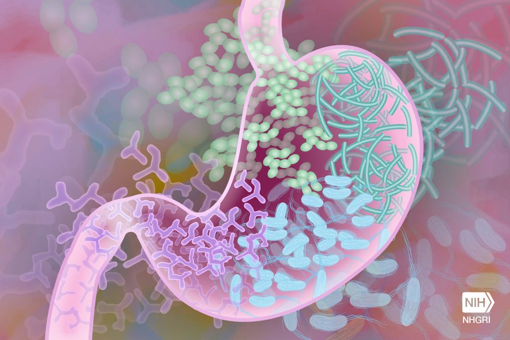
Gut Microbiome. Credit Darryl Leja National Human Genome Research Institute National Institutes Of Health

In research published in PLOS Genetics, scientists have shown that the balance of bacteria in the gut can influence symptoms of hypopituitarism in mice. They also showed that aspirin was able to improve hormone deficiency symptoms in mice with this condition.

People with mutations in a gene called Sox3 develop hypopituitarism, where the pituitary gland doesn’t make enough hormones. It can result in growth problems, infertility and poor responses of the body to stress.

The scientists at the at the Francis Crick Institute removed Sox3 from mice, causing them to develop hypopituitarism around the time of weaning (starting to eat solid food).

They found that mutations in Sox3 largely affect the hypothalamus in the brain, which instructs the pituitary gland to release hormones. However, the gene is normally active in several brain cell types, so the first task was to ask which specific cells were most affected by its absence.

The scientists observed a reduced number of cells called NG2 glia, suggesting that these play a critical role in inducing the pituitary gland cells to mature around weaning, which was not known previously. This could explain the associated impact on hormone production.

The team then treated the mice with a low dose of aspirin for 21 days. This caused the number of NG2 glia in the hypothalamus to increase and reversed the symptoms of hypopituitarism in the mice.

Although it’s not yet clear how aspirin had this effect, the findings suggest that it could be explored as a potential treatment for people with Sox3 mutations or other situations where the NG2 glia are compromised.

An incidental discovery revealed the role of gut bacteria in hormone production

When the National Institute for Medical Research (NIMR) merged with the Crick in 2015, mouse embryos were transferred from the former building to the latter, and this included the mice with Sox3 mutations.

When these mice reached the weaning stage at the Crick, the researchers were surprised to find that they no longer had the expected hormonal deficiencies.

After exploring a number of possible causes, lead author Christophe Galichet compared the microbiome – bacteria, fungi and viruses that live in the gut – in the mice from the Crick and mice from the NIMR, observing several differences in its makeup and diversity. This could have been due to the change in diet, water environment, or other factors that accompanied the relocation.

He also examined the number of NG2 glia in the Crick mice, finding that these were also at normal levels, suggesting that the Crick-fed microbiome was somehow protective against hypopituitarism.

To confirm this theory, Christophe transplanted faecal matter retained from NIMR mice into Crick mice, observing that the Crick mice once again showed symptoms of hypopituitarism and had lower numbers of NG2 glia. 

Although the exact mechanism is unknown, the scientists conclude that the make-up of the gut microbiome is an example of an important environmental factor having a significant influence on the consequences of a genetic mutation, in this case influencing the function of the hypothalamus and pituitary gland.

Source: Francis Crick Institute