Respiratory syncytial virus (RSV), a common infection in children and older adults, can also infect nerve cells and trigger inflammation leading to nerve damage, according to a new Tulane University study.
RSV can cause mild symptoms such as coughing, sneezing and fever or lead to more severe conditions such as pneumonia or bronchiolitis. But since the disease was first discovered in 1956, it has been thought to only infect the respiratory tract.
This study, published in The Journal of Infectious Diseases, is the first to prove that RSV can penetrate nerve cells and may provide the clearest link between RSV and reported neurological symptoms in children.
RSV has been previously detected in the spinal fluid of children with seizures. Additionally, 40% of RSV-positive children under the age of 2 have shown acute encephalopathy, brain damage that can result in confusion, memory loss or cognitive difficulties.
The findings underscore the potential long-term impacts of the disease, as well as the importance of preventative measures such as the two RSV vaccines approved by the FDA in 2023.
“This is the most common respiratory virus in the first years of life as well as an impactful virus among the elderly,” said Dr Giovanni Piedimonte, Tulane University vice president for research and professor of pediatrics, biochemistry and molecular biology.
“This adds a new dimension to the importance of RSV vaccines for both the elderly and mothers to protect their babies.”
Researchers studied the virus using 3D peripheral nerve cultures grown from stem cells and rat embryos.
After finding they can be infected by RSV, researchers found RSV induced the release of chemokines – proteins that fight infections by controlling immune cells – and caused significant inflammation.
With low levels of RSV infection, the nerves became hyperreactive to stimulation. At higher levels, they observed a progressive degeneration of the nerve and increased neurotoxicity due to excess inflammation.
“Until this study, the theory was that the inflammatory response was indirectly activating the nerves,” Piedimonte said.
“This study shows that not only does that happen, but the virus can penetrate directly into the nerves.”
The nerve hyperreactivity could explain why children who get RSV are later more likely to have asthmatic symptoms, Piedimonte said.
The study also found that RSV could enter the spinal cord via peripheral nerves despite not having the ability to enter the spinal neurons directly.
More research is needed to explore that mechanism, but Piedimonte theorises that by using the peripheral nerves to enter the spinal cord, RSV can bypass the blood-brain barrier, enter the central nervous system and infect the brain.
If confirmed, it could signal a connection between RSV and other neurological or developmental disorders, Piedimonte said.
“If indeed it’s confirmed in future studies that viruses like this are able to access the central nervous system, that opens a huge Pandora’s box,” Piedimonte said.
Yale researchers report in the journal Nature that they have identified a drug target that may alleviate joint degeneration associated with osteoarthritis.
The most common therapies for the degenerative disease have been pain relievers and lifestyle changes, to reduce pain and stiffness, but there is a pressing need for therapies that can prevent joint breakdown that occurs in osteoarthritis, which occurs as a result of the breakdown of cartilage in the joints.
Sodium channels found in cell membranes produce electrical impulses in “excitable” cells within muscles, the nervous system, and the heart. And in previous research, Yale’s Stephen G. Waxman identified the key role of one particular sodium channel, called Nav1.7, in the transmission of pain signals.
Now, the labs of Chuan-Ju Liu, professor of orthopaedics, and Waxman, professor neurology, neuroscience and pharmacology, have found that the same Nav1.7 channels are also present in non-excitable cells that produce collagen and help maintain the joints in the body. These channels can be targeted by existing drugs to block them.
In the new study, the researchers deleted Nav1.7 genes from these collagen-producing cells and significantly reduced joint damage in two osteoarthritis models in mice.
They also demonstrated that drugs used to block Nav1.7 – including carbamazepine, a sodium channel blocker currently used to treat epilepsy and trigeminal neuralgia – also provided substantial protection from joint damage in the mice.
“The function of sodium channels in non-excitable cells has been a mystery,” Waxman said.
“This new study provides a window on how small numbers of sodium channels can powerfully regulate the behaviour of non-excitable cells.”
“The findings open new avenues for disease-modifying treatments,” added Wenyu Fu, a research scientist in the Liu laboratory and first author of the study.
The United Nations International Day of Persons with Disabilities (IDPD) is celebrated annually on 3 December, aiming to promote an understanding of disability issues and to mobilise support for the dignity, rights and well-being of persons with disabilities. An estimated 1.3 billion people experience significant disability.1a This represents 16% of the world’s population, or 1 in 6 of us.1a In South Africa, that figure is 15%, or 8,9 million.2a
Persons with disabilities face many health inequities, including stigma, discrimination, poverty, and exclusion from education and employment. They also face barriers in all aspects of the health system, such as negative attitudes and discriminatory practices and lack of information or data collection and analysis on disability.1b+c
“Disability inclusion is critical to achieving the Sustainable Development Goals (SDGs) and global health priorities to achieve health for all, as envisioned in the 2030 Agenda for Sustainable Development,” says Prudence Selani, Head of Corporate Affairs at Sanofi South Africa. On this International Day of Persons with Disabilities, Sanofi is celebrating its commitment to the 2023 theme, ‘United in Action to Rescue and Achieve the Sustainable Development Goals (SDGs) For, With, and By Persons with Disabilities,’ through several initiatives.
In collaboration with its implementation partners, Sanofi has launched a unique external training programme for persons with disabilities, especially those from disadvantaged communities. This programme is designed to break barriers to education post-matriculation, offering management training and entrepreneurship skills in areas like financial literacy and marketing. This initiative also supports people with post-matric qualifications striving for employment, enhancing their employability and professional growth.
“As part of our commitment to Broad-Based Black Economic Empowerment (B-BBEE), 10% of learners on our Youth Employment Service “Y.E.S.” programme are persons with disabilities, underlining our commitment to diversity and inclusion,” says Selani.
Sanofi’s Diversity, Equity and Inclusion is bolstered through Employee Resource Groups (ERGs). The Ability+ ERG promotes a safe environment for employees to declare their disabilities, offering support and resources. Sanofi is also offering employees the chance to enrol in South African Sign Language courses, to transform its workplace into a disability-friendly space.
“Our partnerships with local and global organisations that are focused on disabilities will enable us to conduct workshops with leaders and employees, fostering a culture of understanding and empathy.”
“Sanofi also emphasises employee wellness and mental health, offering extensive support and wellness programmes,” says Selani. “These initiatives underscore our dedication to the well-being of all our employees.”
“As we mark IDPD 2023, Sanofi encourages organisations across all sectors to join us in these efforts. Together, we can make significant strides towards a more inclusive society and achieving the SDGs for, with, and by persons with disabilities,” concludes Selani.
Together, we are making a difference. Join us in our journey towards an inclusive future.
For decades, the standard way to prevent people who were exposed to tuberculosis (TB) from falling ill with the disease was to offer them a medicine called isoniazid, taken daily for six or more months. That changed in the last decade with the development of new preventive therapy regimens that are taken for four, three, or even just one month.
One complexity, however, is that both isoniazid and the new regimens are much better at preventing normal drug-sensitive TB than they are at preventing drug-resistant forms of TB. This is not surprising. As explained by Paediatric Infectious Disease doctor and Professor of Global Child Health at Imperial College London, Dr James Seddon, the two drugs that have mainly been used to prevent drug-susceptible TB are isoniazid and rifampicin (rifampicin’s sister drug rifapentine is also used). Now, by definition, he explains multidrug-resistant (MDR) TB is resistant to both these drugs so it’s unlikely to have any impact.
The situation is particularly tricky when it comes to children. In a 2020 statement the World Health Organization (WHO) says that it estimated that worldwide between 25 000 and 32 000 children develop MDR-TB each year, and mainly acquire it through transmission from close contact with an adult or adolescent who has MDR-TB. According to Seddon, while there is some emerging observational evidence on the use of drugs other than isoniazid and rifampicin to prevent MDR-TB, there has been no clinically tested regimen to give to children following MDR-TB exposure.
Now, much anticipated results from a phase three trial has shown that a single antibiotic pill, given daily for six months, is safe and effective to use in children who have been exposed to MDR-TB.
Results from TB CHAMP
The trial, called TB-CHAMP, looked at the efficacy and safety of using the antibiotic levofloxacin to prevent TB in children exposed to MDR-TB. Top-line findings from the study was presented last week at the Union World Lung Conference held in Paris, France.
“The paediatric population is probably the most neglected of all the populations affected by MDR-TB,” Dr Anneke Hesseling, Director of the Paediatric TB Research Programme at Stellenbosch University, told the conference. “Fewer than 20% who develop MDR-TB disease are actually diagnosed and treated, and so to find more cases and prevent more cases is really, really critical…So prevention is really key, and the TB-CHAMP trial is really a phase three efficacy trial looking at levofloxacin to prevent new cases of TB in children and also looking at the safety of levofloxacin.”
Hesseling, who is the Principal Investigator of the study, says that TB-CHAMP is the first trial to provide clinical data on what drug might be used to prevent TB in children who have been exposed to MDR-TB. It was conducted at five sites across South Africa, all with high MDR-TB burdens. The study was led by Stellenbosch University and the Desmund Tutu TB Centre. The findings have not yet been published in a peer-reviewed journal.
922 children were randomised to receive either levofloxacin or a placebo for six months. 453 children got levofloxacin and 469 got the placebo. The primary efficacy data featured data from 916 of those children, with 451 in the levofloxacin arm and 465 in the placebo arm.
Hesseling says that only children who were exposed to an adult in their household with confirmed MDR-TB were included in the study. At first children below the age of five were recruited, regardless of their TB infection status. Later children between the ages of five and 17 were included, but they had to either have a TB infection or be living with HIV. The majority of the children, 90%, were younger than five years. TB infection was confirmed with a blood test.
By 48 weeks, Hesseling says five children in the levofloxacin arm versus 12 in the placebo arm developed TB, which amounts to an incidence rate of 1.1% in the levofloxacin arm, and 2.6% in the placebo arm.
Implication of results
“While TB preventive therapy (TPT) has long been recommended and available for young child contacts of people with drug-susceptible TB, there has not been sufficient evidence to make strong recommendations for treatment that could prevent DR-TB. Therefore, the TB-CHAMP findings are critically important for a number of reasons,” says Professor Guy Marks, President and Interim Executive Director, International Union Against Tuberculosis and Lung Disease (The Union).
“The study provides the first high-quality evidence that DR-TB can be prevented in children by using six months of daily levofloxacin, and that this is a safe medication. Furthermore, this will encourage more community-based contact screening, which will also lead to early detection of children and contacts of all ages who already have disease, and initiate treatment,” he adds.
“The impact [of the TB-CHAMP results] is potentially tremendous as it would prevent DR TB among child contacts. DR TB is more complex to treat and cure and often children are marginalised, so this study puts the spotlight on an effective way to protect children. This is not just about the life and health of the child but the social, economic and mental health implications for the caregiver and the entire family,” says Dr Priashni Subrayen, Technical Director for TB at The Aurum Institute.
Seddon, who is also one of the Co-PIs for the study, tells Spotlight that it was important to establish the safety of levofloxacin since it belongs to a class of drugs called the fluoroquinolones, which were thought to have terrible side effects when used in children.
Results from TB-CHAMP show that this is not the case.
The side effects were mild, and the regimen was well tolerated, according to Hesseling, with only eight children having a grade one or higher adverse event in the levofloxacin arm compared to four in the placebo arm. Two deaths were reported, one in each study arm, but were unrelated to the study. Overall, six children in the levofloxacin arm discontinued treatment or left the study early.
Researchers from TB-CHAMP collaborated with researchers from the V-QUIN trial – a phase three study that looked at levofloxacin as TB prevention in adults in Vietnam – in order to combine their data which allowed them to show data for levofloxacin across different age groups. Seddon explains: “They’ve applied a novel analytic approach, which uses a Bayesian, or probabilistic, framework, where we take the results of TB-CHAMP and we say well, if we actually use some of the information from V-QUIN to inform the TB-CHAMP results, we can make that a slightly more confident estimate,” he says.
The combined results, according to Hesseling were able to also show that levofloxacin reduced the risk of TB by about 60% across the age spectrum but with a tighter confidence interval, indicating a more precise estimate of the effect.
Seddon tells Spotlight that the combined data showed that there were no serious adverse events, but the adult population experienced more grade one and grade two side effects than the children, but these went away either over time or when the drug was stopped. The side effects included inflammation in the joints and tendons, which is a known side effect of this class of drug.
Not a silver bullet
While the findings could be a game-changer and potentially inform MDR-TB prevention guidelines, particularly in children, the regimen is by no means a silver bullet. Seddon says that while the regimen was safe, when participants were asked whether they liked the medicine, more people said they didn’t like it in the levofloxacin group versus the placebo. Another downside is that the pill was an adult formulation and thus needed to be cut and/or crushed for the kids to swallow.
Seddon explains that the WHO, who have been provided with the data from both studies and expected to meet in early December, would need to consider a variety of factors before deciding what to recommend about the use of levofloxacin for prevention. That includes the fact that you need to treat a lot of children for six months who might not have TB despite being exposed in order to prevent a few cases.
“You have to weigh up the benefits versus the risks and the risks are low, but it is still giving a drug for six months to children and most of them don’t need it. But the consequences of getting MDR-TB are so bad that we really want to prevent that,” he says.
There is also the question around what effect using a broad antibiotic as preventive treatment will have on the microbiome of children and how this might drive resistance to the fluoroquinolones. Seddon says stool samples were collected from the study participants to determine how the drug affected a child’s microbiome and the potential for driving resistance. These data will also be provided to the WHO.
“I think that the evidence base is now very strong on the basis of these two trials. I think you can really say the issue of whether levofloxacin prevents MDR-TB, we’ve put that to bed,” he says. “Are there going to be other studies? Yes. I think that this is not over, levofloxacin is not the perfect drug for preventive therapy.”
Marks adds to this saying: “An important next step for TPT in DR-TB contacts will be studies that evaluate regimens that are shorter than six months – a long time to take medication every day, which can often be challenging. Effective and safe shorter regimens are now being used for child contacts of drug-susceptible TB and we hope the same progress can be made for contacts of DR-TB.”
As Marks has already stated, currently there are no strong recommendations for MDR-TB prevention by the WHO. In the 2020 TB prevention guidelines, it recommends that the preventative treatment for MDR-TB should be either a fluroquinolone or other second-line agent. It does however caution that these recommendations are based on low-quality evidence. Because of this, it recommends that the preventative treatment for MDR-TB should be individualised, and it be based on the drug resistance profile of the presumed contact. The drugs levofloxacin and moxifloxacin- both fluoroquinolones – may be used unless resistance is suspected. For levofloxacin a dosing schedule for both adults and children are proposed in the document.
Subrayen says that in South Africa the 2019 guidelines for the management of Rifampicin Resistant-TB (RR-TB) does indicate the use of levofloxacin as prevention treatment. The guidelines state that for prevention treatment a fluoroquinolone-based, multidrug regimen is preferred (either levofloxacin and high-dose isoniazid or levofloxacin, high-dose isoniazid and ethambutol). And if exposed to fluoroquinolone-resistant RR-TB, then high-dose isoniazid could be given. Delamanid could be considered as a potential option in very select cases. A training manual published this year by the Department of Health suggests that levofloxacin can be given on its own – but also stresses that the evidence base is weak, something that TB-CHAMP has presumably now changed.
Future of TPT
Seddon says that in a perfect world the ideal TB preventive regimen would be a so-called Pan regimen that could be given for a short period of time, to someone who has been exposed to TB and it works regardless of whether they had been exposed to drug-susceptible or drug-resistant TB.
“There are studies planned to use other drugs for prevention. There’s a study planned to use bedaquiline for a month or two and potentially using injectables that you just have to give once every couple of weeks. So, I think although this [levofloxacin] is a good option now, and it’s probably the best option we have now, this is not perfect,” Seddon says.
The study Seddon is referring to is the BREACH-TB study, a phase three trial that will look at whether a one-month treatment regimen of oral bedaquiline could prevent all forms of tuberculosis. It would be given to people exposed to both drug-resistant and drug-susceptible TB, and in people with HIV infection, including pregnant women and children.
Responding to questions from Spotlight earlier this year when this study was announced in the press, Sonya Krishnan, Assistant Professor of Medicine at Johns Hopkins University and Eric Nuermberger, Professor of Medicine at Johns Hopkins University, said that they anticipate recruiting between 1600 and 2 00 people to take part in the study – they expect around 400 to 500 of these will be people living with HIV. They also said that the control arm will receive the current standard of care in the country rather than placebo.
When asked whether any South African study sites will be included in the clinical trial, they said, “We very much plan to partner with study sites in South Africa. South Africa has a long-standing history of research excellence in TB.”
“A shorter regimen that fights both drug-resistant and drug-susceptible TB would be a game-changer for those living with TB and get us closer to our shared goal of ending the epidemic by 2030,” said Dr. Atul Gawande, USAID assistant administrator for Global Health, in a statement on the study. “This clinical trial will lay the foundation for a remarkable innovation in our fight against TB: a single-dose, long-acting injectable medicine.”
Indeed, if the science and development pans out as Gawande suggests it might, the future of TB preventive therapy might well be an entire course of therapy delivered through a single injection rather than a month or more of pills. As indicated in an article in the journal Clinical Infectious Diseases, work is already underway on the development of bedaquline, isoniazid, and rifapentine long-acting injections – though the research is for now still only in mice.
‘Communities need to be involved’
Hesseling raises the point that when treating or preventing TB, more than just the latest research advancement is needed to improve TB outcomes.
“For me treatment follows diagnosis, actually strengthening healthcare services, making communities more aware and creating demand for kids accessing diagnosis, preventive treatment and appropriate treatment, is actually where it starts,” she says. “So tools are amazing, but we actually need to have strong, effective healthcare services and knowledgeable, empowered communities.”
Seddon adds to this saying that results like those from TB-CHAMP are “a bit irrelevant if it is all kind of top down, paternalistic coming from the researchers, coming from the health system”.
“We really need to generate a community demand for this, where individuals living in communities where this is a problem are calling for this and getting angry about this and demanding it in a way that I think we’ve achieved very well with the HIV community,” he says. “It’s all well and good doing the science and then even better to get it [levofloxacin] into a guideline, but until there’s real demand for from the end user, I think it’s only going to have a certain amount of reach.”
Note: The terms DR-TB and MDR-TB are used somewhat interchangeably – Spotlight uses DR-TB to refer to drug-resistant forms of TB in general and MDR-TB to refer specifically to TB that is resistant to isoniazid and rifampicin.
One of the primary chlorine disinfectants currently used for hospital infection control does not kill off spores of the notorious cause of hospital-acquired infection Clostridioides difficile, according to a new study published in the journal Microbiology.
Research by the University of Plymouth has shown that C. Diff spores are completely unaffected despite being treated with high concentrations of bleach used in many hospitals.
In fact, the chlorine chemicals are no more effective at damaging the spores when used as a surface disinfectant – than using water with no additives.
The study’s authors say susceptible people working and being treated in clinical settings might be unknowingly placed at risk of contracting the superbug.
As a result, and with incidence of biocide overuse only serving to fuel rises in antimicrobial resistance (AMR) worldwide, they have called for urgent research to find alternative strategies to disinfect C. diff spores in order to break the chain of transmission in clinical environments.
Dr Tina Joshi, Associate Professor in Molecular Microbiology at the University of Plymouth, carried out the study with Humaira Ahmed, a fourth year Medicine student from the University’s Peninsula Medical School.
Dr Joshi, said: “With incidence of anti-microbial resistance on the rise, the threat posed by superbugs to human health is increasing. But far from demonstrating that our clinical environments are clean and safe for staff and patients, this study highlights the ability of C. diff spores to tolerate disinfection at in-use and recommended active chlorine concentrations. It shows we need disinfectants, and guidelines, that are fit for purpose and work in line with bacterial evolution, and the research should have significant impact on current disinfection protocols in the medical field globally.”
C. diff causes diarrhoea, colitis and other bowel complications, causing around 29 000 deaths per year in the USA, and almost 8500 in Europe, with the most recent data showing that, in the UK, incidence of C. diff infection was increasing prior to the start of the COVID pandemic.
Previously, Dr Joshi and colleagues had demonstrated the ability of C. diff spores to survive exposure to recommended concentrations of sodium dichloroisocyanurate in liquid form and within personal protective fabrics such as surgical gowns.
The new study examined spore response of three different strains of C. diff to three clinical in-use concentrations of sodium hypochlorite. The spores were then spiked onto surgical scrubs and patient gowns, examined using scanning electron microscopes to establish if there were any morphological changes to the outer spore coat.
Dr Joshi, who is on the Microbiology Society Council and Co-Chairs their Impact & Influence Committee, added: “Understanding how these spores and disinfectants interact is integral to practical management of C. diff infection and reducing the burden of infection in healthcare settings. However, there are still unanswered questions regarding the extent of biocide tolerance within C. diff and whether it is affected by antibiotic co-tolerance. With AMR increasing globally, the need to find those answers – both for C. diff and other superbugs – has never been more pressing.”
Red Blood Cell Infected with Malaria Parasites
Colourised scanning electron micrograph of red blood cell infected with malaria parasites (teal). The small bumps on the infected cell show how the parasite remodels its host cell by forming protrusions called ‘knobs’ on the surface, enabling it to avoid destruction and cause inflammation. Uninfected cells (red) have smoother surfaces. Credit: NIAID
New compounds are continuously required due to the risk of malaria parasites becoming resistant to the medicines currently used. A team of researchers at Friedrich-Alexander-Universität Erlangen-Nürnberg (FAU) combined the anti-malaria drug artemisinin with coumarin, and developed a compound from both bioactive plant-derived substances. This compound is also autofluorescent, making it particularly useful as it can be used for imaging in live cells.
The working group, led by Prof Dr Svetlana B. Tsogoeva, also discovered that the autofluorescent artemisinin-coumarin hybrids are able to destroy a certain drug-resistant malaria pathogen called Plasmodium palcifarum. They published their findings in the journalChemical Science.
Artemisinin is a highly-effective and common ingredient for the manufacture of malaria medication gained from a plant called sweet wormwood (Artemisia annua L.). Coumarin is a secondary plant compound found in various plants.
In the development of drugs against malaria, active substances such as artemisinin are labelled with fluorescent substances in order to identify how they act against malaria pathogens in precise chronological order using imaging techniques.
Combining substances to achieve autofluorescence
A significant disadvantage of labeling with fluorescent substances is the fact that they alter how the medication works.
For example, this means that in certain circumstances cells infected with malaria absorb a drug like artemisinin differently after fluorescent marking than previously.
The solubility of the drug can also change. This was avoided by the development of autofluorescent hybrids, which are compounds made of two or more basic compounds that are inherently fluorescent and whose mode of action can be precisely observed using imaging techniques.
Active agent with special skills
The team decided to combine artemisinin with bioactive coumarins because coumarin derivatives also possess anti-malaria properties. They can also be easily chemically altered so that they become extremely fluorescent.
The researchers discovered that it was not only possible to observe the mode of action of this first autofluorescent artemisinin-coumarin hybrid in living red blood cells infected with P. falciparum.
In conjunction with Prof. Barbara Kappes (Department of Chemical and Biological Engineering, FAU) and Dr. Diogo R. M. Moreira (Instituto Gonçalo Moniz, Fiocruz Bahia, Brazil), they also discovered that the active agent was highly effective against P. falciparum strains in vitro that are resistant to chloroquin and other malaria drugs.
Above all, however, the new compound also proved highly effective against the malaria pathogens in vivo in mouse models.
With the creation of the first autofluorescent artemisinin-coumarin hybrid, the FAU researchers hope that they have laid the foundation for the development of further autofluorescent agents for treating malaria and have made significant process in overcoming multi-drug resistance in the treatment of malaria.
People who drink more than three litres of fluid a day may be suffering from a rare condition known as vasopressin deficiency. For many, however, it is just a harmless habit. Failing to differentiate the two correctly can be fatal, so researchers have been investigating what kind of test delivers a reliable diagnosis.
In most cases, drinking excessive amounts of fluid, known as polyuria-polydipsia syndrome, either arises out of habit over time or is concomitant with a psychological illness. In rare cases, however, it can be caused by vasopressin deficiency. This hormone, released by the pituitary gland, regulates the body’s water and salt content. People with vasopressin deficiency cannot concentrate their urine, causing them to lose large quantities of fluid and to feel very thirsty.
It is extremely important to differentiate between a “harmless” form of excessive fluid ingestion and vasopressin deficiency: in the first case, those affected receive behavioural therapy to help them reduce their fluid intake gradually. People with vasopressin deficiency, however, are given the hormone vasopressin. If a patient is mistakenly treated with vasopressin, it can lead to water intoxication, which can be life-threatening.
Test with salt or arginine?
Over the past few years, the two research group leaders Professor Mirjam Christ-Crain and PD Dr Julie Refardt, together with a number of national and international centres, have been working intensively on testing methods to distinguish between these two disorders. They have found that a test that stimulates vasopressin release via a highly concentrated salt infusion is very reliable. “However, due to the resulting increase in salt concentration, constant monitoring is necessary, including half-hourly measurements of the salt levels in the patients’ blood,” explains Professor Christ-Crain.
A simplified and more easily tolerated test uses an infusion of arginine. Arginine, an essential amino acid, also stimulates the release of vasopressin and was shown to deliver a reliable diagnosis.
Clarity in diagnostics
With an international team, Christ-Crain and Refardt have now performed a direct comparison between the two tests and have published the results in the New England Journal of Medicine. The study, involving 158 participants, shows that the salt infusion resulted in a correct diagnosis for over 95% of patients. The test that uses arginine infusion, however, only led to a correct diagnosis in just under 75% of cases. Dr Refardt sums up: “In view of these results, we recommend the salt infusion test as the gold standard for reliable differentiation between polydipsia and vasopressin deficiency.”
In a post hoc analysis of the phase 2 NOBILITY trial, researchers found that treatment with obinutuzumab was superior to placebo for preserving kidney function and preventing flares in patients with lupus nephritis, a kidney condition associated with the autoimmune disease lupus.
Obinutuzumab is a recombinant, humanised type II anti-CD20 IgG1 monoclonal antibody glycoengineered to enhance antibody-dependent cell-mediated cytotoxicity and phagocytosis.
In the analysis, which is published in Arthritis & Rheumatology, compared with standard-of-care treatment alone, the addition of obinutuzumab to lupus nephritis treatment reduced the risk of developing a composite outcome of death, fall in kidney function, or treatment failure by 60%. Adding obinutuzumab also reduced the risk of lupus nephritis relapses by 57% and significantly decreased the rate of decline in kidney function over the trial’s two years duration.
Overall, 38% of obinutuzumab-treated patients compared with 16% of placebo-treated patients achieved a complete remission of lupus nephritis by week 76, with the need for fewer glucocorticoids.
“These data are really important because the ultimate goal of lupus nephritis therapy is to preserve kidney survival so patients never have to face the need for dialysis or transplantation because their kidneys failed,” said corresponding author Brad Rovin, MD, of Ohio State University Wexner Medical Center. “The addition of obinutuzumab to standard lupus nephritis therapy may increase the likelihood of achieving this goal.”
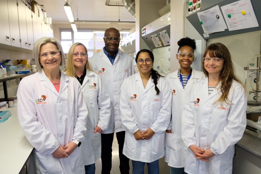
Members of the University of Cape Town’s Holistic Drug Discovery and Development Centre H3D
A formidable disease that has plagued humanity for centuries, malaria has exacted a heavy toll on human lives, disrupting communities and hindering socio-economic progress across some of the most vulnerable regions of the world, particularly the African continent.
With its stealthy transmission through the bites of infected mosquitoes, malaria has earned the dubious reputation of being one of the deadliest vector-borne diseases on the planet. So much so that the World Health Organization’s World Malaria Report reveals that malaria cases are on the rise, with instances rising from 245 million cases in 2020 to over 247 million a year later1.
With an estimated 619,000 people succumbing to the disease in 20211, it remains a defining challenge for global healthcare systems. However, through the unyielding persistence and spirit of medical innovation and scientific ingenuity exemplified by research facilities such as the University of Cape Town’s Holistic Drug Discovery and Development Centre (H3D), solutions to mitigate the severity of malaria are on the horizon.
“As the first and only integrated drug discovery platform on the African continent, H3D’s mission is to discover and develop innovative life-saving medicines for diseases that predominantly affect African patients,” explains Bada Pharasi, CEO of the Innovative Pharmaceutical Association of South Africa (IPASA).
H3D’s focus on building Africa-specific models aims to improve treatment outcomes in African patients and to educate and train a critical mass of skilled African-based drug discovery scientists. H3D’s scientific output and research model includes attracting international investment in local innovative pharmaceutical research and development (R&D) across the African continent to address the disproportionately high global disease burden. Importantly, H3D targets critical infectious diseases, including tuberculosis, antibiotic-resistant microbial diseases, and malaria.
“Given the vulnerability of many of the African populations, the continent accounted for 95% of malaria cases and 96% of malaria deaths in 20211. Accordingly, continued antimalarial drug research and development, such as the studies conducted by H3D, is important to prevent and treat the millions of cases that arise each year, all of which have consequences on both the health and socioeconomic development of the continent,” adds Pharasi.
Since the official launch of H3D’s programs in April 2011, there have been notable advances in innovative drug discovery projects. The centre has demonstrated a strong track record with multiple chemical series discovered and being progressed at H3D in each stage of the drug development pipeline.
A significant achievement reached by H3D was the discovery of the malaria clinical candidate, MMV390048, which reached phase II human trials in African patients. This was the first ever small molecule clinical candidate, for any disease, researched on African soil by an African drug discovery research unit.
According to Dr Candice Soares de Melo, Chief Investigator at H3D, the centre’s current anti-malarial programmes will focus on the identification of quality leads suitable for optimisation and candidate selection as potential agents for the treatment of uncomplicated Plasmodium falciparum malaria, ideally with additional activity against liver-stage parasites to offer protection and prevent relapses (in case of malaria caused by the species Plasmodium vivax), as well as blocking the transmission of the disease.
“A critical component of the research conducted at H3D is to develop medicines that are safe and sufficiently tolerated to be given to the widest range of recipients, including infants and pregnant women,” says Soares de Melo.
Besides the potential benefits of providing a new cure for malaria, H3D serves as a catalyst for training scientists in infectious disease research and influencing the R&D environment in Africa. As part of its partnership with the South African Medical Research Council, H3D has worked to mentor and develop scientists at other African universities, including those at Historically Disadvantaged Institutions (HDIs) within South Africa.
Furthermore, apart from strengthening drug discovery innovation at UCT, the centre has also taken a lead role in partnership with the Bill & Melinda Gates Foundation in catalysing drug discovery across sub-Saharan Africa, with upwards of 16 university research groups working on malaria and tuberculosis drug discovery.
“An example of this is the Phase 1 clinical trial for the H3D clinical candidate MMV390048, which was carried out at the UCT Division of Clinical Pharmacology,” adds Soares de Melo.
Another is the MATRIX independent special project, which has the potential to transform local drug manufacturing across the continent. Funded by the United States Agency for International Development (USAID), the project aims to pilot cost-effective local manufacture of antiretroviral Active Pharmaceutical Ingredients using flow reactor technology.
“Should Africa intend on a path to self-sufficiency, it’s important to drive continued investment in health innovations developed for and by Africa.
“We support the research efforts of H3D, and strongly believe that now is the time to take a deliberate and systematic approach to develop new capabilities, transfer technologies, leverage partnerships and networks, and train scientists, all while delivering on drug discovery projects to help address the continent’s, and the world’s, greatest health challenges,” concludes Pharasi.
A woman with Systemic Lupus Erythematosus. Source: Wikimedia CC0
The autoimmune condition lupus occurs in women at a rate nine times higher than in men. Some of the factors that cause the disease’s high prevalence in women have eluded discovery, but in a new study published in the Journal of Clinical Investigation Insight, Johns Hopkins Medicine researcher investigated the immune system processes in lupus and the X chromosome, and uncovered answers about the disease’s frequency in females.
A number of dysregulated genetic and biological pathways contribute to the development of lupus and its varied symptoms of muscle and joint pain, skin rashes, kidney problems and other complications throughout the body. One such pathway involves a protein in the immune system called toll-like receptor 7 (TLR7), which, in lupus, reacts to the body’s own RNA, molecules that act as messengers of genetic information. TLR7’s reaction to RNA triggers an immune response that damages healthy tissue.
In the full article, researchers honed in on this TLR7 immune response in lupus, looking specifically at how a piece of genetic material only found in women, known as X-inactive specific transcript (XIST), could trigger TLR7’s immune system response. XIST is a type of RNA that plays a crucial role in inactivating one of the two X chromosomes found in female cells so that females do not have imbalanced gene expression.
“XIST has previously been implicated in autoimmunity, but more as something that could prevent autoimmune conditions like lupus, rather than drive the disease’s development,” says study author and lead researcher Erika Darrah, PhD. “Our findings show the opposite, that XIST actually plays a role in promoting autoimmunity – increasing the susceptibility to lupus and its severity in women.”
The research team first tested whether XIST could bind to TLR7 and initiate the receptor’s immune response using cellular experiments. They observed that XIST could strongly bind to TLR7 and trigger the production of molecules called interferons, an immune system protein seen at high levels in lupus that contributes to tissue damage in this disease. Rather than protect from TLR7 and interferon’s negative effects on the body, these tests illustrated that XIST drove the process of an overactive immune response and therefore contributed to lupus development.
“XIST has now taken on a different role, an alarm signal related to autoimmunity,” says study author Brendan Antiochos, MD. “The immune system activation through XIST and TLR7 is female-specific, helping explain the observation that lupus is so much more common in women compared to men.”
To further study XIST’s role in lupus, researchers also examined XIST levels in patients from two lupus cohorts. The team tested blood samples from patients at the Johns Hopkins Lupus Center for XIST levels, and also used publicly available data from another study that showed XIST and interferon levels in white blood cells taken from the kidneys of people with lupus. They assessed that not only did the levels of XIST in the kidney correlate with higher interferon levels, but also, those with more XIST in their blood cells experienced greater disease severity and worsened lupus symptoms.
Darrah and Antiochos say these findings may implicate XIST in other autoimmune conditions that are more often seen in women, and that more research should be conducted to investigate this female-specific process.
Researchers also say that understanding XIST’s role in lupus development may lead to creative therapies that target the XIST-TLR7 pathway, as well as offer an additional explanation for patients who may wonder about the origins of their disease.