Category: Diseases, Syndromes and Conditions

Tofacitinib Could Treat Rare but Deadly Systemic Sclerosis

Photo by Louise Reed on Unsplash

Systemic sclerosis, or scleroderma that affects the skin and internal organs, is one of the rarest autoimmune diseases, affecting roughly 100 000 people in the US, mostly women. It has the highest mortality rate among rheumatic diseases.

There are no licensed treatments available for this subset of scleroderma patients, and rheumatology researchers are constantly searching for opportunities to use resources and technology that have proven beneficial in treating other autoimmune and rheumatic diseases.

In a new study published in JCI Insightresearchers found that tofacitinib, a drug approved for rheumatoid arthritis, was well tolerated among patients with early systemic sclerosis, and discovered the drug primarily affected the protein, interferon, both in fibroblasts and keratinocytes cells.

Dinesh Khanna MBBS., MSc, director of the Michigan Medicine Scleroderma Program, explained: “We wanted to understand first, if there was any clinical benefit of tofacitinib to patients, but we were also asking, what are the differences in the cells of healthy skin versus systemic sclerosis cells…how does the drug work?”

The study sample size consisted of 15 patients with early diffuse cutaneous systemic sclerosis — patients with skin hardening and issues with organs. Of the total participants, 10 patients received 5mg of tofacitinib twice a day, and the remaining received placebo in a double-blind randomised placebo-controlled trial.

Over the course of the 24-week trial period, researchers found no patients who exhibited severe adverse effects at or before the trial ended. Measures included the modified Rodnan skin score (mRSS).

These results showed that the average mRSS score and other measures improved over the course of the trial. In addition, patients on placebo went on open label tofacitinib after 24 weeks and there was ongoing improvement during the next 24 weeks, indicating improvement in the measure.

“We are delighted to find that the drug is safe to use and can possibly be repurposed for systemic sclerosis treatment,” said Khanna, “but what made this study innovative was the use of single cell technology.”

Participants in the study had a skin biopsy at the start of the trial and then again six weeks after they received tofacitinib or placebo. Then, clinicians used the relatively new technology — single cell RNA sequencing — to watch the mechanism of tofacitinib at work in the trial participants’ skin cells.

“This work highlights the ability of single-cell RNA-sequencing to determine how disease states are maintained and how various cell populations in the skin, both fibroblasts, skin cells, and immune cells communicate, providing unparallelled power to address disease mechanisms, and how drugs, like tofacitinib, work in a disease where they have not previously been used,” said Johann Gudjonsson MD, PhD, professor of dermatology and a collaborator on this study.

Along with discovering how tofacitinib inhibits fibroblasts and keratinocytes, researchers found that the drug had minimal effect on T cells.

“Because we found that the drug was working on one part (the mechanism of fibroblasts and keratinocytes), we are now considering if we can combine tofacitinib with another drug with complementary mechanism in action, in order to treat early systemic sclerosis without causing toxicity,” explained Khanna.

To understand more about the drug, researchers will need to conduct a more robust study and trial to see if their recent discoveries hold true.

“From this combined effort between Michigan Medicine and University of Pittsburgh, we know that the drug is safe, and we know that the technology (RNA sequencing) is feasible, now we can start to utilise the technology and find out what type of therapies we can mix and match that will add benefit to patients,” Khanna said.

Source: Michigan Medicine – University of Michigan

Strong Link of Type 1 Diabetes to Enterovirus Infection

A 3D map of the islets in the human pancreas. Source: Wikimedia

A meta-analysis of molecular studies found a strong link between enterovirus infection and type 1 diabetes. Across 48 abstracts on the topic, people with type 1 diabetes were nearly eight times more likely to have an enterovirus infection than those with normal pancreatic function, according to a presentation at the European Association for the Study of Diabetes (EASD) meeting.

For this review, researchers examined 56 studies using molecular methods to identify enterovirus infection by looking for viral nucleic acid or protein in a human clinical sample.

People with islet autoimmunity were twice as likely to have had an enterovirus infection at any point in time, based on data from 18 studies.

Subgroup analysis of people who were diagnosed with the enterovirus within the past month found 16.22-fold higher odds of recent-onset type 1 diabetes, based upon 28 studies.

“This is very recent onset type 1 diabetes, and that was the highest risk group that we detected,” reported Sonia Isaacs, PhD candidate, of University of New South Wales in Australia, during a press conference.

Looking more closely at other subgroup criteria revealed associations.

First, those who had any islet autoimmunity and eventually progressed to full-blown type 1 diabetes were over five times more likely to have an enterovirus infection than controls; this was a higher risk than those who had islet autoimmunity and never progressed to type 1 diabetes. When it came to timing, only infections at the time of or after islet autoimmunity seroconversion carried higher risk (OR 5.1), whereas the increased risk wasn’t significant for infections prior to seroconversion.

Isaacs noted that those with a familial risk of type 1 diabetes (ie in a first-degree relative) also had a much higher risk for an enterovirus infection (OR 9.8), higher than the subgroup recruited for having a high-risk HLA gene. Those who had the high-risk HLA gene and a familiar risk of type 1 diabetes had 141.1-times higher odds of prior enterovirus infection.

Having several enterovirus infections was also linked with a doubled chance of islet autoimmunity.

Specific type of enterovirus linked to risk of type 1 diabetes included:

  • Enterovirus A: OR 3.7
  • Enterovirus B: OR 12.7
  • Enterovirus C: OR 13.8

“This is where the coxsackieviruses come from,” Isaacs pointed out. “Coxsackievirus B1 and B4 stood out.”

Isaacs suggested the possibility of enterovirus vaccinations and antivirals as a prevention strategy for type 1 diabetes.

Source: MedPage Today

Lassa Virus Endemic Area may Grow in Coming Decades

Pictured are projections of the ecological niche suitability for Lassa virus based on climate models and other data. Credit: Scripps Research and University of Brussels

Analysing decades of environmental data associated with Lassa virus outbreaks, researchers projected that areas hospitable to Lassa virus spread may extend from West Africa into Central and East Africa in the next several decades. With this expansion and expected African population growth, the human population living in the areas where the virus should theoretically be able to circulate may rise by more than 600 million.

“Our analysis shows how climate, land use, and population changes in the next 50 years could dramatically increase the risk of Lassa fever in Africa,” says first author Raphaëlle Klitting, PhD, a postdoctoral researcher at Scripps Research during the study, which is published in Nature Communications.

Lassa virus is a zoonotic pathogen found in the Natal multimammate rat (Mastomys natalensis), most likely transmitted to humans via its droppings. While an estimated 80% of infections are mild or asymptomatic, the remaining cases are more severe, with signs and symptoms that can include haemorrhaging from the mouth and gut, hypotension, and potentially permanent hearing loss. Mortality rate in hospitalised patients can be up to 80%.

An estimated several hundred thousand infections occur each year, chiefly in Nigeria and several other West African countries. So far there is no approved vaccine or highly effective drug treatment.

Although the primary animal reservoir for Lassa virus is known, the virus spreads in only a subset of the areas where the animal is found. Thus, it is possible that environmental factors also help determine whether and where significant viral transmission can occur. In the study, the researchers developed an ‘ecological niche’ model of Lassa virus transmission, using environmental data at sites of known spread.

Combining the model with projections of climate and land-use changes in Africa in the next several decades, as well as the known range of the Natal multimammate rat, the researchers estimated the areas of Africa that could support Lassa virus transmission currently, and in the years 2030, 2050, and 2070. The projected current areas corresponded well to known endemic areas in West Africa, but the estimates for future decades suggested a vast expansion within and beyond West Africa.

“We found that several regions will likely become ecologically suitable for virus spread in Central Africa, including in Cameroon and the Democratic Republic of the Congo, and even in East Africa, in Uganda,” Klitting said.

Currently Africa’s population is undergoing rapid growth; the researchers therefore considered projections of this population growth for the areas of current and potential future Lassa virus circulation. They found that the number of people potentially exposed to the virus could increase from about 92 million today to 453 million by 2050, and 700 million by 2070 – an increase of over 600%.

More hopefully, the researchers examined the dynamics of the spread of Lassa virus using data on sequenced viral genomes sampled at various locations in West Africa and found that virus dispersal appeared to be slow. They concluded that, unless transmission dynamics change drastically in the new location where the virus circulates, the virus’s spread into new ecologically suitable areas in the coming decades may also be slow.

The authors say that the findings should inform African public health policies, for example, by encouraging officials to add Lassa virus to lists of viruses under epidemiologic surveillance in parts of Central and East Africa.

“With the ongoing climate change and increasing impact of human activities on the environment, further comprehensive studies of the ecology and spread of zoonotic and vector-borne diseases are needed to anticipate possible future changes in their distribution as well as their impact on public health,” said senior author Simon Dellicour, PhD, of the University of Brussels.

Source: Scripps Research Institute

Viruses may ‘Watch’ for the Right Moment to Strike

Lassa virus scanning electron micrograph
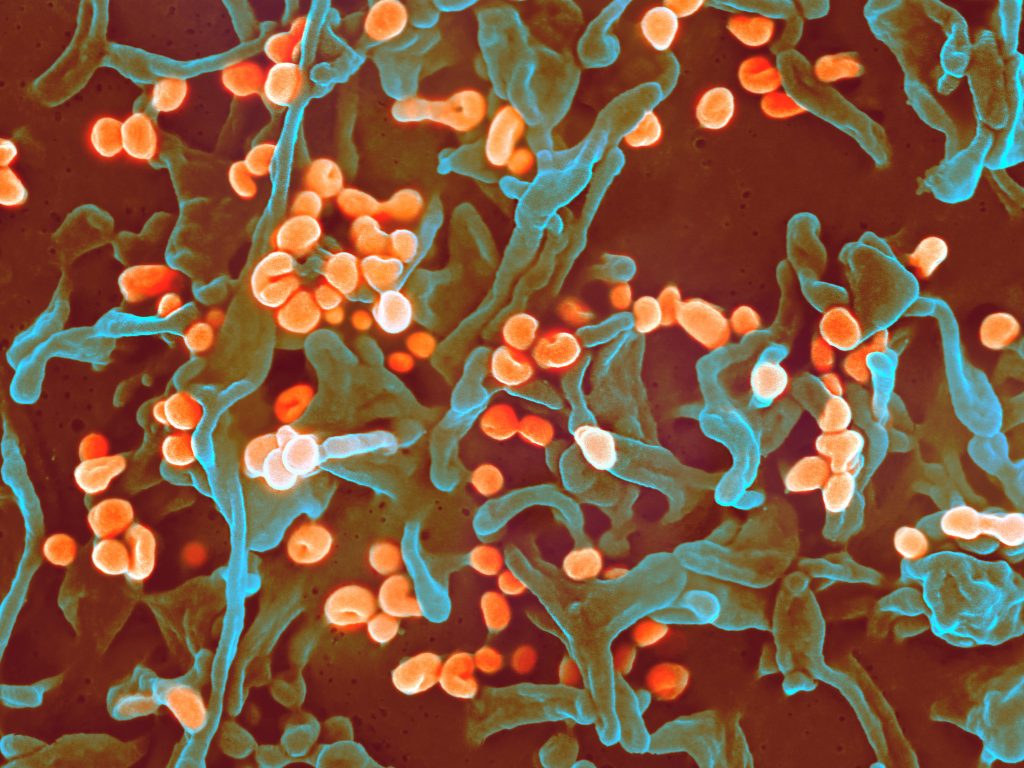
Scanning electron micrograph of Lassa virus budding off a Vero cell. Image credit: National Institute of Allergy and Infectious Diseases, NIH

Scientists have found that viruses lurking inside cells may be on the ‘watch’ for information from their environment to choose when to multiply and burst out. The work, published in Frontiers in Microbiology, has implications for antiviral drug development.

A virus’s ability to sense its environment, including elements produced by its host, adds “another layer of complexity to the viral-host interaction,” said senior author Ivan Erill, professor of biological sciences. Currently, viruses use that ability to their benefit. But in the future, he says, “we could exploit it to their detriment.”

Not a coincidence

The new study focused on bacteriophages, viruses which infect bacteria – also known as ‘phages.’ The phages in the study can only infect their hosts when the bacterial cells have special appendages, called pili and flagella, that help the bacteria move and mate. The bacteria produce a protein called CtrA that controls when they generate these appendages. The new paper shows that many appendage-dependent phages have patterns in their DNA where the CtrA protein can attach, called binding sites. A phage having a binding site for a protein produced by its host is unusual, explained Prof Erill.

Even more surprising, Erill and the paper’s first author Elia Mascolo, a PhD student in Erill’s lab, found through detailed genomic analysis that these binding sites were not unique to a single phage, or even a single group of phages. Many different types of phages had CtrA binding sites – but they all needed their hosts to have pili and/or flagella to infect them. It couldn’t be a coincidence, they decided.

The ability to monitor CtrA levels “has been invented multiple times throughout evolution by different phages that infect different bacteria,” Prof Erill said. This convergent evolution indicates that the trait is useful.

Timing is everything

Another wrinkle in the story: The first phage in which the research team identified CtrA binding sites infects a particular group of bacteria called Caulobacterales. Caulobacterales are an especially well-studied group of bacteria, because they exist in two forms: a free-swimming ‘swarmer’ form which has pili/flagella, and a ‘stalked’ form that attaches to a surface and lacks those appendages. In these bacteria, CtrA also regulates the cell cycle, determining whether a cell will divide evenly into two more of the same cell type, or divide asymmetrically to produce one swarmer and one stalk cell.

Because the phages can only infect swarmer cells, it’s in their best interest only to burst out of their host when there are many swarmer cells available to infect. Generally, Caulobacterales live in nutrient-poor environments, and they are very spread out. “But when they find a good pocket of microhabitat, they become stalked cells and proliferate,” Prof Erill said, eventually producing large quantities of swarmer cells.

“We hypothesise the phages are monitoring CtrA levels, which go up and down during the life cycle of the cells, to figure out when the swarmer cell is becoming a stalk cell and becoming a factory of swarmers,” Prof Erill said, “and at that point, they burst the cell, because there are going to be many swarmers nearby to infect.”

Listening in

Unfortunately, the method to prove this hypothesis is labour-intensive and extremely difficult, so that wasn’t part of this latest paper — although Erill and colleagues hope to tackle that question in the future. However, the research team sees no other plausible explanation for the proliferation of CtrA binding sites on so many different phages, all of which require pili/flagella to infect their hosts. Even more interesting, they note, are the implications for viruses that infect humans.

“Everything that we know about phages, every single evolutionary strategy they have developed, has been shown to translate to viruses that infect plants and animals,” he said. “It’s almost a given. So if phages are listening in on their hosts, the viruses that affect humans are bound to be doing the same.”

There are a few other documented examples of phages monitoring their environment in interesting ways, but none include so many different phages employing the same strategy against so many bacterial hosts.

Prof Erill predicts that more examples of this will be found, and his lab is already discovering more.

New therapeutic avenues

The key takeaway from this research is that “the virus is using cellular intel to make decisions,” Erill says, “and if it’s happening in bacteria, it’s almost certainly happening in plants and animals, because if it’s an evolutionary strategy that makes sense, evolution will discover it and exploit it.”

For example, to optimize its strategy for survival and replication, an animal virus might want to know what kind of tissue it is in, or how robust the host’s immune response is to its infection. While it might be unsettling to think about all the information viruses could gather and possibly use to make us sicker, these discoveries also open up avenues for new therapies.

“If you are developing an antiviral drug, and you know the virus is listening in on a particular signal, then maybe you can fool the virus,” Erill said. “We are just starting to realise how actively viruses have eyes on us – how they are monitoring what’s going on around them and making decisions based on that. “It’s fascinating.”

Source: University of Maryland Baltimore County

Eye Infection Risk Greatly Increased for Wearers of Reusable Contact Lenses

Photo by Lensabl on Unsplash

People who wear reusable contact lenses are nearly four times as likely as those wearing daily disposables to develop a rare sight-threatening eye infection, Acanthamoeba keratitis (AK), according to new research. The study, published in Ophthalmology, identifies multiple factors that increase the risk of AK, including reusing lenses or wearing them overnight or in the shower.

AK is a type of microbial keratitis – a condition that results in inflammation of the cornea.

Eye infection from Acanthamoeba. Credit: CDC

Lead author, Professor John Dart (UCL Institute of Ophthalmology and Moorfields Eye Hospital NHS Foundation Trust) said: “In recent years we have seen an increase of Acanthamoeba keratitis in the UK and Europe, and while the infection is still rare, it is preventable and warrants a public health response.

“Contact lenses are generally very safe but are associated with a small risk of microbial keratitis, most commonly caused by bacteria, and which is the only sight threatening complication of their use. Given that an estimated 300 million people across the globe wear contact lenses, it is important that people know how to minimise their risks for developing keratitis.”

Contact lens use is now the leading cause of microbial keratitis in patients with otherwise healthy eyes in countries in the global north. While sight loss resulting from microbial keratitis is uncommon, Acanthamoeba, is one of the most severe causes and is responsible for about half of those contact lens users who develop sight loss after keratitis. Avoidable risks are associated with 90% of AK cases. The infection remains rare, affecting fewer than 1 in 20 000 contact lens wearers per year in the UK.

In AK, ,infection by Acanthamoeba, a cyst-forming microorganism, causes the cornea to become painful and inflamed. The most severely affected patients (a quarter of the total) end up with less than 25% of vision or become blind following the disease and face prolonged treatment. Overall, 25% of people affected require corneal transplants to treat the disease or restore vision.

For the study, the researchers recruited over 200 patients of Moorfields Eye Hospital who completed a survey, including 83 people with AK, and compared them with 122 participants who came to eyecare clinics with other conditions, who acted as a control group.

People who wore reusable soft contact lenses (such as monthlies) had 3.8 times the odds of developing AK, compared to people who wore daily disposable lenses. Showering with lenses increased AK risk by 3.3 times, while wearing lenses overnight increased risk by 3.9 times. Among daily disposable wearers, reusing their lenses increased their infection risk. Having had a recent contact lens check with a health professional reduced the risk.

With further analysis, the researchers estimated that 362% of cases in the UK, and potentially in many other countries, could be prevented if people switched from reusable to daily disposable lenses.

A recent study led by Prof Dart found that AK is increasing in prevalence in the UK. By reviewing incidence data from Moorfields Eye Hospital from 1985 to 2016, he and his team found an increase starting in 2000–2003, when there were eight to 10 cases per year, to between 36 and 65 annual cases at the end of the study period.

First author, Associate Professor Nicole Carnt (UNSW, Sydney, UCL Institute of Ophthalmology and Moorfields Eye Hospital) said: “Previous studies have linked AK to wearing contact lenses in hot tubs, swimming pools or lakes, and here we have added showers to that list, underlining that exposure to any water when wearing lenses should be avoided. Public pools and coastal authorities could help reduce this risk by advising against swimming in contact lenses.”

Prof Dart added: “Contact lens packaging should include information on lens safety and risk avoidance, even as simple as ‘no water’ stickers on each case, particularly given that many people buy their lenses online without speaking to a health professional.

“Basic contact lens hygiene measures can go a long way in avoiding infections, such as by thoroughly washing and drying your hands before putting in your lenses.”

Source: EurekAlert!

New Study Explains Diabetes and UTI Link

Huge clumps of E. coli (red) infecting diabetic mouse bladder. Photo: Soumitra Mohanty

Lower immunity and recurring infections are common in type 1 and type 2 diabetes. Research has shown that the immune system of people with diabetes has lower levels of the antimicrobial peptide psoriasin, which compromises the urinary bladder’s cell barrier, increasing the risk of urinary tract infection. The study is published in Nature Communications.

One effect of diabetes is that it compromises the innate immune system, leaving many people with increased susceptibility to regular infections, such as urinary tract infections (UTI)s caused by E. coli bacteria. In people with diabetes, these are more likely to lead to general blood poisoning, sepsis, originating in the urinary tract.

An endogenous antibiotic

Karolinska Institutet researchers investigated whether glucose levels in people with diabetes (type 1, type 2, or prediabetes) are linked with psoriasin, an endogenous antibiotic which is a part of the innate immune system.

Using samples of urine, bladder cells and blood serum from patients, the researchers analysed levels of psoriasin and other peptides necessary for ensuring that the bladder mucosa remains intact and protects against infection. The findings were then verified in mice and urinary bladder cells with and without infection.

“We found that high glucose concentrations reduce the levels of the antimicrobial peptide psoriasin, while insulin has no effect,” said Professor Annelie Brauner, who led the study. “People with diabetes have lower levels of psoriasin, which weakens the cells’ protective barrier function and increases the risk of bladder infection.”

Oestrogen therapy reduced bacterial population

Professor Brauner’s research group has previously shown that oestrogen restores the protective function of bladder cells in humans and mice and thereby help to regulate the immune response to a UTI. The researchers therefore tested how oestrogen treatment affects infected cells exposed to high glucose concentrations. They found that the treatment boosted levels of psoriasin and reduced bacterial populations, indicating that the treatment may have an effect also among patients with diabetes.

“We now plan to probe deeper into the underlying mechanisms of infections in individuals with diabetes,” said lead author Soumitra Mohanty. “The ultimate goal is to reduce the risk of infection in this growing patient group.”

Source: Karolinska Institutet

International Gaucher Day on 1 October Highlights Need For Greater Attention on Rare Diseases

Photo by Cottonbro on Pexels

Currently, there are an estimated 6000–8000 rare diseases, which affect 350 million people worldwide.One such rare condition is Gaucher Disease (GD) – a lysosomal storage disorder (LSD).  GD is a rare genetic disorder, passed down from parents to children (inherited) in an autosomal recessive manner. 

GD is one of the most common LSDs with a prevalence in the general population of ~1 per 100 000 and ~1/855 in the Ashkenazi Jewish population.2 As with many rare conditions, diagnosis of GD may present a significant challenge to non-GD specialities, owing to the wide variability in age, severity, type of clinical manifestation and lack of awareness of the early signs and symptoms of GD among non-specialist physicians.3 One in 6 patients with GD reported a diagnosis delay of 7 years or more after first consulting a doctor.3

International Gaucher Day on 1 October, therefore, aims to improve patients’ quality of life through greater awareness and earlier diagnosis of GD amongst healthcare professionals.

GD arises from an inherited deficiency of an enzyme called glucocerebrosidase, normally found within the lysosomes of cells, due to mutations in the GBA gene.4 This enzyme is responsible for breaking down a fatty substrate, glucocerebroside, into glucose and a simpler fat molecule (ceramide).4 Patients with GD have a progressive build-up of glucocerebroside within the lysosomes, particularly of macrophages, resulting in enlarged cells known as ‘Gaucher’ cells.4

These ‘Gaucher’ cells accumulate in organs throughout the body, predominately affecting the bone marrow, liver, and spleen.4 There are three types of GD, based on the presence and severity of neurological involvement.4 Type 1, known as the non-neuronopathic GD  characterised by haematological abnormalities such as thrombocytopenia, leukopenia and anaemia, hepatomegaly and/or splenomegaly, bone crises and/or osteoporosis, and fatigue. Phenotypically, there is a wide spectrum of disease manifestations, ranging from asymptomatic to severe type 1 child-onset disease.5,6 Type 2, the acute neuronopathic form, is the rarest and most severe form of GD.  It includes the rapid progression of severe neurological abnormalities early in life, leading to death in infancy or early childhood.4,6 Type 3, the chronic form, encompasses multiple phenotypes.  Type 3 typically occurs during the paediatric years and varies in severity: patients have the same symptoms as in type 1, plus some neurological involvement that generally appear later in life, such as abnormal eye movement, ataxia, seizures and dementia.4

Anaemia, thrombocytopenia, enlargement of the liver and/or spleen, and skeletal abnormalities (osteopenia, lytic lesions, pathological fractures, chronic bone pain, bone crisis, bone infarcts, osteonecrosis and skeletal deformities) are typical manifestations of type 1 GD, the most prevalent form of the disease.However, the severity and coexistence of different symptoms are highly variable, and GD patients are often misdiagnosed as having other malignant haematological conditions.4

Although GD is rare, clinicians are encouraged to maintain a high index of suspicion with patients presenting with atypical symptoms, and should consider testing for rare diseases where other haematological pathologies have been excluded4 or when testing for them. Such patients may be referred to a GD specialist or be tested through North West University (NWU), where global pharmaceutical company Sanofi and the NWU Centre of Human Metabolomics, headed by Prof Chris Vorster, have partnered to test for the most common lysosomal storage disorders in South Africa, including GD, using dried blood spot samples.

Says Prof. Vorster: “Rare conditions such as GD require the cooperation of a multidisciplinary team in order to find and treat them. Interventions can improve a patient’s quality of life through improvement or restoration of their physical function, so that they may carry out regular daily activities. The NWU Centre of Human Metabolomics provides internationally competitive metabolomic analytic services, and electronic results may be sent by high priority straight to healthcare practitioners, speeding up diagnosis.”

Monique Nel, Medical Advisor – Rare Diseases at Sanofi, says: “We understand the difficulty that healthcare professionals face when it comes to diagnosing patient with GD. It requires a coordinated approach to diagnosis and care for people living with the condition. Early diagnosis of GD, and the initiation of treatment will delay the occurrence of irreversible complications, and improve the patient’s quality of life. We therefore direct the attention of healthcare providers to the RD Nexus platform, which is Sanofi’s dedicated platform for rare diseases, at www.RDNexus.com. This platform offers educational materials, road maps to a differential diagnosis and how to test a patient for these conditions.”

For more information on GD and other rare diseases, visit: www.RDNexus.com

  1. https://www.ncbi.nlm.nih.gov/pmc/articles/PMC6505568/pdf/EMMM-11-e10486.pdf Accessed September 2022.
  2. Burrow TA et al. Prevalence and management of Gaucher disease. Paediatric Health, Medicine and Theraeutics 2011;2:59-73.
  3. Revel-Wilk S, et al. How we manage Gaucher Disease in the era of choices. British Journal of Haematology 2018;182:467-480.
  4. CPD Gaucher. Gaucher Disease. Medical Chronicle June 2020:30-32.
  5. Linari S, Castaman G. Clinical manifestations and management of Gaucher disease. Clinical Cases in Mineral and Bone Metabolism 2015;12(2):157-164.
  6. Roshan Lal T and Sidransky E. The spectrum of neurological manifestations associated with Gaucher Disease. Diseases 2017;5,10.

New Way to Improve Outcomes in Kids with Eosinophilic Oesophagitis

Children
Photo by Ben Wicks on Unsplash

Researchers have determined the threshold for a new measure of early scarring in the oesophagus of children with eosinophilic oesophagitis (EoE), which allows immediate intervention during endoscopy to halt further damage and prevent food from getting stuck. Their findings were published in the journal Clinical Gastroenterology and Hepatology.

EoE is a chronic immune-mediated disorder of the oesophagus that affects adults and children with a prevalence of 0.5 to 1 per 1000. Left untreated, chronic inflammation promotes scarring of the oesophagus, and the development of oesophageal rings and stricture, which interferes with passage of solid food and can cause impaction (when food is stuck in the oesophagus and cannot dislodge).

The researchers used the Endoscopic Functional Luminal Impedance Probe (EndoFLIP) in the study to measure the “distensibility index,” which is a functional measure of how much force is required to stretch open the oesophagus. Previously, the extent of scar tissue in the oesophagus could only be evaluated visually during endoscopy, making it challenging to detect the early changes and intervene before the damage becomes more extensive.

“This is a gamechanger in how we care for kids with EoE,” said senior author Joshua Wechsler, MD, MSCI. “Now, if distensibility is low, we can dilate the oesophagus during the same procedure, and because we can pinpoint exactly where the scarring is, our intervention is more targeted and takes much less time. We are seeing improvements in symptoms, which is incredibly exciting.”

Source: Ann & Robert H. Lurie Children’s Hospital of Chicago

Oxygen Responses Control How T Cells Respond to TB

Tuberculosis bacteria
Tuberculosis bacteria. Credit: CDC

A new publication in Nature Communications explains how T cell protection against tuberculosis is controlled by their oxygen responses.

In 2021, 10 million people fell ill and 1.5 million died of Tuberculosis (TB), caused by infection with the intracellular Mycobacterium tuberculosis bacteria. Proper CD4 T cell responses are critical for the control of M. tuberculosis infection by activating intracellular bacterial killing.

Professor Martin Rottenberg and PhD student Ruining Liu at the Karolinska Institutet, explained how they discovered that hypoxia-inducible factors (HIF-1 and HIF-2) control T cell metabolism as well as activation and differentiation in response to hypoxia or during inflammation.

“We showed that genetically modified mice, in which HIF-1 expression T cells was stabilised by genetic manipulation, were highly susceptible to the infection with M. tuberculosis and did not respond to vaccination. CD4 T cells from these mice were profoundly weakened in their early responses to mycobacteria-specific antigens, said Prof Rottenberg. “By impairing and or controlling HIF-1 stabilisation in T cells, responses to vaccines and protection against infections might be improved”.

The studies were carried out on mouse models of M. tuberculosis infection. The mice used were genetically modified to either lack or overexpress HIF-1 in T cells.

“The infection with M. tuberculosis, and the immune responses it generates in man, is fairly mimicked in the mouse infection. Our next step is to identify the molecular targets HIF-1 in T cells that account for their impaired activation, which could be targeted for improving T cell responses,” Prof Rottenberg concluded.

Source: Karolinska Institutet

Early Sensing of Malaria in the Brain Leads to Cerebral Malaria

Colourised scanning electron micrograph of red blood cell infected with malaria parasites, which are colourised in blue. The infected cell is in the centre of the image area. To the left are uninfected cells with a smooth red surface. Credit: National Institute of Allergy and Infectious Diseases, NIH

A recent study published in PNAS revealed that endothelial cells in the brain are able to sense the infection by the malaria parasite at an early phase, triggering the inflammation underlying cerebral malaria. This discovery identified new targets for adjuvant therapies that could restrain brain damage in initial phases of the disease and avoid neurological sequelae.

Cerebral malaria is a severe complication of infection with Plasmodium falciparum, the most lethal of the parasites causing malaria. This form of the disease manifests through impaired consciousness and coma and affects mainly children under 5, being one of the main causes of death in this age group in countries of Sub-Saharan Africa. Survivors are frequently affected by debilitating neurological sequelae, such as motor deficits, paralysis, and speech, hearing, and visual impairment.

To prevent certain molecules and cells from reaching the brain, which would disturb its normal functioning, endothelial cells forming a tight barrier between the blood and this organ. Cerebral malaria results from an unrestrained inflammatory response to infection which leads to significant alterations in this barrier and, consequently, neurological complications.

Over the last years, specialists in this field have turned their attention to a molecule, named interferon-β, which seems to be associated with this pathological process. So called for interfering with viral replication, this highly inflammatory molecule has two sides: it can either be protecting or cause tissue destruction. It is known, for example, that despite its antiviral role in COVID-19, at a given concentration and phase of infection, it can cause lung damage. A similar dynamic is thought to occur in cerebral malaria. However, we still don’t know what leads to the secretion of interferon-β, nor the main cells involved.

The present study revealed that endothelial cells in the brain play a crucial role, being able to sense the infection by the malaria parasite at an early phase. These detect the infection through an internal sensor which triggers a cascade of events, starting with the production of interferon-β. Next, they release a signalling molecule that attracts cells of the immune system to the brain, initiating the inflammatory process.

To reach these conclusions, researchers used mice that mimic several symptoms described in human malaria and a genetic manipulation system that allowed them to delete this sensor in several types of cells. When they deleted this sensor in brain endothelial cells, the animals’ symptoms were not as severe with lower mortality. They then realised these brain cells contributed greatly to the pathology of cerebral malaria. “We thought brain endothelial cells acted in a later phase, but we ended up realising that they are participants from the very beginning”, explained Teresa Pais, a post-doctoral researcher at the IGC and first author of the study. “Normally we associate this initial phase of the response to infection with cells of the immune system. These are already known to respond, but cells of the brain, and maybe other organs, also have this ability to sense the infection because they have the same sensors.”

But what really surprised the researchers was the factor activating the sensor and triggering this cell response. This factor is nothing more nothing less than a by-product of the activity of the parasite. Once in the blood, the parasite invades the host’s red blood cells, where it multiplies. Here, it digests haemoglobin, a protein that transports oxygen, to get nutrients. During this process, a molecule named haeme is formed and it can be transported in tiny particles in the blood that are internalised by endothelial cells. When this happens, haeme acts as an alarm for the immune system. “We weren’t expecting that haeme could enter cells this way and activate this response involving interferon-β in endothelial cells”, the researcher confessed.

This six-year project allowed the researchers to identify a molecular mechanism that is critical for the destruction of brain tissue during infection with the malaria parasite and, with that, new therapeutic targets. “The next step will be to try to inhibit the activity of this sensor inside the endothelial cells and understand if we can act on the host’s response and stop brain pathology in an initial phase,” explained principal investigator Carlos Penha Gonçalves. “If we could use inhibitors of the sensor in parallel with antiparasitic drugs maybe we could stop the loss of neuronal function and avoid sequelae which are a major problem for children surviving cerebral malaria.”

Source: Instituto Gulbenkian de Ciência (IGC)