Exposure to polycyclic aromatic hydrocarbons (PAH), formed from burning various substances such as coal, wood or tobacco, or from grilled meat, is strongly linked to a person’s risk of developing rheumatoid arthritis, suggests research published in the open access journal BMJ Open.
These chemicals also seem to account for most of smoking’s impact on risk of the disease, the findings indicate. Growing evidence links several environmental toxicants with various long term conditions. But few studies have looked at their association with inflammatory conditions, such as rheumatoid arthritis, which is thought to arise from an interplay between genes, sex, and age, and environmental factors, including smoking, nutrition, and lifestyle.
To try and shed some light on the potential role of environmental exposure on rheumatoid arthritis risk, the researchers drew on responses to the nationally representative US National Health and Nutrition Examination Survey (NHANES) between 2007 and 2016.
NHANES evaluates a wide variety of toxicants, including PAH; chemicals used in the manufacture of plastics and various consumer products (PHTHTEs); and volatile organic compounds (VOCs), derived from paints, cleaning agents, and pesticides, among other things; along with data related to health, nutrition, behaviours and the environment.
The study included 21 987 adults, 1418 of whom had rheumatoid arthritis and 20 569 of whom didn’t. Blood and urine samples were taken to measure the total amount of PAH (7090 participants), PHTHTEs (7024), and VOCs (7129) in the body.
The odds of rheumatoid arthritis were highest among those in the top 25% of bodily PAH levels, irrespective of whether or not they were former or current smokers.
After accounting for potentially influential factors, including dietary fibre intake, physical activity, smoking, household income, educational attainment, age, sex, and weight (BMI), only one PAH, 1-hydroxynaphthalene, was strongly associated with higher odds (80%) of the disease.
PHTHTE and VOC metabolites weren’t associated with heightened risk after accounting for potentially influential factors.
Somewhat surprisingly, however, smoking wasn’t associated with heightened rheumatoid arthritis risk either, after accounting for PAH levels in the body.
And further analysis to separate out the influences of PAH and smoking showed that bodily PAH level accounted for 90% of the total effect of smoking on rheumatoid arthritis risk.
This is an observational study, and as such, can’t determine cause. And the researchers acknowledge various limitations to their findings, including that measurements of environmental toxicants in fat (adipose) tissue weren’t available.
Nor did they measure heavy metal levels which have previously been linked to rheumatoid arthritis risk. Cigarettes are a major source of the heavy metal cadmium.
But they write: “To our knowledge, this is the first study to demonstrate that PAH not only underlie the majority of the relationship between smoking and [rheumatoid arthritis], but also independently contribute to [it].
“This is important as PAH are ubiquitous in the environment, derived from various sources, and are mechanistically linked by the aryl hydrocarbon receptor to the underlying pathophysiology of [rheumatoid arthritis].”
They add: “While PAH levels tend to be higher in adults who smoke…other sources of PAH exposure include indoor environments, motor vehicle exhaust, natural gas, smoke from wood or coal burning fires, fumes from asphalt roads, and consuming grilled or charred foods.
“This is pertinent as households of lower socioeconomic status generally experience poorer indoor air quality and may reside in urban areas next to major roadways or in high traffic areas.” These people may therefore be particularly vulnerable, they suggest.
The cooler months bring with them a surge in cases of influenza or ‘flu’. Flu infection causes up to 650 000 deaths globally each year, and the highest numbers occur in sub-Saharan Africa.1
Seasonal flu is characterised by a sudden onset of fever, cough (usually dry), headache, muscle and joint pain, severe malaise, sore throat and a runny nose. The cough can be severe and can last two or more weeks.2
South Africa’s seasonal flu usually has its highest number of recorded cases between May and September each year, with over 11 000 flu-related deaths occurring in the country annually.1 It is therefore important that healthcare professionals (HCPs) and high-risk population groups such as those living with chronic illnesses do not delay getting their flu shot this winter season.
Who is at risk of contracting severe flu, and of experiencing complications?
According to the National Institute for Communicable Diseases (NICD), the people most at risk for severe/complicated influenza include:1
Pregnant women and women up to 2 weeks postpartum
Young children (particularly those under 2 years of age)
Persons over the age of 65 years
Individuals who are morbidly obese (body mass index ≥40)
immunosuppression (e.g. those on immunosuppressive medication, or who have cancer)
heart disease (e.g. congestive cardiac failure), except for hypertension
metabolic disorders (e.g. diabetes mellitus)
kidney or liver disease
neurological and neurodevelopmental conditions
abnormal production or structure of haemoglobin (e.g. sickle cell disease)
Those under 18 years receiving chronic aspirin therapy
HCPs are particularly vulnerable for contracting flu: a systematic review comparing the incidence of flu in healthy adults and HCPs found a significantly higher incidence in HCPs, since they are exposed to the virus via their patients.3 The World Health Organization (WHO) adds that “Because healthcare workers are dedicated individuals, they often come to work when they are sick, increasing the risk of transmission,” and therefore recommends that all HCPs are vaccinated against seasonal flu every autumn.3
The NICD indicates that it is particularly important to protect HCPs and ensure that they are able to continue to work and to reduce any additional burden on the health system.1
Most people recover from fever and other symptoms within a week of contracting the flu, without requiring medical attention. However, among high-risk groups, and those with serious medical conditions, flu can cause severe illness or death.2
Complicated influenza includes cases requiring hospital admission and/or with symptoms and signs of lower respiratory tract infection (hypoxaemia, dyspnoea, tachypnoea, lower chest wall indrawing and inability to feed), central nervous system involvement and/or a significant exacerbation of an underlying medical condition.1
When is the best time to get vaccinated?
Dr Lourens Terblanche, Medical Head: South Africa, Sanofi Vaccines, says: “People should ideally get vaccinated against flu before the flu season begins for the most effective coverage, although vaccination at any time during the flu season can still help protect against flu infections.”
“Influenza viruses evolve constantly, so twice a year the WHO makes recommendations to update vaccine compositions. HCPs and patients who are known to be at high risk for developing severe or complicated illness as a result of contracting the flu, should prioritise immunisation against flu every year, as recommended by the NICD,” says Dr Terblanche. “The vaccine is however available to any individual from the age of 6 months to help prevent influenza infection.”
How vaccination could protect beyond flu
Flu can impact many systems in the body, so flu vaccination can provide protection where these systems would have been affected. For example, complications of flu include a 10x higher risk of having a heart attack,4 an 8x higher risk of stroke,4 and an 8x greater risk of pneumonia in children under the age of 14,5 while persons with diabetes experience a 75% increase in glycaemic events.6
According to the US Centers for Disease Control (CDC), during the 2019-2020 season, flu vaccination averted 7.5 million cases of flu, 3.7 million medical visits, 105 000 flu-associated hospitalisations, and 6 300 deaths.7
A 2021 study by the CDC also showed that among adults hospitalised with flu, vaccinated patients had a 26% lower risk of having to go into the ICU and a 31% lower risk of death from flu, compared with those who were unvaccinated.7
“Flu vaccination is also essential considering the possible co-circulation of both the flu and SARS-Cov-2 or other respiratory pathogens. However, it is important to remember that the flu vaccine will not prevent COVID-19 and vice versa; therefore, it is important to ensure that HCPs and their patients are vaccinated against both. Simultaneous infection with flu and COVID-19 canresult in severe disease,8” says Dr Terblanche.
Current guidance from the Department of Health regarding administering flu and COVID-19 vaccinations at the same time is that this may be done, if they are given in different arms.9
The WHO reports that there are still a number of myths about the flu vaccine10 – myths to which HCPs are not immune – including that ‘Flu is not serious, so I don’t need the vaccine’. The WHO responds as follows: “As many as 650 000 people a year can die of the flu. This only represents respiratory deaths, so the likely impact is even higher. Even healthy people can get the flu, but especially people whose immune systems are vulnerable. Most people will recover within a few weeks, but some can develop complications including sinus and ear infections, pneumonia, heart or brain inflammation.”
“It is good to be aware of the myths surrounding flu vaccination in order to encourage high-risk individuals to have their flu vaccine timeously,” says Dr Terblanche.
The quadrivalent Vaxigrip Tetra vaccine produced by Sanofi Pasteur complies with the WHO’s Southern Hemisphere recommendations for the 2023 season11 and protects against the following strains:
∙ an A/Sydney/5/2021 (H1N1)pdm09-like virus;
∙ an A/Darwin/9/2021 (H3N2)-like virus;
∙ a B/Austria/1359417/2021 (B/Victoria lineage)-like virus; and
∙ a B/Phuket/3073/2013 (B/Yamagata lineage)-like virus.
4. Warren-Gash C, et al. Laboratory-confirmed respiratory infections as triggers for acute myocardial infarction and stroke: a self-controlled case series analysis of national linked datasets from Scotland. Eur Respir J. 2018; DOI: 10.1183/13993003.01794-2017
5. Kubale J, et al. Individual-level Association of Influenza Infection With Subsequent Pneumonia: A Case-control and Prospective Cohort Study. Clin Inf Dis. 2021; 73(11): e4288–e4295.
6. Samson SI, et al. Quantifying the Impact of Influenza Among Persons With Type 2 Diabetes Mellitus: A New Approach to Determine Medical and Physical Activity Impact. J Diabetes Sci Technol. 2019; 15(1):44-52.
8. Stowe J, et al. Interactions between SARS-CoV-2 and influenza, and the impact of coinfection on disease severity: a test-negative design. International Journal of Epidemiology 2021;1-10. doi: 10.1093/ije/dyab081.
Scanning electron microscope image of Vibrio cholerae bacteria, which infect the digestive system. Zeiss DSM 962 SEM T.J. Kirn, M.J. Lafferty, C.M.P Sandoe and R.K. Taylor, 2000, “Delineation of pilin domains required for bacterial association into microcolonies and intestinal colonization”, Molecular Microbiology, Vol. 35(4):896-910 Copyright: Darthmouth College Electron Microscope Facility / These images are in the public domain
The natural ability of bacteria to adapt to various environmental stimuli can also make them resistant to drugs that would kill or slow their growth. In an article published in PLoS Genetics, microbiologist Dr Salvador Almagro-Moreno uncovers the evolutionary origins of antimicrobial resistance (AMR) in bacteria. His studies on the cholera-causing bacterium Vibrio cholerae show that mutations in a bacterial membrane protein, OmpU, are linked to developing antimicrobial resistance.
These findings provide insight into deciphering what conditions must occur for infectious agents to become resistant.
Dr Almagro-Moreno studied genetic variants of a protein found in bacterial membranes called OmpU. Using computational and molecular approaches, his team found that several OmpU mutations in the cholera bacteria led to resistance to numerous antimicrobial agents. This resistance included antimicrobial peptides that act as defences in the human gut. The researchers found that other OmpU variants did not provide these properties, making the protein an ideal system for deciphering the specific processes that occur to make some bacteria resistant to antimicrobials.
By comparing resistant and antibiotic sensitive variants, the researchers were able to identify specific parts of OmpU associated with the emergence of antibiotic resistance. They also discovered that the genetic material encoding these variants, along with associated traits, can be passed between bacterial cells, increasing therisk of spreading AMR in populations under antibiotic pressure.
By understanding how mutations occur, researchers can better understand and develop therapeutics to combat resistant infections. Dr Almagro-Moreno is also looking at environmental factors such as pollution and warming of the oceans, as possible causes of resistant bacteria. “We are studying the genetic diversity of environmental populations, including coastal Florida isolates, to develop a new approach to understanding how antimicrobial resistance evolves,” he explained.
Understanding the bacteria that causes cholera, an acute diarrhoeal illness linked to infected water and foods, has global implications. The disease sickens up to 4 million people worldwide and severe cases can cause death within hours.
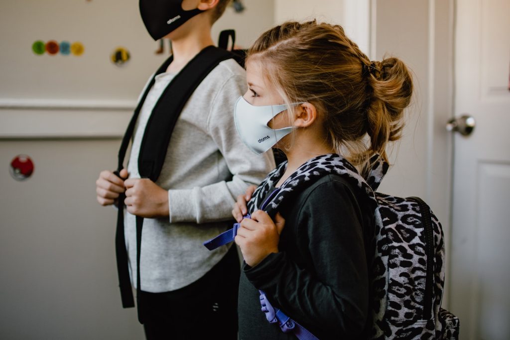
In a new study published in Pediatric Investigation, researchers demonstrate that face masks reduce the release of exhaled particles when used by school-aged children, helping to slow the spread of various respiratory viruses. While there was little difference between no protection and masking in exhaled particles from breathing, sneezing saw a significant reduction in the number of particles produced.
Respiratory viruses, including SARS-CoV-2, are transmitted via respiratory droplets and aerosols generated by all activities that involve exhalation, including tidal breathing, speaking, singing, coughing, and sneezing. Droplets, large particles subject to gravitational forces, are rapidly deposited from air and form fomites on surfaces. Aerosols, fine solid or liquid particles which remain suspended in the air, can travel long distances (> 6m) and reach high concentrations in poorly-ventilated areas. The relative contribution of the various modes of infection (direct contact, indirect contact via fomite, large droplet, or aerosol) for various respiratory viruses is difficult to determine, but survival of infectious viruses has been demonstrated in aerosols.
For the study, 23 healthy children were asked to perform activities that ranged in intensity (breathe quietly, speak, sing, cough, and sneeze) while wearing no mask, a cloth mask, or a surgical mask.
The production of exhaled particles that were 5μm or smaller, which is the dominant mode of transmission of many respiratory viruses, increased with coughing and sneezing. Face masks, especially surgical face masks, effectively reduced the release of these and other sized particles.
“Understanding the factors that affect respiratory particle emission can guide public health measures to prevent the spread of respiratory infections, which are a leading cause of death and hospitalisation among young children worldwide,” said corresponding author Peter P. Moschovis, MD, MPH, of Massachusetts General Hospital and Harvard Medical School.
A new study has identified potential broad-spectrum antiviral agents that can target multiple families of RNA viruses with pandemic potential. The study, published in Cell Reports Medicine, tested an array of innate immune agonists that work by targeting pathogen recognition receptors, and found several agents that showed promise, including one that exhibited potent antiviral activity against members of RNA viral families.
The authors say recent epidemics as well as global climate change and the continuously evolving nature of the RNA genome indicate that arboviruses, viruses spread by arthropods such as mosquitoes, are prime candidates for the next pandemic after COVID. These include Chikungunya virus (CHIKV), Dengue virus, West Nile virus and Zika virus. The researchers write: “Given their already-demonstrated epidemic potential, finding effective broad-spectrum treatments against these viruses is of the utmost importance as they become potential agents for pandemics.”
Led by Gustavo Garcia Jr. in the UCLA Department of Molecular and Medical Pharmacology, researchers found that several antivirals inhibited these arboviruses to varying degrees. “The most potent and broad-spectrum antiviral agents identified in the study were cyclic dinucleotide (CDN) STING agonists, which also hold promise in triggering an immune defence against cancer,” said senior author Vaithi Arumugaswami, Associate Professor in the UCLA Department of Molecular and Medical Pharmacology.
“A robust host antiviral response induced by a single dose treatment of STING agonist cAIMP is effective in preventing and mitigating the debilitating viral arthritis caused by Chikungunya virus in a mouse model. This is a very promising treatment modality as Chikungunya virus-affected individuals suffer from viral arthritis years and decades from the initial infection,” Arumugaswami added.
“At molecular level, CHIKV contributes to robust transcriptional (and chemical) imbalances in infected skin cells (fibroblasts) compared to West Nile Virus and ZIKA Virus, reflecting a possible difference in the viral-mediated injury (disease pathogenesis) mechanisms by viruses belonging to different families despite all being mosquito-borne viruses,” said senior author Arunachalam Ramaiah, Senior Scientist in the City of Milwaukee Health Department.
“The study of transcriptional changes in host cells reveals that cAIMP treatment rescues (reverses) cells from the harmful effect of CHIKV-induced dysregulation of cell repair, immune, and metabolic pathways,” Ramaiah added.
The study concludes that the STING agonists exhibited broad-spectrum antiviral activity against both arthropod-borne- and respiratory viruses, including treaded SARS-CoV-2 and Enterovirus D68 in cell culture models.
Garcia notes, “The next step is to develop these broad-spectrum antivirals in combination with other existing antivirals and be made readily available in the event of future respiratory and arboviral disease outbreaks.”
Methicillin-resistant Staphylococcus aureus (MRSA). Image by CDC on Unsplash
Researchers reported in Cell Host & Microbe that early tests of a bioengineered drug candidate were successful in countering Staphylococcus aureus, a bacteria particularly dangerous to hospitalised patients.
Experiments demonstrated that SM1B74, an antibacterial biologic agent, was superior to a standard antibiotic drug at treating mice infected with S. aureus, including its treatment-resistant form known as MRSA.
The researchers tested mAbtyrins, a combination molecule based on an engineered version of a human monoclonal antibody (mAb), a protein that clings to and marks S. aureus for uptake and destruction by immune cells. Attached to the mAb are centyrins, small proteins that prevent these bacteria from boring holes into the human immune cells in which they hide. As the invaders multiply, these cells die and burst, eliminating their threat to the bacteria.
Together, the experimental treatment targets ten disease-causing mechanisms employed by S. aureus, but without killing it, say the study authors. This approach promises to address antibiotic resistance, say the researchers, where antibiotics kill vulnerable strains first, only to make more space for others that happen to be less vulnerable until the drugs no longer work.
“To our knowledge, this is the first report showing that mAbtyrins can drastically reduce the populations of this pathogen in cell studies, and in live mice infected with drug-resistant strains so common in hospitals,” said lead study author Victor Torres, PhD, the C.V. Starr Professor of Microbiology and director of the NYU Langone Health Antimicrobial-Resistant Pathogen Program.”Our goal was to design a biologic that works against S. aureus inside and outside of cells, while also taking away the weapons it uses to evade the immune system.”
Inside Out
The new study is the culmination of a five-year research partnership between scientists at NYU Grossman School of Medicine and Janssen to address the unique nature of S. aureus.
The NYU Langone team together with Janssen researchers, published in 2019 a study that found that centyrins interfere with the action of potent toxins used by S. aureus to bore into immune cells. They used a molecular biology technique to make changes in a single parental centyrin, instantly creating a trillion slightly different versions of it via automation. Out of this “library,” careful screening revealed a small set of centyrins that cling more tightly to the toxins blocking their function.
Building on this work, the team fused the centyrins to a mAb originally taken from a patient recovering from S. aureus infection. Already primed by its encounter with the bacteria, the mAb could label the bacterial cells such that they are pulled into bacteria-destroying pockets inside of roving immune cells called phagocytes. That is unless the same toxins that enable S. aureus to drill into immune cells from the outside let it drill out of the pockets to invade from the inside.
In a “marvel of bioengineering,” part of the team’s mAbtyrin serves as the passport recognised by immune cells, which then engulf the entire, attached mAbtyrin, along with its centyrins, and fold it into the pockets along with bacteria. Once inside, the centyrins block the bacterial toxins there. This, say the authors, sets their effort apart from antibody combinations that target the toxins only outside of cells.
The team made several additional changes to their mAbtyrin that defeat S. aureus by, for instance, activating chain reactions that amplify the immune response, as well by preventing certain bacterial enzymes from cutting up antibodies and others from gumming up their action.
The researchers tracked the growth of S. aureus strains commonly occurring in US communities in the presence of primary human immune cells (phagocytes). Bacterial populations grew almost normally in the presence of the parental antibody, slightly less well in the presence of the team’s engineered mAb, and half as fast when the mAbtyrin was used.
In another test, 98% of mice treated with a control mAb (no centyrins) developed bacteria-filled sores on their kidneys when infected with a deadly strain of S. aureus, while only 38% of mice did so when treated with the mAbtyrin. Further, when these tissues were removed and colonies of bacteria in them counted, the mice treated with the mAbtyrin had one hundred times (two logs) fewer bacterial cells than those treated with a control mAb.
Finally, the combination of small doses of the antibiotic vancomycin with the mAbtyrin in mice significantly improved the efficacy of the mAbtyrin, resulting in maximum reduction of bacterial loads in the kidneys and greater than 70% protection from kidney lesions.
“It is incredibly important,” said Torres, “that we find new ways to boost the action of vancomycin, a last line of defence against MRSA.”
A global study looking at the benefits of cranberry products has determined that cranberry juice, and its supplements, reduce the risk of repeat symptomatic UTIs in women by more than a quarter. The study researchers, from Flinders University and The Children’s Hospital at Westmead, also found that was reduced in children by more than half, and in people susceptible to UTI following medical interventions by about 53%.
Cranberry juice and healthcare supplements that commonly include the fruit, such as capsules and tablets, have long been promoted as a readily available solution to ward off the infection but the most recent review in 2012, with evidence from 24 trials, showed no benefit from the products.
The medical scientists behind this updated review published in Cochrane Reviews aimed to update these findings, as by looking at 50 more recent trials that included almost 9000 participants.
“This incredible result didn’t really surprise us, as we’re taught that when there’s more and better evidence, the truth will ultimately come out. UTIs are horrible and very common; about a third of women will experience one, as will many elderly people and also people with bladder issues from spinal cord injury or other conditions,” says the study lead author Dr Gabrielle Williams.
“Even back in 1973, my mum was told to try cranberry juice to prevent her horrible and frequent UTIs, and for her it’s been a saviour. Despite me niggling in her ear about evidence, she’s continued to take it daily, first as the nasty sour juice and in recent years, the easy to swallow capsules. As soon as she stops, wham the symptoms are back. As usual, it turns out that mum was right! Cranberry products can help some women prevent UTIs.”
Flinders University epidemiologist Dr Jacqueline Stephens, a co-author of the study, says if the UTI persists untreated it can move to the kidneys and cause pain and more complications, including sepsis in very severe cases, so prevention is the most effective way to reduce risks.
“Most UTIs are effectively, and pretty quickly, treated with antibiotics, sometimes as little as one dose can cure the problem. Unfortunately, in some people UTIs keep coming back. Without being sure if or how it works, some healthcare providers began suggesting it to their patients. It was a harmless, easy option at the time. Even centuries ago, Native Americans reportedly ate cranberries for bladder problems, leading somewhat more recently, to laboratory scientists exploring what it was in cranberries that helped and how it might work.”
“The studies we looked at included a range of methods to determine the benefits of cranberry products. The vast majority compared cranberry products with a placebo or no treatment for UTI and determined drinking cranberries as a juice or taking capsules reduced the number of UTIs in women with recurrent cases, in children and in people susceptible to UTi’s following medical interventions such as bladder radiotherapy.”
“It’s also important to consider that few people reported any side effects with the most common being tummy pain based on the results. We also did not find enough information to determine if cranberry products are more or less effective compared with antibiotics or probiotics in preventing further UTIs.”
The data also doesn’t show any benefit for elderly people, pregnant women or in people with bladder emptying problems.
The study’s senior author, Professor Jonathan Craig, says the real benefits of cranberry products became clear when the researchers expanded the scope of the review to include the most recently available clinical data.
“This is a review of the totality of the evidence and as new evidence emerges, new findings might occur. In this case, the new evidence shows a very positive finding that cranberry juice can prevent UTI in susceptible people,” says Professor Craig.
“We have shown the efficacy of cranberry products for the treatment of UTIs using all the evidence published on this topic since the mid-nineties. The earlier versions of this review didn’t have enough evidence to determine efficacy and subsequent clinical trials showed varied results, but in this updated review the volume of data has shown this new finding.”
The study authors conclude that while cranberry products do help prevent UTIs in women with frequent recurrence, more studies are needed to further clarify who with UTI would benefit most from cranberry products.
Scientists predict that we are entering the era of pandemics.
A sustainable global commitment to pandemic preparedness is instrumental to maintaining the upper hand and winning the battle.
In collaboration with the Abbott Pandemic Defense Coalition, CERI unveiled a new genomics facility to help identify, analyse and test infectious diseases in Africa to enable early detection and rapid responses to potential viral threats.
With the increasing rates of urbanisation, global travel and climate change, infectious disease experts predict the world is entering a new era of pandemics. In response to this, Abbott founded the Abbott Pandemic Defense Coalition which comprises of 20 scientific and public health organisations from across the globe who are committed to detecting and responding to emerging viral threats.
Since its launch in 2021, the global Coalition has partnered with organisations in Africa to build up the network and capabilities in the region. The Centre for Epidemic Response and Innovation (CERI) at Stellenbosch University is one of the recent partners, who recently unveiled a new genomics facility that will enable early detection and the rapid response to emerging threats in Africa in collaboration with the Abbott Pandemic Defense Coalition.
“No one organisation, network or country is strong enough to effectively fight against viral pathogens,” says Mary Rodgers, principal scientist at Abbott’s diagnostics business. “We therefore must have an ongoing global commitment to pandemic preparedness – and key to that is collaboration across the private and public sectors to detect and have a rapid response to emerging threats. Our partnership with CERI will expand testing capacity to continue research in understanding how known viruses are spreading in order to identify new viral outbreaks, so that we can stop them from becoming the next pandemic.”
New Technology to Detect Viral Threats in Africa
The state-of-the-art genomics facility is equipped with new technology such as the Metagenomics Next Generation Sequencing which is revolutionising how viruses are discovered – creating a genome that once took years to complete can now be sequenced and analysed in a day or two. The genomics center will also have access to Abbott’s diagnostics molecular lab testing capabilities to provide fast and scalable molecular testing, as well as provide researchers the ability to create molecular tests for new and emerging viral threats.
Professor Tulio de Oliveira, director of CERI concludes, “The partnership with the Abbott Pandemic Defense Coalition will help train Africa’s next generation of virus hunters and public health experts in cutting-edge sequencing, bioinformatics, and other technologies so that they can track and identify viruses faster and smarter. The launch of this new genomics facility is a testament to our shared commitment to advancing scientific knowledge and protecting public health in Africa.”
Colourised transmission electron micrograph of hepatitis B virus particles (colourised red and yellow). Credit: NIAID and CDC (Transmission electron micrograph image courtesy of CDC; colourisation by NIAID).
In 2020, bulevirtide (BLV) was conditionally approved for treating chronic hepatitis delta (CHD), an inflammation of the liver caused by hepatitis D virus (HDV). Now, as reported in the Journal of Hepatology, real-world studies confirm that long-term suppressive therapy with BLV monotherapy reduces viral replication and improves liver tests of these difficult-to-treat patients.
Two of the studies, led by Pietro Lampertico, MD, PhD, were designed to assess the effectiveness and safety of patients with advanced HDV-related compensated cirrhosis being treated with BLV 2mg monotherapy and the consequences of discontinuing this treatment.
“HDV is the most severe form of chronic viral hepatitis,” explained Dr Lampertico. “For many years, the only therapeutic option was the off-label administration of pegylated-interferon-alpha (PegIFNa), an approach characterised by suboptimal efficacy, an unfavourable safety profile and several contraindications.”
In a study of 18 patients with HDV-related advanced cirrhosis treated with BLV 2mg/day for 48 weeks, Dr. Lampertico and colleagues demonstrated significant virological, biochemical and combined response rates associated with improvement of liver function.
“The efficacy and safety of BLV monotherapy in patients with advanced compensated cirrhosis were unknown before this study. Virological and biochemical responses to BLV monotherapy that we observed in our difficult-to-treat patients with HDV-related compensated cirrhosis were similar to those shown in the phase III registration study,” Dr Lampertico noted.
In a case report, Dr Lampertico and colleagues demonstrated that HDV could be successfully eradicated from both serum and liver following a three-year course of BLV monotherapy. This was despite the persistence of HBsAg, in a patient with HDV-related compensated cirrhosis and oesophageal varices. During the 72-week off-BLV follow-up, liver biopsy, intrahepatic HDV RNA and hepatitis D antigen were undetectable, less than 1% of hepatocytes were HBsAg positive and all were negative for hepatitis B core antigen.
“We were surprised to demonstrate that HDV can be eradicated following a finite course of an entry inhibitor administered as monotherapy such as BLV 2mg/day, despite the persistence of HBsAg positivity,” commented Dr Lampertico.
In a study in JHEP Reports led by Katja Deterding, MD, investigators report the first data from the largest multicentre cohort of patients to date who were treated with BLV under real-world conditions. This included 50 patients with signs of significant portal hypertension, elevated pressure in the major vein that leads to the liver.
The retrospective analysis of 114 cases covered 4289 patient weeks of BLV treatment. Viral response was observed in 87 cases while hepatic inflammation improved, and treatment was well tolerated. More than 50% of patients showed a virologic response with less than 10% of patients not achieving an HDV RNA drop of at least 90% after 24 weeks. An improvement of biochemical hepatitis activity as measured by the liver enzyme alanine transaminase (ALT) values was observed regardless of virologic response. Investigators concluded that treatment was safe and well tolerated and associated with improvements in liver cirrhosis and portal hypertension with prolonged treatment.
“In line with other real-world cohorts and clinical trials our real-world study confirms the antiviral activity of BLV,” noted Dr Deterding. “We were surprised to see an improvement in biochemical hepatitis activity even in cases without viral response. Potential explanations for this phenomenon include anti-inflammatory properties of BLV.”
“This is the first time that patients with HDV-related chronic advanced liver disease can be treated with an antiviral therapy since 1977 when HDV was discovered. Long-term suppressive therapy with BLV 2mg/day has the potential to improve survival, of these difficult-to-treat patients for the first time in 45 years,” concluded Dr Lampertico. “We also found that BLV treatment can be successfully discontinued in some HDV patients who achieved long-term viral suppression while on therapy.”
HDV infection occurs when people become infected with both hepatitis B and D virus either simultaneously (co-infection) or acquire the hepatitis D virus after first being infected with hepatitis B (super-infection). According to the World Health Organization, HDV affects nearly 5% of individuals with a chronic infection resulting from hepatitis B virus (HBV). Populations that are more likely to have HBV and HDV co-infection include indigenous populations, haemodialysis recipients and individuals who inject drugs.
Pompe disease (PD) is an autosomal-recessively inherited neuromuscular disease that can be fatal if it is not diagnosed and treated early.1 Due to lack of acid alpha-glucosidase (GAA), there is progressive intracellular accumulation of glycogen, which can severely damage the muscles and heart.1
PD can present from early infancy into adulthood, with variable rates of disease progression.1 Severity is determined by age of onset, organ involvement, including the degree of muscle involvement (skeletal, respiratory, and cardiac), and rate of progression.1
Classification1
PD is classified into two groups: infantile and late-onset.
Infantile form:
• Classic infantile PD is most severe and rapidly progressive, and is characterised by prominent cardiomegaly, hepatomegaly, muscular weakness and hypotonia. Death results from cardiorespiratory failure in <1 year, if not treated.
• Infantile variant form (non-classic, in the <1-year group that has slower progression and less severe or absent cardiomyopathy).
Late-onset form:
• Childhood/juvenile or muscular variant (heterogeneous group) presenting later than infancy and typically excluding cardiomyopathy.
• Adult-onset form characterised by slowly progressive myopathy predominantly involving skeletal muscle and presenting as late as the 2nd – 6th decade of life.
Signs and symptoms
In infants, symptoms begin in the first months of life, with feeding problems, poor weight gain, breathing difficulties, profound hypotonia, and cardiomegaly.2 Many infants with PD also present with macroglossia.2
Kelly du Plessis, CEO and Founder of non-profit organisation, Rare Diseases SA (RDSA), says that the difficulty for both parents and healthcare professionals is that PD shows itself in many ways. “There is not one specific thing that you can pinpoint. My child, who is a PD sufferer, took longer to reach his milestones, and got slower as time progressed. It is better to be overcautious than under-cautious because early identification is critical to a positive outcome, and the damage done up until diagnosis cannot be undone.”
Du Plessis says that RDSA is also seeing many more adults being diagnosed with PD lately, and describes a few of the signs and symptoms: “In adults these include difficulty walking, particularly up stairs or inclines, recurring chest infections, being very fatigued, finding that their arms are getting weaker when they try to reach something on a top shelf, and falling over quite often owing to lower muscle tone and foot drop. Healthcare professionals need to be aware of this link with PD – because early intervention is critical to outcomes.”
Diagnosis
While making an early diagnosis is imperative to optimise disease management and outcomes,1 many patients experience a diagnostic odyssey.3
Monique Nel, Medical Advisor – Rare Diseases at Sanofi, says: “The diagnostic odyssey for PD can be quite long and complicated, as the symptoms can be similar to those of other conditions, and the disease is quite rare. The journey to diagnosis can take years, and many patients go through a battery of tests and specialists before finally receiving a correct diagnosis.”
In the United States it was reported that before implementation of newborn screening, there was, on average, a 3-month delay in diagnosing infantile-onset PD after the onset of symptoms.3 In late-onset PD, symptoms may begin any time from infancy to adulthood.3 In paediatric onset cases, on average: symptom onset occurs at approximately 6 years of age, yet diagnosis is generally made around 18 years of age, with a potential 12-year delay in diagnosis.3 The average age of symptom onset in adult-onset PD is 35 years, with a 7-year delay in diagnosis after symptom onset.3
Adds Nel: “In South Africa, we do enzyme activity testing via a dried blood spot test to measure the activity of the alpha-glucosidase enzyme. If the enzyme activity is low, it suggests that the individual may have PD. Genetic testing is currently performed abroad. This involves analysing a person’s DNA to look for mutations in the GAA gene. If two mutated copies of the GAA gene are found, it confirms a diagnosis of PD.”
Treatment
Enzyme replacement therapy (ERT) is available for all forms of PD, and has dramatically changed patient outcomes.3 This life-changing therapy is more effective when started before the onset of symptoms.3
Since the end of 2012, ERT (as alglucosidase alfa) has been registered with the South African Health Products Regulatory Authority (SAHPRA) for use in PD patients.1 Patients with infantile-onset PD who receive ERT have significantly prolonged survival, decreased cardiomegaly, and improved cardiac and skeletal muscle function.1 Cardiac response appears to be good, irrespective of the stage of disease at initiation of ERT, while the skeletal muscle response appears more variable.1 The best skeletal muscle response occurs when ERT is administered prior to skeletal muscle damage.1
Says Nel: “Early screening for PD and prompt treatment is crucial to prevent or delay the onset of disease complications. Therefore, healthcare providers must consider PD as a potential differential diagnosis when evaluating patients with muscle weakness, respiratory difficulties, and other related symptoms.”
Says du Plessis: “With medication, you see a difference in the patients within weeks, and they have a lot more energy. RDSA advocates as much as is necessary to get patients approved for medication, since this treatment changes their lives and quality of life – and in fact saves their lives. We need to do everything we can now, with the treatments we have today, to keep these patients as healthy as possible, so that they can benefit from the treatments that come tomorrow.”