Beyond the Smile: South Africa Must Prioritise Oral Health as a Public Health Imperative
South Africa’s burden of oral diseases is not only inextricably linked to non-communicable diseases but also presents an urgent public health challenge, with rising concern over its impact on mental health.
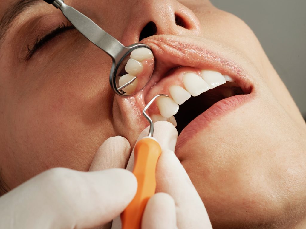
Oral diseases are a major health concern for many countries and negatively impacts people throughout their lives. Oral diseases lead to pain and discomfort, social isolation and loss of self-confidence, and they are often linked to other serious health issues. And yet, there is no reason to suffer: most oral health conditions are preventable and can be treated in their early stages.
Globally, every year on March 20, World Oral Health Day is commemorated with the aim to empower people with the knowledge, tools, and confidence to secure good oral health.
This year, the Day’s focus shifts to the mind-mouth connection, with the tagline from the FDI World Dental Federation: “A Happy Mouth Is… A Happy Mind”. This campaign aims to raise awareness of how poor oral health can negatively impact quality of life, highlighting the importance of a healthy mouth for mental well-being.
Macelle Erasmus, Head of Expert at Haleon South Africa – a leader in consumer health and self-care, says, “Oral health is not just about bright smiles and good-looking teeth – it is a critical component of overall well-being. In South Africa, the high prevalence of oral diseases, particularly among children and vulnerable communities, reinforces the urgent need for improved oral health education and preventive care.”
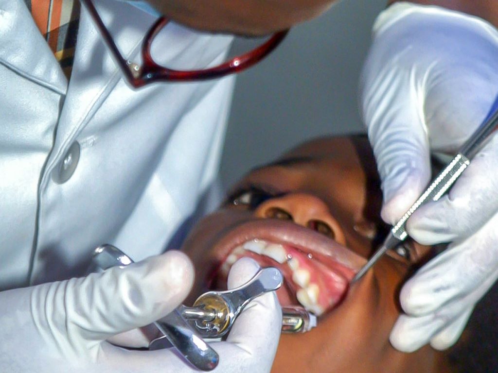
Haleon’s leading oral health brands Aquafresh and Sensodyne, are committed to improving oral health education and access across the country.
Over the course of just three months, we have conducted more than 39,000 gum health screenings across 16 clinics. In 2025, our expansion aims to reach 100,000 underserved communities as part of Haleon’s oral health care outreach programs.
According to the South African Dental Association (SADA), 41% of children aged 1-9 years and close to 28% of people aged 5 years and over experienced untreated tooth decay in milk and permanent teeth respectively, while nearly 25% of people aged 15 years and over experienced severe periodontal disease in 2019. The country also saw 1,933 new cases of lip and oral cavity cancer in 2020.
The World Health Organisation’s Global Strategy and Action Plan on Oral Health 2023–2030, explains that oral health encompasses a range of diseases and conditions. The most prevalent public health issues include dental caries, severe periodontal (gum) disease, complete tooth loss (edentulism), oral cancer, oro-dental trauma, noma and congenital malformations such as cleft lip and palate, most of which are preventable.
The main oral diseases and conditions are estimated to affect close to 3.5 billion people worldwide. These conditions combined have an estimated global prevalence of 45%, which is higher than the prevalence of any other NCD.
However, oral diseases and conditions share risk factors common to the leading NCDs, including all forms of tobacco use, harmful alcohol use, high intake of free sugars and lack of exclusive breastfeeding.
The Department of Health’s National Oral Health Policy and Strategy 2024-2034 acknowledges that oral health is poorly integrated in other health programmes, “though it is an integral part of general health.” It further recognises that: “Its role in management and care of communicable diseases, genetic disorders, trauma, injury, and violence is often overlooked.”
This integration is particularly important as more than three million patients are treated in the country’s public primary healthcare facilities annually, at a cost of R650 million. Addressing oral health holistically – within the broader healthcare system – can significantly reduce this burden.