Newborns with heart complications can rely on their newly developed immune systems to regenerate cardiac tissues, but adults aren’t so lucky. After a heart attack, most adults struggle to regenerate healthy heart tissue, leading to scar-tissue buildup and, often, heart failure.
A new Northwestern Medicine study in experimental animals reveals a critical difference in how macrophages help repair the heart in newborns versus adults after a heart attack. The study highlights a fundamental difference in how the immune system drives healing based on age.
“Understanding why newborns can regenerate their hearts while adults cannot will open the door to developing treatments that could ‘reprogram’ adult macrophages,” said first and co-corresponding author Connor Lantz, lead scientist of the bioinformatics core at the Comprehensive Transplant Center at Northwestern University Feinberg School of Medicine.
In newborns, macrophages perform a process called efferocytosis, which recognizes and eats dying cells. This process triggers the production of a bioactive lipid called thromboxane, signaling nearby heart muscle cells to divide, and allowing the heart to regenerate damaged heart muscle, the study found. In adults, macrophages produce much less thromboxane, leading to a weaker repair signal.
“By mimicking the effects of thromboxane, we might one day improve tissue repair after a heart attack in adults,” Lantz said.
How the study worked
The study examined how the immune system responds to heart injury in mice of different ages, including newborn mice (one day old) and adult mice (eight weeks old). The researchers found the ability of macrophages to engulf dying cells was enhanced in newborn mice due to increased expression of MerTK, a receptor that recognizes dying cells. Therefore, when the scientists blocked this key receptor, newborn mice lost their ability to regenerate their hearts, resembling adult hearts after a heart attack.
Engulfment of dying cells by newborn macrophages triggered a chemical chain reaction that produced a molecule called thromboxane A2, which unexpectedly stimulated heart muscle cells to multiply and repair the damage, the study found. Additionally, nearby muscle heart cells in newborns are primed to respond to thromboxane A2, leading them to change their metabolism to support their growth and healing. But in adults, this process did not work the same way – after an injury, their macrophages did not produce enough thromboxane A2, limiting their ability to regenerate heart tissue.
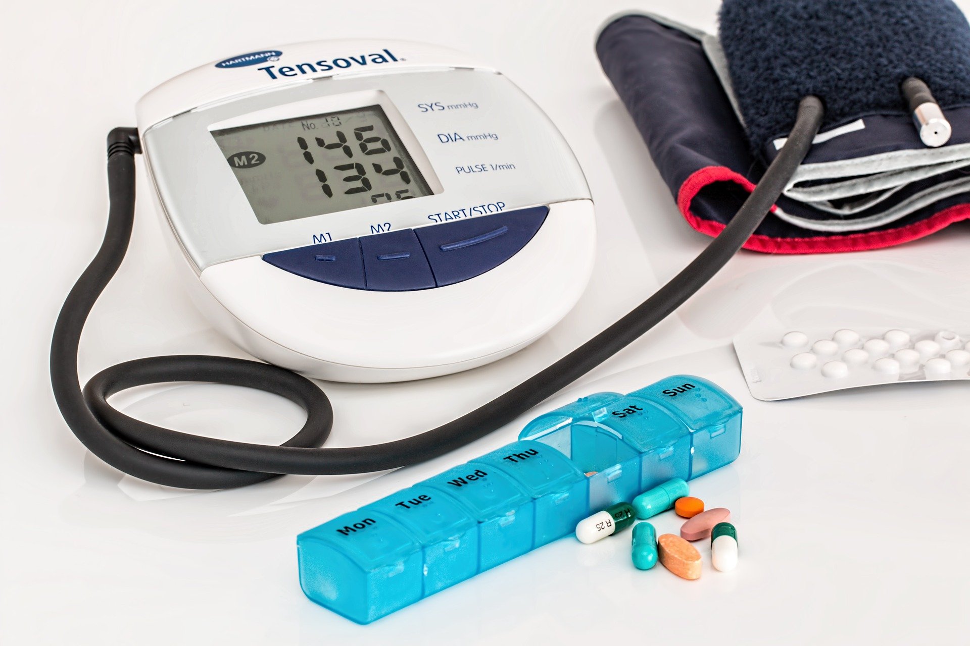
Doctors at Queen Mary University of London, Barts Health NHS Trust and University College London have developed a groundbreaking, minimally invasive treatment, Triple T, offering hope for millions of people with high blood pressure caused by a commonly overlooked condition.
The treatment, which could transform blood pressure management, has been published in The Lancet.
Triple T, also known as endoscopic ultrasound-guided radiofrequency ablation, is poised to change the way we address primary aldosteronism (PA) – a hormonal disorder that causes high blood pressure in one in 20 patients with blood pressure yet is often undiagnosed and untreated. This treatment has shown promising results in clinical trials and could become an accessible alternative to surgery, offering relief to those who suffer from this condition.
The hidden cause of high blood pressure
High blood pressure, affecting one in three adults, has several underlying causes, with PA being one of the most common yet underdiagnosed. In this condition, benign nodules in the adrenal glands produce excess aldosterone, a hormone that raises blood pressure by increasing salt levels in the body. Patients with PA often do not respond to standard medications and face increased risks of heart attacks, strokes and kidney failure.
Until now, the only effective treatment for PA has been the surgical removal of the affected adrenal gland. However, this procedure requires general anaesthesia, a hospital stay, and weeks of recovery, causing many patients to go untreated. Triple T provides a faster, safer, and less invasive alternative by targeting and destroying the malfunctioning adrenal nodule without removing the gland.
How Triple T works
The procedure uses a combination of radiofrequency or microwaves and ultrasound to deliver targeted heat to the adrenal nodule. A fine needle is inserted through the stomach to the adrenal gland, guided by real-time ultrasound imaging, where short bursts of heat are used to destroy the problematic tissue. This targeted approach ensures minimal damage to surrounding healthy tissues. The entire procedure lasts only 20 minutes and requires no incision.
Triple T’s success stems from recent advances in diagnostic scans, which use molecular dyes to accurately locate even the smallest adrenal nodules. These breakthroughs, combines with the ability to directly target nodules adjacent to the stomach, have enabled this minimally invasive approach.
Successful trial and promising results
The Feasibility study of radiofrequency endoscopic ABlation, with ULtrasound guidance (FABULAS) trial which tested Triple T on 28 patients with PA, showed excellent results. The procedure was found to be safe and effective, with most patients experiencing normalised hormones levels within six months. Many participants were able to stop all blood pressure medications, and the condition did not recur.
Professor Morris Brown, co-senior author of the study and Professor of Endocrine Hypertension at Queen Mary University of London, reflected on the significance of this milestone: “It is 70 years since the discovery in London of the hormone aldosterone, and, a year later, of the first patient in USA with severe hypertension due to an aldosterone-producing tumour. This patient’s doctor, Jerome Conn, predicted, with perhaps only minor exaggeration, that 10-20% of all hypertensions might one day be traced to curable nodules in one or both glands. We are now able to realise this prospect, offering 21st-century breakthroughs in diagnosis and treatment.”
One trial participant, Michelina Alfieri, shared her experience: “Before the study, I suffered from debilitating headaches for years despite multiple GP visits. As a full-time worker and single parent, my daily life was severely affected. This non-invasive treatment provided an immediate recovery—I was back to my normal routine straight away. I’m incredibly grateful to the team for giving me this choice.”
What’s next?
The success of the FABULAs trial has led to a larger study, WAVE, which will compare Triple T with traditional surgery in 120 patients. Results are expected in 2027.
Professor Stephen Pereira, Chief Investigator of FABULAS, emphasised the potential global impact of Triple T. he said: “This less invasive technique could be widely offered in endoscopy units across the UK and internationally.”
Clinical Endocrinology Lead at Addenbrooke’s Hospital and Professor of Clinical Endocrinology at the University of Cambridge, Professor Mark Gurnell, said: “Thanks to this work, we may finally be able to diagnose and treat more people with primary aldosteronism, lowering their risk of developing cardiovascular diseases and other complications, and reducing the number of people dependent on long-term blood pressure medication.”
The research was primarily supported by Barts Charity, National Institute for Health and Care Research (NIHR) through the Barts and Cambridge Biomedical Research Centres (BRCs), and the British Heart Foundation.
It is being followed by a larger randomised trial, called ‘WAVE’, which will compare TTT to traditional surgery in 120 patients. The results are expected in 2027.
National Institute of Health (NIH) scientists have made a significant breakthrough in understanding how “bad” cholesterol, known as low-density lipoprotein-cholesterol or LDL-C, builds up in the body. The researchers were able to show for the first time how the main structural protein of LDL binds to its receptor – a process that starts the clearing of LDL from the blood – and what happens when that process gets impaired.
The findings, published in Nature, further the understanding of how LDL contributes to heart disease, the world’s leading cause of death, and could open the door to personalising LDL-lowering treatments like statins to make them even more effective.
“LDL is one of the main drivers of cardiovascular disease which kills one person every 33 seconds, so if you want to understand your enemy, you want to know what it looks like,” said Alan Remaley, MD, PhD, co-senior author on the study who runs the Lipoprotein Metabolism Laboratory at NIH’s National Heart, Lung, and Blood Institute.
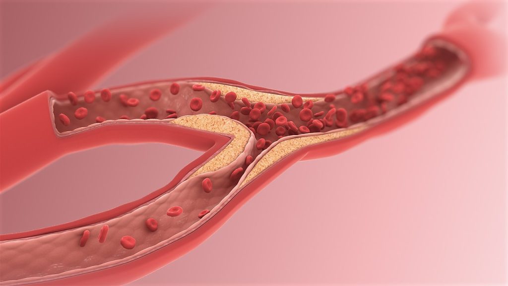
Until now scientists have been unable to visualise the structure of LDL, specifically what happens when it links up with its receptor, a protein known as LDLR. Typically, when LDL binds to LDLR, the process of clearing LDL from the blood begins. But genetic mutations can prevent that work, causing LDL to build up in the blood and get deposited into the arteries as plaque, which can lead to atherosclerosis, a precursor for heart disease.
In the new study, the researchers were able to use high-end technology to get a view of what’s happening at a critical stage of that process and see LDL in a new light.
“LDL is enormous and varies in size, making it very complex,” explained Joseph Marcotrigiano, PhD, chief of the Structural Virology Section in the Laboratory of Infectious Diseases at NIH’s National Institute of Allergy and Infectious Diseases and co-senior author on the study. “No one’s ever gotten to the resolution we have. We could see so much detail and start to tease apart how it works in the body.”
Using cryo-electron microscopy, the researchers were able to see the entirety of the structural protein of LDL when it bound to LDLR. Then, with AI-driven protein prediction software, they were able to model the structure and locate the known genetic mutations that result in increased LDL.
The researchers found that many of the mutations that mapped to the location where LDL and LDLR connected, were associated with familial hypercholesterolaemia (FH). FH is marked by defects in how the body uptakes LDL into its cells, and people with it have extremely high levels of LDL and can have heart attacks at a very young age. They found that FH-associated variants tended to cluster in particular regions on LDL.
The study findings could open new avenues to develop targeted therapies aimed at correcting these kinds of dysfunctional interactions caused by mutations. But, as importantly, the researchers said, they could also help people who do not have genetic mutations, but who have high cholesterol and are on statins, which lower LDL by increasing LDLR in cells. By knowing precisely where and how LDLR binds to LDL, the researchers say they may now be able to target those connection points to design new drugs for lowering LDL from the blood.
These results give hope to stroke patients worldwide who may not be able to access thrombolytic medications within the approved time window, which in China is within 4.5 hours, said the trial’s principal investigator Min Lou, MD, PhD, a professor at the Second Affiliated Hospital of Zhejiang University’s School of Medicine in China.
In the US, alteplase is approved to treat stroke within three hours of symptom onset and is recommended for use up to 4.5 hours for select patients. Other research has indicated it may also work well in some patients 4.5 to 9 hours after stroke onset.
Researchers enrolled 372 stroke patients whose symptoms began 4.5 hours to 24 hours earlier. They used widely available CT perfusion imaging (advanced brain scanning) to confirm that these patients still had brain tissue that could recover with treatment. Participants were randomised to receive alteplase, while the other received standard stroke care of antiplatelet therapy at the discretion of the investigator, based on the Chinese Guidelines for Diagnosis and Treatment of Acute Ischemic Stroke 2018. Functional recovery was assessed at 90 days.
“We believe these findings mean more people may return to normal or near-normal lives after a stroke, even if they receive treatment later than originally thought beneficial,” Lou said. “This method of treatment could become the new standard, especially in hospitals that use CT perfusion imaging. This technology helps health care professionals see how blood flows in different parts of the brain after an ischemic stroke. This could extend treatment eligibility to millions more patients across the globe.”
The study found:
40% of participants treated with alteplase had little to no disability after 90 days, compared to 26% of those who received standard care – a 54% higher chance of functional recovery.
Less than 3% of participants in either group received rescue mechanical clot removal as an additional treatment.
Rates of death were the same (10.8%) for both groups.
The risk of brain bleeding was higher among those who received alteplase than among participants who did not (3.8% vs. 0.5%), but researchers believe this is a manageable risk.
“We also need to look more closely at how safe and effective other clot-dissolving medications, like tenecteplase, are when given after a stroke, especially beyond the usual time frames. It’s also important to learn if our findings apply to other groups of people, especially in areas with different stroke risks and health care resources,” Lou explained.
Study limitations include the that both participants and researcher knew which treatment was being given, which could have introduced bias, and results may not be generalizable to patients outside of China.
Study design, background and details:
The study enrolled 372 stroke patients in a multicenter, prospective, randomized trial at 26 stroke centers in China.
The patient’s average age was 72 years, and 43% were women.
The trial used widely available CT perfusion imaging software to gauge salvageable brain tissue, making the findings more applicable to real-world clinical settings.
Enrolled patients were assigned to the alteplase group or a standard medical treatment group.
The primary outcome was a score of 0 or 1 on the modified Rankin scale, which scores disability from 0 (no symptoms) to 6 (death) at 90 days.
Study co-authors, funding and disclosures are available in the abstract.
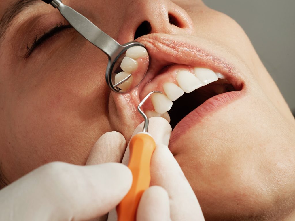
Flossing your teeth at least once a week may be linked to a lower risk of stroke caused by blood clotting and atrial fibrillation, according to a preliminary study to be presented at the American Stroke Association’s International Stroke Conference 2025. The meeting is in Los Angeles, Feb. 5-7, 2025, and is a world premier meeting for researchers and clinicians dedicated to the science of stroke and brain health.
“A recent global health report revealed that oral diseases – such as untreated tooth decay and gum disease – affected 3.5 billion people in 2022, making them the most widespread health conditions,” said study lead author Souvik Sen, MD, MS, MPH, chair of the Department of Neurology, Prisma Health Richland Hospital and the University of South Carolina School of Medicine in Columbia, South Carolina. “We aimed to determine which oral hygiene behaviour – dental flossing, brushing or regular dentist visits – has the greatest impact on stroke prevention.”
The Atherosclerosis Risk in Communities (ARIC) study, one of the first large-scale investigations of this kind in the US, assessed the home use of dental floss through a structured questionnaire of more than 6000 people. Among those who reported flossing, 4092 had not experienced a stroke, and 4050 had not been diagnosed with atrial fibrillation (AFib).
Participants were asked about their status regarding high blood pressure, diabetes, high cholesterol, smoking, body mass index, education, regular brushing and dentist visits. During the 25 years of follow-up, 434 participants were identified as having strokes, of which 147 were larger artery brain clots, 97 were heart-driven clots and 95 were hardening of the smaller arteries. Additionally, 1291 participants were noted to have experienced AFib.
The analysis found:
Flossing was associated with a 22% lower risk of ischaemic stroke, 44% lower risk of cardioembolic stroke (blood clots traveling from the heart) and 12% lower risk of AFib.
The associated lower risk was independent of regular brushing and routine dental visits or other oral hygiene behaviours.
Increasing the frequency of flossing had a greater chance of stroke risk reduction.
Flossing was also associated with a lower chance of cavities and periodontal disease.
Researchers were surprised by the reduction of irregular heartbeats, or AFib. AFib is the most common form of irregular heartbeat. It can lead to stroke, heart failure or other cardiovascular complications.
“Oral health behaviours are linked to inflammation and artery hardening. Flossing may reduce stroke risk by lowering oral infections and inflammation and encouraging other healthy habits,” Sen said. “Many people have expressed that dental care is costly. Flossing is a healthy habit that is easy to adopt, affordable and accessible everywhere.”
Study limitations include that data were based on answers to a questionnaire, and the 25-year follow-up appears to have focused on stroke and heart outcomes only. There was no follow-up concerning flossing or other oral behaviours over the years, Sen said.
Propranolol, a drug often used to treat hypertension and prevent migraines was associated with a reduction in ischaemic stroke risk among women – but not men – using the drug for migraine prevention, according to a preliminary study to be presented at the American Stroke Association’s International Stroke Conference 2025. The meeting is in Los Angeles, Feb. 5-7, 2025, and is a world premier meeting for researchers and clinicians dedicated to the science of stroke and brain health.
The beta blocker propranolol had a stronger protective effect for ischaemic stroke risk in women with migraine, particularly those without aura. The medication did not have the same protective effect on men.
Migraine headaches are common in the general population, but they occur three times more often in women than in men. This debilitating condition is associated with an increased risk of stroke. While the beta blocker propranolol can be used to prevent migraines, its effectiveness in reducing overall stroke risk is still uncertain.
“Migraine is an often-ignored risk factor for cardiovascular issues. Until recently, preventive treatments for people who have migraines were not available,” said lead study author Mulubrhan Mogos, PhD, MSc, FAHA, an assistant professor at Vanderbilt University School of Nursing. “Many women suffer from migraines, and it’s important to note that propranolol may be beneficial for these women, particularly those who experience migraine without aura. This is an important discovery for those dealing with migraines.”
Mogos also noted that migraine disproportionately affects women from historically under-resourced communities, and this disparity may impact the ability to achieve education goals or maintain stable employment, creating a vicious cycle. While new treatments have proven effective, they may not be accessible to women in these groups due to high costs.
For the study, researchers reviewed more than 3 million electronic health records from two large databases. In separate analyses, researchers identified people with migraine who developed stroke and a control group of those with migraine who did not develop stroke. They then assessed whether the individuals were treated with propranolol for migraine and whether that treatment had impacted stroke risk.
“We initially looked at overall stroke and then ischemic stroke specifically. We refined our analysis further by controlling for possible confounders and found the association is significant and stronger for ischaemic stroke,” Mogos said.
After adjusting for potential variables, such as demographics (age, sex, race), other conditions (high blood pressure, diabetes, etc) and hormonal factors (use of birth control, pregnancy – considered separately for each woman) that might affect results the analysis found:
Propranolol was significantly associated with a reduced risk of ischaemic stroke in women with migraine, particularly in those without aura. The risk of developing a stroke was 52% lower for women taking the medication in one database analysis and 39% lower in the other. No stroke risk reduction was seen in men in either analysis group.
The protective effect of propranolol was stronger for ischaemic stroke and in women with migraine without aura. Migraine aura can include disturbances, such as flashing lights, blind spots, zigzag patterns or seeing coloured spots. Other symptoms include tingling or numbness in the face or hands, difficulty speaking, dizziness or confusion.
Secondary analyses showed lower overall stroke rates in women taking propranolol at multiple time points in both databases.
“Our findings indicate that women and health care professionals should discuss the advantages of preventive migraine interventions. For under-resourced individuals who bear a greater burden from this condition and may lack access to new treatments, we must ensure these treatments are available to them. This approach can help reduce health disparities,” Mogos said.
The main limitation is that this was a review of past data using electronic health records, which may introduce biases, such as misclassification errors from reliance on ICD codes (codes used to classify and report health conditions and diseases). These findings highlight the need for studies that look forward in time to confirm these results.
Right side heart failure. Credit: Scientific Animations CC4.0
Pathways to new treatments for heart failure take time – as long as four decades for two now accepted therapies. So, new attempts to repair scar tissue in infarcted hearts using cells or cell products need more time to develop clinical therapies that can reduce risk of death from heart failure after a heart attack.
This message is part of a critical review of cell-based and cell product-based therapies for the treatment of heart failure. The review details 20 years of completed and ongoing clinical trials. While none has gained medical approval, they have proven safe and some have shown beneficial effects.
More importantly, the reviewers note, it took longer, nearly 40 years, to optimise two current therapies to reduce mortality in heart failure: implantable cardioverter–defibrillators and guideline-directed medical therapy.
“The history of the development of life-saving medical therapies for heart failure serves as an important lesson that we should remain hopeful of the promise of cell therapy in heart failure,” Jianyi “Jay” Zhang, MD, PhD, and colleagues write in the review, “Trials and tribulations of cell therapy for heart failure: an update on ongoing trials,” published in Nature Reviews Cardiology. Zhang is professor and chair of the University of Alabama at BirminghamDepartment of Biomedical Engineering.
Heart failure is responsible for 13% of deaths worldwide. Half of patients with heart failure die within five years. The most common cause of heart failure is blockage of coronary arteries leading to death of the cardiomyocyte heart muscle cells. When that muscle tissue is replaced by dense scar tissue with little blood circulation, the infarcted heart loses contractile power, leading to heart enlargement, progressive loss of pumping ability, increased chance of ventricular arrhythmias and clinical end-stage heart failure.
The problem is that shortly after birth, human heart muscle cells lose their ability to divide, so a damaged infarcted heart cannot repair itself by growing new muscle cells. Thus, the simple idea behind initial cell therapies was to add or inject replacement cells to the scar area to restore muscle tissue.
The two decades since has been a long road, with bumps and turns. The three parts of the Nature Reviews Cardiology paper describe the journey.
First is a history of the slow development, obstacles, setbacks and scepticism for two current heart failure therapies, implantable cardioverter–defibrillators and guideline-directed medical therapy. The next two sections, and main focus of the review, survey 13 completed clinical trials published in the last 12 years and 10 very recently initiated and ongoing clinical trials that are based on the lessons learned from the past 20 years of research, to assess the safety and efficacy of cell- and cell products-based therapy approaches.
While several randomised, double-blind, multicentre phase II or III trials published in the past 20 years support the concept that even a single dose of cell products has beneficial effects in patients with heart failure on optimal medical therapy, the ongoing trial are taking novel directions, Zhang says.
These include:
New cell types — pluripotent stem cell-derived cardiomyocytes/ spheroids and umbilical cord-derived mesenchymal stem cells
Repeated intravenous injections as a noninvasive cell delivery method
New cell products, such as engineered epicardial cardiomyocyte patches
Novel cell-free products — extracellular vesicle-enriched or exosome-enriched secretomes.
“The results of these trials will continue to define and refine our understanding of cell and cell product therapy as a novel addition in the treatment of patients with heart failure,” Zhang said.
The review acknowledges scientific criticism during the slow but consistent progress and evolution of cell therapy. Some have questioned the use of public funding to support cell therapy research for heart failure treatment, due to poorly designed or underpowered clinical trials and very modest improvements in cardiac function in preclinical studies that are not always substantiated in large-scale clinical trials.
“These criticisms must be addressed in future trials that are adequately powered and rigorously designed to ensure continued progress of the field,” Zhang said. “Critique is an essential part of science, and the basis for growth, innovation and evolution – this is no less true for the field of cell therapy.”
Yet Zhang is confident that current research will yield clinical translation. “In the past 20 years, cell therapy has emerged and evolved as a promising avenue for cardiac repair and regeneration,” he said. “Cell therapy has encountered substantial barriers in both preclinical studies and clinical trials, but the field continues to progress and evolve through lessons learned from such research.”
Ischaemic and haemorrhagic stroke. Credit: Scientific Animations CC4.0
A new study by researchers at the Department of Molecular Medicine at SDU sheds light on one of the most severe consequences of stroke: damage to nerve fibres – the brain’s “cables” – which leads to permanent impairments. The study, which is published in the Journal of Pathology, used unique tissue samples from Denmark’s Brain Bank located at SDU, may pave the way for new treatments that help the brain repair itself.
The brain tries to repair damage
Following an injury, the brain tries to repair the damaged nerve fibres by re-establishing their insulating myelin sheaths. Unfortunately, the repair process often succeeds only partially, meaning many patients experience lasting damage to their physical and mental functions. According to Professor Kate Lykke Lambertsen, one of the study’s lead authors, the brain has the resources to repair itself. “We need to find ways to help the cells complete their work, even under difficult conditions,” Prof Lykke said.
The researchers have thus focused on how inflammatory conditions hinder the rebuilding. The study has identified a particular type of cell in the brain that plays a key role in this process. These cells work to rebuild myelin, but inflammatory conditions often block their efforts.
How researchers used the brain collection
-Using the brain collection, we can precisely map which areas of the brain are most active in the repair process, explains Professor Kate Lykke Lambertsen.
This mapping has enabled researchers to analyse tissue samples from Denmark’s Brain Bank and gain a deeper understanding of the mechanisms that control the brain’s ability to heal itself.
Through advanced staining techniques, known as immunohistochemistry, the researchers have been able to detect specific cells that play a central role in the reconstruction of myelin in the damaged areas of the brain.
The samples were analysed to distinguish between different areas of the brain, including the infarct core (the most damaged area), the peri-infarct area (surrounding tissue where rebuilding is active), and tissue that appears unaffected.
The analysis provided insight into where repair cells accumulate and how their activity varies depending on gender and time since the stroke.
Women and men react differently
An interesting discovery in the study is that women’s and men’s brains react differently to injuries.
-The differences underscore the importance of future treatments being more targeted and taking into account the patient’s gender and individual needs, says Kate Lykke Lambertsen.
In women, it seems that inflammatory conditions can prevent cells from repairing damage, while men have a slightly better ability to initiate the repair process. This difference may explain why women often experience greater difficulties after a stroke.
The brain collection at SDU is key to progress
The researchers behind the study emphasise that the discoveries could not have been made without the Danish Brain Bank at SDU. The collection consists of tissue samples from humans, used to understand brain diseases at a detailed level.
With access to this resource, researchers can investigate the mechanisms behind diseases like stroke and develop new treatment strategies.
People whose parents divorced during their childhood may be at a greater risk of stroke later in life, according to a new study published January 22, 2025 in the open-access journal PLOS One by Esme Fuller-Thomson of University of Toronto, Canada, and colleagues.
Each year, approximately 795 000 individuals in the U.S. have a stroke. Previous work has established many sociodemographic risk factors for stroke, as well as connections between adverse childhood events and stroke. In the new study, researchers looked specifically at the impact of childhood parental divorce among adults with no history of childhood abuse. They used data on 13 205 adults aged 65 and over from the 2022 Behavioral Risk Factor Surveillance System.
The study found that people who had experienced parental divorce before they were 18 years old had 1.61 times higher odds of having a stroke when compared to respondents who did not experience parental divorce (AOR=1.61, 95% CI=1.15-2.24). The association did not vary by sex, and remained even after controlling for known risk factors such as diabetes, depression, and small social support networks.
The current study was not designed to analyse the potential mechanism of this association, nor to prove causation. The conclusions may not be generalisable to younger generations, who have experienced overall higher rates of parental divorce. In addition, several potential confounding factors – including blood pressure, cholesterol, contraceptive use, age at parents’ divorce, and types of strokes – were not available in the data.
However, the authors say that their data supports an association between parental divorce during childhood and increased stroke risk, even in the absence of childhood abuse and other trauma.
Senior author Esme Fuller-Thomson adds: “It is extremely concerning that older adults who grew up in divorced families had 60% higher odds of stroke, even after excluding those who had been physically or sexually abused as children. The magnitude of the association between parental divorce and stroke was comparable to well-established risk factors for stroke such as male gender and having diabetes.”
People with intermuscular fat are at a higher risk of dying or being hospitalised from a heart attack or heart failure, regardless of their body mass index, according to research published in the European Heart Journal.
This intermuscular fat is highly prized in beef steaks for cooking but little is known about it in humans, and its impact on health. This is the first study to comprehensively investigate the effects of fatty muscles on heart disease.
The new finding adds evidence that existing measures, such as body mass index or waist circumference, are not adequate to evaluate the risk of heart disease accurately for all people.
The new study was led by Professor Viviany Taqueti, Director of the Cardiac Stress Laboratory at Brigham and Women’s Hospital and Faculty at Harvard Medical School, Boston, USA. She said: “Obesity is now one of the biggest global threats to cardiovascular health, yet body mass index – our main metric for defining obesity and thresholds for intervention – remains a controversial and flawed marker of cardiovascular prognosis. This is especially true in women, where high body mass index may reflect more ‘benign’ types of fat.
“Intermuscular fat can be found in most muscles in the body, but the amount of fat can vary widely between different people. In our research, we analyse muscle and different types of fat to understand how body composition can influence the small blood vessels or ‘microcirculation’ of the heart, as well as future risk of heart failure, heart attack and death.”
The new research included 669 people who were being evaluated at the Brigham and Women’s Hospital for chest pain and/or shortness of breath and found to have no evidence of obstructive coronary artery disease (where the arteries that supply the heart are becoming dangerously clogged). These patients had an average age of 63. The majority (70%) were female and almost half (46%) were non-white.
All the patients were tested with cardiac positron emission tomography/computed tomography (PET/CT) scanning to assess how well their hearts were functioning. Researchers also used CT scans to analyse each patient’s body composition, measuring the amounts and location of fat and muscle in a section of their torso.
To quantify the amount of fat stored within muscles, researchers calculated the ratio of intermuscular fat to total muscle plus fat, a measurement they called the fatty muscle fraction.
Patients were followed up for around six years and researchers recorded whether any patients died or were hospitalised for a heart attack or heart failure.
Researchers found that people with higher amounts of fat stored in their muscles were more likely to have damage to the tiny blood vessels that serve the heart (coronary microvascular dysfunction or CMD), and they were more likely to go on to die or be hospitalised for heart disease. For every 1% increase in fatty muscle fraction, there was a 2% increase in the risk of CMD and a 7% increased risk of future serious heart disease, regardless of other known risk factors and body mass index.
People who had high levels of intermuscular fat and evidence of CMD were at an especially high risk of death, heart attack and heart failure. In contrast, people with higher amounts of lean muscle had a lower risk. Fat stored under the skin (subcutaneous fat) did not increase the risk.
Professor Taqueti said: “Compared to subcutaneous fat, fat stored in muscles may be contributing to inflammation and altered glucose metabolism leading to insulin resistance and metabolic syndrome. In turn, these chronic insults can cause damage to blood vessels, including those that supply the heart, and the heart muscle itself.
“Knowing that intermuscular fat raises the risk of heart disease gives us another way to identify people who are at high risk, regardless of their body mass index. These findings could be particularly important for understanding the heart health effects of fat and muscle-modifying incretin-based therapies, including the new class of glucagon-like peptide-1 receptor agonists.
“What we don’t know yet is how we can lower the risk for people with fatty muscles. For example, we don’t know how treatments such as new weight-loss therapies affect fat in the muscles relative to fat elsewhere in the body, lean tissue, and ultimately the heart.”
Professor Taqueti and her team are assessing the impact of treatments strategies including exercise, nutrition, weight-loss drugs or surgery, on body composition and metabolic heart disease.
In an accompanying editorial, Dr Ranil de Silva from Imperial College London and colleagues said: “Obesity is a public health priority. Epidemiologic studies clearly show that obesity is associated with increased cardiovascular risk, though this relationship is complex.
“In this issue of the Journal, Souza and colleagues hypothesise that skeletal muscle quantity and quality associate with CMD and modify its effect on development of future adverse cardiovascular events independent of body mass index (BMI).
“In this patient population who were predominantly female and had a high rate of obesity, the main findings were that increasing levels of intermuscular adipose tissue (IMAT) were associated with a greater occurrence of CMD, and that the presence of both elevated IMAT and CMD was associated with the highest rate of future adverse cardiovascular events, with this effect being independent of BMI.
“The interesting results provided by Souza et al are hypothesis generating and should be interpreted in the context of several limitations. This is a retrospective observational study. Whilst a number of potential mechanisms are suggested to explain the relationship between elevated IMAT and impaired coronary flow reserve, these were not directly evaluated. In particular, no details of circulating inflammatory biomarkers, insulin resistance, endothelial function, diet, skeletal muscle physiology, or exercise performance were given.
“The data presented by Souza et al. are intriguing and importantly further highlight patients with CMD as a population of patients at increased clinical risk. Their work should stimulate further investigation into establishing the added value of markers of adiposity to conventional and emerging cardiac risk stratification in order to identify those patients who may benefit prognostically from targeted cardiometabolic interventions.”