Category: Ageing

Joining Circulatory Systems of Old and Young Mice Slows Aging

Photo by Kanashi ZD on Unsplash

By surgically joining together the circulatory systems of a young and old mouse, scientists were able to slow the aging process at the cellular level and lengthens the lifespan of the older animal by up to 10%. Published in Nature Aging, the Duke Health-led team also found that that the longer the animals shared circulation, the longer the anti-aging benefits lasted once the two were separated.

The findings suggest a cocktail of components and chemicals in the blood of the young contributes to vitality, and these factors could potentially be isolated as therapies to speed healing, rejuvenate the body and add years to an older individual’s life. (Joining up the circulatory systems of young and old humans should hopefully remain the stuff of dystopian science fiction novels).

“This is the first evidence that the process, called heterochronic parabiosis, can slow the pace of aging, which is coupled with the extension in lifespan and health,” said senior author James White, PhD, assistant professor at Duke University School of Medicine.

White and colleagues set out to determine whether the benefits of heterochronic parabiosis, surgically fusing two animals of different ages to enable a shared circulatory system, were fleeting, or more long-lasting.

Earlier studies at Duke and elsewhere documented anti-aging benefits in tissues and cells of the older mice after three weeks of parabiosis. These studies found that the older mice became more active and animated, and their tissue showed evidence of rejuvenation.

“Our thought was, if we see these anti-aging effects in three weeks of parabiosis, what happens if you bring that out to 12 weeks,” White said. “That’s about 10% of a mouse’s lifespan of three years.”

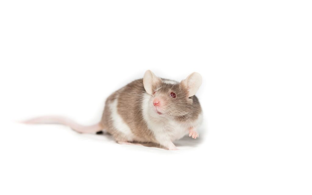
White said the ages of the mice were also important, with the young mouse aged four months, and the older mouse aged two years.

With follow-up during a two-month detachment period, the older animals exhibited improved physiological abilities and lived 10% longer than animals that had not undergone the procedure.

At the cellular level, parabiosis drastically reduced the epigenetic age of blood and liver tissue, and showed gene expression changes opposite to aging, but akin to several lifespan-extending interventions such as calorie restriction.

The rejuvenation effect persisted even after two months of detachment.

In human terms, the parabiosis exposure would be the equivalent of pairing a 50 year-old with an 18-year-old for about eight years, with the effects adding eight years to the person’s lifespan.

White said the experiment was designed to study if long-term exposure of young blood will cause lasting effects in the old mouse. Pairing humans for heterochronic parabiosis is obviously not practical or even ethical, he said. He also noted that other anti-aging strategies, such as calorie restriction, work better to extend longevity in mice.

“Our work points to a need to explore what factors in the circulation of youthful blood cause this anti-aging phenomenon” White said. “We have demonstrated that this shared circulation extends life and health for the older mouse, and the longer the exposure, the more permanent the changes.

“The elements that are driving this are what’s important, and they are not yet known,” White said. “Are they proteins or metabolites? Is it new cells that the young mouse is providing, or does the young mouse simply buffer the old, pro-aging blood? This is what we hope to learn next.”

Source: Duke University Medical Center

Metformin Also Seems to Protect Against Muscle Atrophy and Fibrosis

Photo by Barbara Olsen on Pexels

Diabetes and muscle function might seem like they don’t have much to do with each other. But University of Utah Health researchers have discovered that metformin can also prevent muscle atrophy and muscular fibrosis – which can help the elderly bounce back faster from injury or illness. Their findings were published in the journal Aging Cell.

Metformin, the researchers found, actually has surprising applications on a cellular level. It can target senescent cells which impact muscle function. Senescent cells secrete factors associated with inflammation that may underlie fibrotic tissue, a hardening or scarring of tissues. They also discovered that metformin also reduces muscle atrophy.

“We’re interested in clinical application of this research,” says Micah Drummond, PhD, senior author of the study and professor of physical therapy and athletic training at the College of Health. “For example, knee surgeries in the elderly are notoriously hard to recover from. If we give a metformin-type agent during the recovery period, could we help the muscles get back to normal faster?”

Reinvigorating muscle recovery

Ageing comes with the risks of falls, hospitalisation, or developing chronic disease, which are more likely with muscle disuse. The research team wanted to find a therapeutic solution that could properly target both disuse atrophy and muscle recovery.

There’s an optimal level of senescent cells that are beneficial, no matter your age. In younger, healthier people, short-term senescence is required for a proper recovery from injury, and completely blocking the senescent effect impedes the body’s efforts to heal. Typically, a younger person can bounce back more easily after muscle disuse without the use of an intervention such as Metformin.

“In the case of aging, we know that there’s immune dysfunction,” says Drummond. “As you get older, it becomes harder for your body to clear senescent cells and they accumulate. That’s one reason recovery is much slower for the elderly after periods of disuse.”

Metformin’s anti-senescent properties have been demonstrated through pre-clinical studies. To test the intervention in humans, the team recruited 20 healthy male and female older adults for a multi-week study. They had participants undergo a muscle biopsy and MRI before the intervention, which involved five days of bed rest. One group of 10 received metformin and the other 10 received placebo pills during a two-week run-in period, then each group continued the placebo or metformin treatment during bed rest.

After the bed rest, participants received another muscle biopsy and MRI, then ceased treatments. All patients completed a seven-day re-ambulation period followed by a final muscle biopsy.

“We saw two things in our study,” Drummond says. “When participants took Metformin during a bed rest, they had less muscle atrophy. During the recovery period, their muscles also had less fibrosis or excessive collagen. That build-up can make it harder for the muscle to properly function.”

Tying these results to senescence, the research team examined muscle biopsies from study participants. They found that the participants who took Metformin had fewer markers of cellular senescence.

“This is the first paper that has made the direct connection between a therapy targeting cellular senescence and improved muscle recovery following disuse in aging,” says lead author Jonathan Petrocelli, PhD He explains that metformin helps muscle cells better remodel and repair tissue during periods of recovery after inactivity.

“Our real goal is to have patients maintain their muscle mass and function as they age, because atrophy and weakness are some of the strongest predictors of disease development and death,” he says.

Drummond’s team is following up on these findings by examining combining the drug with leucine, an amino acid that promotes growth and could accelerate recovery even further. They’ve already demonstrated the potency of this combination in preclinical animal studies.

“Metformin is cheap, effective and quite safe, so it’s exciting to see that we can use it to accelerate recovery for older individuals,” adds Drummond.

Source: University of Utah Health

Rethink Preventative Aspirin for Older Adults, Researchers Say

Photo by cottonbro studio: https://www.pexels.com/photo/person-holding-white-round-medication-pill-4661296/

Low-dose aspirin is used as primary prevention for ischaemic stroke, but its protective effect weighed against the increased risk of bleeding events is controversial. A new secondary analysis of daily aspirin in older people found that, in this population, aspirin failed to reduce the risk for ischaemic stroke but increased it for intracranial bleeding. The findings were presented in JAMA Network Open.

The researchers analysed data from the ASPREE randomised clinical trial, the first large-scale trial to study the risks and benefits of 100mg daily aspirin in an older population, where increased bleeding risk may alter the balance of risks and benefits of aspirin. This is particularly relevant to intracerebral events because intracranial haemorrhage is harder to treat than ischaemic events and more frequently fatal or disabling. With previous aspirin trials in mostly younger participants, excess intracerebral haemorrhagic events was seen, though usually few in number and non-significant.

Cloud et al. performed a secondary analysis of the ASPREE trial, which included 19 114 older adults, and found a statistically significant 38% increase in intracranial bleeding resulting from a combination of haemorrhagic stroke and other causes of intracerebral haemorrhage among individuals randomised to aspirin. The difference in incidence of ischaemic stroke was not statistically significant.

While aspirin did not cause a statistically significant reduction in ischaemic stroke (hazard ratio [HR], 0.89), there was a a statistically significant 38% increase in intracranial bleeding. Rates of intracranial bleeding from those assigned to aspirin (1.1%) were higher than placebo (0.8%). This came from an increase in a combination of subdural, extradural, and subarachnoid bleeding with aspirin (0.6%) compared with placebo (0.4%). Haemorrhagic stroke was recorded in 0.5% of those assigned to aspirin compared with 0.4% for placebo.

Absolute numbers of haemorrhagic and non-haemorrhagic events were small. Among 1000 individuals taking 100mg/day of low-dose aspirin over five years, there were 2.5 fewer ischaemic strokes at the expense of 3.5 cases of intracranial haemorrhage, but not statistically significant. No difference would be expected for overall stroke incidence or stroke mortality, but haemorrhagic stroke was associated with a mortality rate of nearly a third, compared to 7.7% for ischaemic stroke. Major extracranial haemorrhage was driven by the increased risk of upper gastrointestinal bleeding with aspirin compared with placebo, as previously found (Hazard Ratio, 1.87).

The researchers concluded that “there was no statistically significant benefit from aspirin in preventing stroke or any conventional stroke etiological subtype. However, aspirin significantly increased the overall risk of intracranial bleeding.”

Black Adults Experience Less Cognitive Decline after Retirement

Photo by Barbara Olsen on Pexels

A study published in the Journal of the American Geriatrics Society found that immediately after retirement, white adults tended to experience a significant decline in cognitive function, whereas Black adults experienced minimal cognitive decline. White men showed the steepest post-retirement cognitive decline across sex/race combinations, whereas Black women showed the least decline.

White women performed better cognitively at retirement than other race/sex subgroups, and after retirement, their cognitive functioning declined at a rate that was slightly less than the average for this study. Results were adjusted for sociodemographics and physical and mental health indicators.

The study, which included 2226 US participants followed for up to 10 years, revealed greater post-retirement cognitive decline among individuals who attended college compared with those who did not.

“The results seem to point to the possibility that better job opportunities could lead to greater cognitive losses after retirement whereas exposure to lifelong structural inequalities may actually ease transition to retirement with respect to cognitive aging,” said lead author Ross Andel, PhD, of Arizona State University’s Edson College of Nursing and Health Innovation.

Source: Wiley

Scientists Discover That a Key Protein Boosts Cell Repair and Healthy Ageing

Photo by Ravi Patel on Unsplash

Researchers have found an anti-ageing function in a protein deep within human cells. They discovered that a protein called ATSF-1 controls a fine balance between the creation of new mitochondria and the repair of damaged mitochondria. Their findings were published in Nature Cell Biology.

Mitochondria create toxic by-products during their energy production process, which contributes to the rate at which the cell ages.

Associate Professor Steven Zuryn and Dr Michael Dai at the Queensland Brain Institute made the discovery of a key repair protein. “In conditions of stress, when mitochondrial DNA has been damaged, the ATSF-1 protein prioritises repair which promotes cellular health and longevity,” Dr Zuryn said.

As an analogy, Dr Zuryn likened the relationship to a race car needing a pitstop.

“ATSF-1 makes the call that a pitstop is needed for the cell when mitochondria need repairs,” he said.

“We studied ATFS-1 in C. elegans, or round worms and saw that enhancing its function promoted cellular health, meaning the worms became more agile for longer. They didn’t live longer, but they were healthier as they aged.”

“Mitochondrial dysfunction lies at the core of many human diseases, including common age-related diseases such as dementias and Parkinson’s. Our finding could have exciting implications for healthy ageing and for people with inherited mitochondrial diseases.”

Understanding how cells promote repair is an important step towards identifying possible interventions to prevent mitochondrial damage.

“Our goal is to prolong the tissue and organ functions that typically decline during ageing by understanding how deteriorating mitochondria contribute to this process,” Dr Dai said. “We may ultimately design interventions that keep mitochondrial DNA healthier for longer, improving our quality of life.”

Source: University of Queensland

Mouse Study Highlights Potential Therapeutic for Metabolic Syndrome

Photo by Diana Polekhina on Unsplash

Mopping up free radicals with antioxidants was a major health fad in the 1970s. In an effort to supposedly blunt the effects of aging and stave off chronic disease, people took huge amounts of antioxidants in the form of minerals and vitamins. Not only was this ineffective, it sometimes caused harm because untargeted antioxidants also compromised beneficial cellular signalling pathways. As theories of mitochondrial causes of disease fell out of favour, this health fad disappeared along with bell bottoms and disco.

Now, research recently published in Free Radical Biology and Medicine suggests a new way of dealing with free radicals: rather than mop them up, take a pill that selectively keeps them from being produced in the first place. Building on this work, collaborative research between the Buck and Calico Labs shows that specifically inhibiting free radical production at a particular mitochondrial site prevents and treats metabolic syndrome in mice, by preventing and reversing insulin resistance.

“We think that mitochondrial radical production drives many chronic diseases of aging, and that blocking the production of free radicals is a viable disease-treating and anti-aging intervention,” said Martin Brand, PhD, Buck Professor Emeritus and senior investigator of the study. “We’ve found a way to selectively keep problematic free radicals in check without compromising normal energy production in the mitochondria. These compounds act like a cork in a wine bottle. They plug a specific site so that it doesn’t produce free radicals, without hindering the mitochondria’s critical function of energy metabolism. We look forward to continuing this groundbreaking area of research.”

The orally bioavailable compound that has been developed, S1QEL1.719 (a new “S1QEL” – Suppressor of site IQ Electron Leak), was given both prophylactically and therapeutically to mice fed a high-fat diet that causes metabolic syndrome. Treatment decreased fat accumulation, strongly protected against decreased glucose tolerance and prevented or reversed the increase in fasting insulin levels by protecting against the development of insulin resistance.

Acting on mitochondrial complex I highlights potential interventions for other conditions

S1QEL1s act on site IQin mitochondrial complex I. (The mitochondrial electron transport chain consists of four protein complexes integrated into the inner mitochondrial membrane. Together they carry out a multi-step process, oxidative phosphorylation, through which cells derive 90% of their energy.)

First author and Buck staff scientist Mark Watson, Ph.D., says current literature strongly implicates complex I in a number of different diseases, from metabolic syndrome to Alzheimer’s, fatty liver disease, and noise-induced hearing loss, as well as the underlying aging process itself.

“S1QELs don’t sequester oxidants or radicals. Rather, they specifically inhibit radical production at the IQ site on complex I without interfering with other sites,” Watson said. “So the normal redox signaling that we require in our cells will continue. S1QELs just modulate that one site. They are very clean, very specific, and do not disrupt mitochondrial functioning like inhibitors of mitochondria do.”

Brand says the data shows that free radical production from complex I is an essential driver of insulin resistance and metabolic syndrome, a major disease of poor lifestyle choices and of aging. He says this feature is a strong reason to revisit the mitochondrial theory of aging. “These compounds fine-tune mitochondrial production of free radicals,” he said. “And it’s really interesting; just inhibiting this specific site improves the whole redox environment and prevents metabolic disease, and that is amazing.”

Source: Buck Institute for Research on Aging

SA Retirement Home Study Reveals the Mental Health Benefits for Residents Interacting with Children

A small South African study published in the open-access journal PLOS ONE suggests that programmes promoting interaction between retirement home residents and children may provide quality of life improvements and could help manage residents’ anxiety and depression.

Among retirement home residents, previous research has shown that common mental health conditions often go undetected and untreated. These conditions, which include anxiety and depression, are typically treated with a combination of drugs and non-pharmacological interventions.

One intervention is the Eden Alternative, which identifies loneliness, helplessness and boredom as key challenges to overcome provide a higher quality of life. Evidence suggests that programmes that enable older adults to regularly interact with children may improve mental health, but these have mostly been done outside of retirement homes and few have looked at such programmes in South Africa.

To deepen the understanding of potential benefits of intergenerational interactions, Elizabeth Jane Earl and Debbie Marais of Stellenbosch University, South Africa, conducted a study at a retirement home in South Africa. Residents were able to regularly interact with children who attend an onsite preschool. Activities include playing games, doing puzzles, reading, or singing with the children.

Ten female residents were recruited and invited to complete a questionnaire evaluating their anxiety and depression levels, as well as asking them to describe their experiences with the children. Four of the participants were screened as possible having anxiety, depression, or both. The participants all took part in the same interactions, though to varying degrees of participation.

Generally, the participants reported positive experiences with the children. Analysing their responses, the researchers found that the interactions fostered a sense of purpose and belonging, fond reminiscences of their own childhood and a positive influence on mood and emotions. Recollections of childhood also sparked a sense of playfulness and positive self-evaluation. They noted that the participants differed in their preconceptions of children, which might have affected their experiences.

The authors wrote that, “Interactions with children promote a sense of belonging and purpose, evoke reminiscence, and positively influence the mental well-being of older persons.”

Based on their findings, Earl and Marais concluded that intergenerational interaction programmes may help manage the mental health conditions that are common for retirement home residents. They suggest that trained staff facilitate the interaction, preparing the children and residents, and should be voluntary, which helps preserve the residents’ agency. Running the interaction as a regular programme should help build bonds and give the residents something to look forward to. Additionally, there should be an educational aspect for the children, giving the residents a sense of purpose.

Looking to the future, they wrote that larger studies would be able to better outline the benefits of such programmes.

Surgical Stabilisation of Odontoid Fractures Linked to Better Outcomes

Photo by Kampus Production on Pexels

In a review of patient treatment data, researchers have found that surgical stabilisation of odontoid fractures was associated with better outcomes than nonsurgical approaches. The article will appear in the September issue of Neurosurgery.

Odontoid fractures (C2 vertebra) are common in elderly patients after a low-energy fall. However, whether the initial treatment should be surgical or nonoperative still isn’t known. Previous studies haven’t accounted for differences in injury severity, or the presence or absence of neurologic impairment, which can affect patients’ results.

Michael B. Cloney, MD, MPH, of the Department of Neurological Surgery at Northwestern University in Chicago, and colleagues have published evidence that surgery should be considered as the initial approach for many patients. Compared with nonoperative approaches to treatment, surgical stabilisation of the fracture was associated with less myelopathy (mobility impairment due to spinal cord damage), and lower rates of fracture nonunion, 30-day mortality, and one year mortality.

“Given the increasing incidence of odontoid fractures with the aging population, we believe our findings could assist with neurosurgical decision-making for an increasingly common and complex problem,” the researchers say.

Accounting for nonrandomised patient groups

Dr Cloney and his colleagues reviewed initial treatment data on 296 patients who were cared for at Northwestern Memorial Hospital between January 1, 2010, and December 31, 2020, because of an odontoid fracture. Their average age was 73. During the hospitalisation, 22% had surgery and 78% had nonoperative treatment (5% were immobilised in a halo-vest and 73% received a cervical collar).

Since the patients weren’t randomised to these treatments, the research team used a type of analysis called propensity score adjustment. They calculated “propensity scores” for each individual – the probability that the patient would have been assigned to receive one of the two treatment approaches based on certain characteristics.

For example, to study the effect of surgery on mortality rates, patients were matched on age, sex, Injury Severity Score, Nurick score (a measure of myelopathy), their number of chronic diseases and chronic conditions such as smoking, and whether they had to be admitted to the intensive care unit.

Surgical stabilisation leads to better results

Follow up with patients lasted an average of 45 weeks. On the propensity score–matched analyses, the group that underwent surgery showed significantly better outcomes than the nonoperative group:

  • Lower rate of fracture nonunion – 39.7% vs 57.3%; treatment effect, 15% less risk of nonunion
  • Lower 30-day mortality rate – 1.7% vs 13.8%; treatment effect, 10% less risk of death
  • Lower one year mortality rate – 7.0% vs 23.7%; treatment effect, 10% less risk of death

Other analyses showed patients in the surgery group were 52% less likely than those in the nonoperative group to have poor Nurick scores at the 26-week postoperative follow-up visit and were 41% less likely to die during the overall follow-up period. Both differences were statistically significant.

“The mortality benefit calculated in the existing literature typically represents an unadjusted mortality rate between two potentially different populations, which leaves it liable to confounding,” the authors note. “Our study represents a relatively large institutional series that suggests a benefit from surgical stabilisation in this population while controlling for confounding factors more thoroughly than existing literature.”

Source: EurekAlert!

Rethink Needed for the Genetic Cause of Very Early Menopause

Photo by Ravi Patel on Unsplash

A new study showed that it may be necessary to rethink the genetic cause previously held to be behind very early menopause. Until now, variants in any one of more than 100 genes were thought to cause premature ovarian insufficiency (POI), which results in menopause before age 40. This affects around 1% of women, making it a leading cause of infertility. Under current guidance, a variation in one of these genes is cause for clinicians to consider a genetic diagnosis of POI.

Now, in the largest study to date, published in Nature Medicine, researchers analysed genetic data from more than 104 733 women in UK Biobank, of whom 2231 reported experiencing menopause before the age of 40.

The study found evidence that 98% of women carrying variations in the genes that were previously considered to be causes of premature menopause in fact had menopause over 40, therefore ruling out a diagnosis of POI in these women.

Anna Murray, Professor of Human Genetics at the University of Exeter Medical School is a senior author on the study. She said: “Our research means rethinking what causes very early menopause. The presence of specific genetic variants in multiple women who experience premature menopause has led to the assumption that they are causing the condition – but we have shown that these gene variations are also found in women with a normal age of menopause and therefore in many cases the link could just be coincidence. It now seems likely that premature menopause is caused by a combination of variants in many genes, as well as non-genetic factors. As genomic medicine evolves, we need to apply this standard of evidence to other conditions, so we can tailor diagnosis, treatment and support.”

Dr Julia Prague, Consultant Endocrinologist and Clinical Academic at the University of Exeter, and an author on the paper, said: “Having a very early menopause is often extremely distressing because it means losing fertility and treatment with hormone replacement is required to prevent negative health consequences. Clinicians need to understand the reasons why premature menopause occurs so that they do not miss the true underlying cause and can counsel patients appropriately. Misinterpreting genetic tests could have negative implications for women, such as suggesting that their relatives may also be at risk of very early menopause due to their genes, when in fact they may not be.”

Stasa Stankovic, of the University of Cambridge’s MRC Epidemiology Unit, and co-lead analyst of the study, said: “Each woman’s unique genetic combination shifts menopause timing, either earlier or later. Although genetic variation in the studied genes were not sufficient to cause very early menopause, we did identify genetic drivers that had a much more subtle impact on reproductive longevity. For example, women carrying genetic variation in TWNK and SOHLH2 genes experienced menopause up to three years earlier than the general population. Our future studies will continue using the power of human genomics to better understand the underlying biology of reproductive ageing in women and key genetic drivers of its extreme forms, including very early menopause. With this knowledge, we are also paving the path towards development of next-generation treatments for reproductive disorders.”

Source: University of Exeter

Losing the Y Chromosome to Age Drives Bladder Cancers but Improves Immunotherapy

Chromosomes. Credit: NIH

As men age, some of their cells lose their Y chromosome and this loss hampers the body’s ability to fight cancer, according to new research from Cedars-Sinai Cancer. The study, published in Nature, found that loss of the Y chromosome helps cancer cells evade the immune system, resulting in aggressive bladder cancer. Somehow, this also renders the disease more responsive to immune checkpoint inhibitors.

Based on their research, investigators are developing a test for loss of the Y chromosome in tumours with the goal of helping clinicians tailor immune checkpoint inhibitor treatment for male patients with bladder cancer.

“This study for the first time makes a connection that has never been made before between loss of the Y chromosome and the immune system’s response to cancer,” said corresponding author Dan Theodorescu, MD, PhD, who initiated the research. “We discovered that loss of the Y chromosome allows bladder cancer cells to elude the immune system and grow very aggressively.”

Lead collaborators on the study also included Johanna Schafer, a postdoctoral fellow, and Zihai Li, MD, PhD, medical oncologist and immunologist, both at The Ohio State University Comprehensive Cancer Center-James Cancer Hospital and Solove Research Institute.

In men, loss of the Y chromosome has been observed in several cancer types, including 10%–40% of bladder cancers. Loss of the Y chromosome also has been associated with heart disease and Alzheimer’s disease.

The Y chromosome contains the blueprints for certain genes. Based on the way these genes are expressed in normal cells in the bladder lining, investigators developed a scoring system to measure loss of the Y chromosome in cancers.  

The investigators then reviewed data on two groups of men. One group had muscle invasive bladder cancer and had their bladders removed, but were not treated with an immune checkpoint inhibitor. The other group participated in a clinical trial and were treated with an immune checkpoint inhibitor. They found that patients with loss of the Y chromosome had poorer prognosis in the first group and much better overall survival rates in the latter.

To determine why this happens, investigators next compared growth rates of bladder cancer cells from laboratory mice.

Cancer cells were grown in vitro and not exposed to immune cells. The researchers also grew the diseased cells in mice that were missing T-cells. In both cases, tumours with and without the Y chromosome grew at the same rate.

In mice with intact immune systems, tumours lacking the Y chromosome grew at a much faster rate than did tumours with the intact Y chromosome.

“The fact that we only see a difference in growth rate when the immune system is in play is the key to the ‘loss-of-Y’ effect in bladder cancer,” Theodorescu said. “These results imply that when cells lose the Y chromosome, they exhaust T-cells. And without T-cells to fight the cancer, the tumor grows aggressively.”

Based on their results derived from human patients and laboratory mice, Theodorescu and his team also concluded that tumours missing the Y chromosome, while more aggressive, were also more vulnerable and responsive to immune checkpoint inhibitors. This therapy, one of the two mainstay bladder cancer treatments available to patients today, reverses T-cell exhaustion and allows the body’s immune system to fight the cancer.

“Fortunately, this aggressive cancer has an Achilles’ heel, in that it is more sensitive than cancers with an intact Y chromosome to immune checkpoint inhibitors,” said co-first author Hany Abdel-Hafiz, PhD, associate professor at Cedars-Sinai Cancer.

Preliminary data not yet published shows that loss of the Y chromosome also renders prostate cancers more aggressive, Theodorescu said.

“Our investigators postulate that loss of the Y chromosome is an adaptive strategy that tumour cells have developed to evade the immune system and survive in multiple organs,” said Shlomo Melmed, MB, ChB, dean of the Medical Faculty at Cedars-Sinai. “This exciting advance adds to our basic understanding of cancer biology and could have far-reaching implications for cancer treatment going forward.”

Further work is needed to help investigators understand the genetic connection between loss of the Y chromosome and T-cell exhaustion.

“If we could understand those mechanics, we could prevent T-cell exhaustion,” Theodorescu said. “T-cell exhaustion can be partially reversed with checkpoint inhibitors, but if we could stop it from happening in the first place, there is much potential to improve outcomes for patients.”

While women do not have a Y chromosome, Theodorescu said these findings could have implications for them as well. The Y chromosome contains a set of related genes, called paralogue genes, on the X chromosome, and these might play a role in both women and in men. Additional research is needed to determine what that role might be.

“Awareness of the significance of Y chromosome loss will stimulate discussions about the importance of considering sex as a variable in all scientific research in human biology,” Theodorescu said. “The fundamental new knowledge we provide here may explain why certain cancers are worse in either men or women, and how best to treat them. It also illustrates that the Y chromosome does more than determine human biologic sex.”

Source: Cedars-Sinai Medical Center